
Abstract
Purpose:
To report our preliminary techniques and experience with transumbilical laparoendoscopic single-site renal pedicle lymphatic disconnection (LESS-RPLD) in seven patients with refractory chyluria.
Patients and Methods:
Between June 2009 and September 2010, seven patients with refractory chyluria underwent LESS-RPLD. In the patients, a 2- to 3-cm single inverted U-shaped supraumbilical incision was made, and a homemade single multichannel port using a surgical glove and three conventional trocars was placed into the abdominal cavity. Flexible electric coagulation hook and pliers were used for renal pedicle dissection. A straight ultrasound knife was used for lymphatic disconnection.
Results:
All the operations were successfully completed without conversion to open surgery, although an additional 3-mm trocar was used to push the liver in one patient. The mean operative time was 125 (96–165) minutes. The mean blood loss was estimated to be 112 (50–250) mL. Chyluria disappeared in all patients after surgery and did not recur during the follow-up period (3–15, mean 8.3 mos).
Conclusion:
LESS-RPLD is safe and feasible, with favorable short-term outcomes and aesthetic effect.
Introduction
To further reduce the number of puncture sites in the abdominal wall and the incidence of complications, and to achieve a better aesthetic effect, laparoendoscopic single-site (LESS) surgery has been adopted by urologists since 2007. To date, the use of LESS for nephrectomies, pyeloplasties, and donor nephrectomies has been reported. 7 –11 There have been no reports on the use of LESS in RPLD, however. In the current study, we present our preliminary techniques and experience with transumbilical LESS -RPLD in seven patients with refractory chyluria.
Patients and Methods
Between June 2009 and September 2010, seven patients (three men and four women) with refractory chyluria underwent LESS-RPLD. The median patient age was 49.3 years (range 31–63 y), and the median body mass index (BMI) was 18.46 kg/m2 (range 14.57–24.52 kg/m2). All patients lived in filariasis-prevalent regions and had a history of chyluria for 2 to 40 years. The main symptoms included chyluria, emaciation, and anemia.
Acute urinary retention developed in four patients because of urethral obstruction by chyle masses, and five patients had various degrees of gross hematuria. All patients had a positive urine chyle test. Based on cystoscopy and collection of renal pelvic urine after a fatty meal, chyluria was determined to arise from the left pelvis in five patients and the right pelvis in two patients. If ureteral catheterization failed to establish a locational diagnosis, retrograde ureteropyelography was used to demonstrate dilated fistulous connections between the urinary system and retroperitoneal lymphatics (Fig. 1).
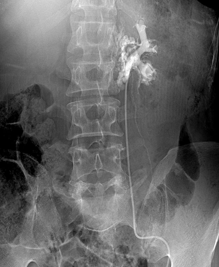
Retrograde ureteropyelography demonstrates dilated fistulous connections between the urinary system and the retroperitoneal lymphatics.
Operative technique
Under general anesthesia and airway management, all patients were placed in the conventional flanked kidney position with the ipsilateral side elevated. A 2- to 3-cm, single, inverted, U-shaped supraumbilical incision was made, and an electrotome was used to cut the abdominal tissues down to the peritoneum. A single working channel was established with a finger, and one 10-mm and two 5-mm trocars were inserted through the channel. A medical rubber glove was used to seal the space between the incision and trocars. A pneumoperitoneum was established with CO2, and the intraperitoneal pressure was maintained at 12 to 15 mm Hg. A 10-mm, 30-degree laparoscope or a 5-mm flexible laparoscope was inserted through the observation channel, and a pair of flexible separating pliers and an electric coagulation hook were inserted through the other two working channels (Autonomy™ Laparo-Angle™; Cambridge Endo, Framingham, MA; Fig. 2).

Overview of the homemade, single-port device.
Liberation of the ureter and inferior pole of the kidney
The lateral peritoneum was incised along the paracolic sulci, and the colon was liberated and pushed away to expose and liberate the upper segment of the ureter to the inferior pole of the kidney. During this process, all lymphatic and fatty tissues that were medial to the ureter and inferior pole of the kidney were cut with an ultrasound knife (Harmonic Scalpel, Ultracision®, Ethicon Endo-Surgery, Cincinnati, OH).
Renal pedicle lymphatic disconnection
On the left side, the splenic flexure of the colon and tail of the pancreas were liberated. On the right side, the hepatic flexure of the colon and duodenum were liberated to expose the renal vein. The vein sheath was incised with an ultrasound knife, then an aspirator was used to bluntly dissect the lymphatic vessels, which were then closed and cut with an ultrasound knife (Fig. 3). The renal artery was then liberated bluntly behind the renal vein with the aspirator, and the artery sheath and surrounding lymphatic vessels were coagulated and cut with an ultrasound knife in a low-speed position. Finally, all lymphatic vessels and fatty tissues around the renal pedicle that connected the renal hilum and abdominal aorta were cut. During the operative procedure, a prebent instrument was used to lift the inferior pole of the kidney to expose the renal pedicle (Fig. 4).

Disconnection of lymphatic vessels around the renal vein.

Intraoperative use of a prebent instrument for lifting the inferior pole of the kidney to expose the renal pedicle.
A 14F silicone tube, which is inserted through the only umbilical incision, was used for abdominal cavity drainage in the first five patients. The drain tube was pulled out 24 hours after surgery if the drainage did not increase in volume.
Results
All of the operations were completed successfully without conversion to open surgery, although an additional 3-mm trocar was used to push the liver in one patient. The mean operative time was 125 minutes (range 96–165 min). The estimated mean blood loss was 112 mL (range 50–250 mL). The chyluria resolved in all patients immediately postoperatively. The mean hospital stay was 3.5 days (range 2–7 d). Lymphatic leakage, which developed in one patient, resolved spontaneously after 7 days of drainage. Because of the prolonged retention of the drainage tube, the patient had delayed healing of the umbilical incision. The complication rate was 14.2% (1 of 7, grade 1) according to the modified Clavien classification system. No complications, such as renal vessel injury, urine leakage, or injury to adjacent organs, occurred in these patients. One month after surgery, only a 2.5-cm scar that was difficult to find formed in the patients' navel (Fig. 5). Chyluria did not recur during the follow-up period (range 3–15 months; mean, 8.3 months).
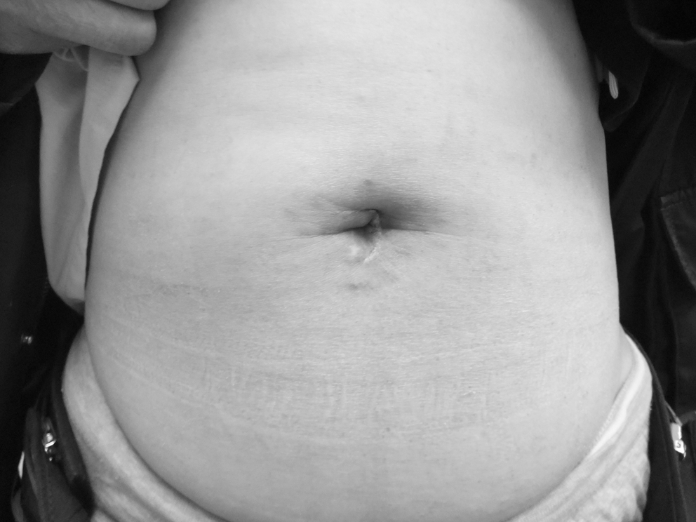
One-month postoperative image of the periumbilical incision.
Discussion
Lymph fluid in the cisterna chyli is called chyle because it contains lipid particles that are absorbed by the intestines. If lymph fluid flows from the cisterna chyli into the lymphatic vessels around the renal pedicle and a lymphopelvic fistula forms, chyle will enter the urine, resulting in ivory white urine—ie, chyluria. Parasitosis is the main underlying cause of chyluria. Patients with Bancroft filariasis usually present with chyluria at a late stage of the disease. Rarely, retroperitoneal lymphatic vessel obstruction resulting from tuberculosis or invasive tumors may cause chyluria. Although rare in the West, chyluria caused by Bancroft filariasis is not uncommon in Asia. Bancroft filariasis is recognized as a tropical disease more prevalent in rural and poverty-stricken populations. The seven patients who were included in this study lived in filariasis-prevalent regions.
The pathogenesis of chyluria is thought to involve dead filariae in lymphatic vessels that lead to mechanical damage, allergic inflammation, and a disturbance in lymphatic return. As a result, chyle flows back into the distal lymphatic vessels and lymphatic vessels around the renal pedicle, and finally lymphopelvic fistulae form. 6
Generally, chyluria resolves spontaneously in most patients after palliative treatment, such as bed rest and a low-fat, low-protein diet. Sclerotherapy, in which the renal pelvis is lavaged with a sclerosing agent, has been used for many years with occasional success. 4 Patients with a long history of serious chyluria usually have complications, such as severe anemia and hypoproteinemia. The seven patients in this group presented with emaciation, anemia, and hypoproteinemia. In addition, multiple episodes of urinary retention developed in four patients because of obstruction by chyle masses. Therefore, surgical intervention is necessary for refractory chyluria.
Traditional open surgical options include nephrectomy, renal capsulectomy, renal autotransplantation, RPLD, transinguinal spermatic lymphangiovenous anastomosis, and inguinal lymph node-saphenous vein anastomosis. 3 RPLD is thought to be the best surgical option based on a cure rate of up to 99%. 1 Nevertheless, conventional open surgery necessitates a long lumbar incision and may interfere with the kidney blood supply. Furthermore, because of limited visualization, fine lymphatic vessels may be missed, which directly causes early recurrence. 3
Chiu and colleagues 2 were the first to report the success of laparoscopic RPLD. Further reports have demonstrated that laparoscopic RPLD via peritoneal or retroperitoneal access has the same cure rate as open surgery, and laparoscopic RPLD has advantages, such as a magnified view, better identification of lymphatics, better anastomatic results, minimal morbidity, shorter hospital stay, excellent cosmesis, and early return to work. 3 –6
With the aim of preventing port-site complications, further decreasing discomfort associated with traditional laparoscopic surgery, and improving aesthetics, natural orifice translumenal endoscopic surgery (NOTES), and LESS have recently been developed. 7 NOTES, however, is still under investigation because of technical limitations. LESS is similar to traditional laparoscopic surgery in terms of the visual field and procedures. Moreover, the incisional scar can be concealed within the umbilicus. Therefore, LESS has been rapidly accepted by surgeons and has proved to be immediately applicable clinically.
Transumbilical LESS has been used successfully for nephrectomies since 2007. 8 LESS is also used for pyeloplasties, adrenalectomies, living donor nephrectomies, and radical nephrectomies. Indeed, clinical experience has demonstrated the safety and feasibility of LESS. 9 –13 LESS is especially indicated for patients with a low BMI who have a high demand for incision aesthetics, and operations do not need to extend the initial umbilical incision for specimen extraction. 11,13
Patients with refractory chyluria always suffer malnutrition; therefore, the questions involving BMI and extension of the umbilical incision do not exist in RPLD. LESS is a good choice for these patients; however, there have been no reports supporting the use of LESS in RPLD. Based on LESS in >100 patients, we have gained experience in the establishment of single ports and the use of flexible instruments through LESS. In particular, we performed 13 nephrectomies using LESS and mastered the management of the renal pedicle in LESS surgery.
Considering that the costs of single-port devices limit the wide application of LESS, 7 we used homemade, single-port devices in these patients within this group. Although homemade, single-port devices were not as good as commercially available devices, such as the Tri-Port, in terms of air-proof performance, homemade, single-port devices were easy to prepare and economical. Han and coworkers 14 also reported the use of homemade, single-port devices.
Flexible instruments and traditional straight instruments can be used interchangeably in LESS-RPLD, just as in LESS nephrectomy. The force applied on the tip of the flexible instruments, however, dissipates along the flexible shaft and makes dissection more demanding. 13 In nephrectomy or RPLD, it is difficult to lift the inferior pole of the kidney to fully expose the renal pedicle using the currently available flexible instruments. To solve this problem, we suspend the inferior pole of kidney onto the abdominal wall with sutures during LESS nephrectomies. In this group of patients, after having liberated the ureter and inferior pole of the kidney, we used a prebent instrument to lift the inferior pole of the kidney, thus fully exposing the renal pedicle.
Hemal and Gupta 4 divided the procedure of RPLD under traditional laparoscopy into five steps: Nephrolympholysis; hilar stripping; ureterolympholysis; fasciectomy; and nephropexy. In our experience involving 55 cases of traditional laparoscopic RPLD, we think that only the first three steps are indispensable, and that hilar stripping is key to the entire procedure.
In patients with chyluria, it is usually difficult to dissect lymphatic vessels of the renal pedicle because the lymphatic vessels are thickened from congestion and inflammation. To manage lymphatic vessels, we usually lift the inferior pole of kidney and ureter first, and use an aspirator to bluntly liberate lymphatic vessels along the blood vessels of the renal pedicle, and then use an ultrasound knife to cut the lymphatic vessels. Thick lymphatic vessels can be coagulated and cut with an ultrasound knife in a low-speed position effectively and safely. Titanium clamps were not used, because titanium clamps may impede further dissection and disconnection. Hilar stripping should be performed carefully in different views with a flexible laparoscope, and renal vessels and upper ureteral segment should be completely skeletonized on the hilar plane. Given complete dissection of perirenal fat and medial tissues on the hilar plane, it is not necessary to dissect the kidney from the perirenal fat. Intact splenorenal and hepatorenal ligaments will prevent nephroptosis after surgery. The right renal pedicle is covered by the liver, which makes exposure difficult. If necessary, a 3-mm trocar may be applied beneath the costal margin to push away the liver. If lymph vessels are ligated completely, the placement of drain tubes is not a must. Small amounts of lymph fluid can be absorbed by the peritoneum.
Currently, the case number of LESS is limited worldwide. Thus, it seemed that the benefits of LESS are not evident at present, with the only claimed advantage being cosmetic. 15 Nevertheless, LESS is readily accepted by patients for the aesthetic effect. With more cases of LESS, more potential benefits of LESS, such as less psychic trauma, should be confirmed. LESS should have great potential as a new variant of laparoscopic procedures with fewer scars.
Conclusion
We report our initial clinical experience with LESS-RPLD. All operations were completed successfully. LESS-RPLD is safe and feasible for refractory chyluria treatment, and short-term follow-up has demonstrated the efficacy of LESS-RPLD. Nevertheless, more clinical experience is needed to show whether LESS can replace traditional laparoscopic surgery in the treatment of patients with refractory chyluria.
Footnotes
Disclosure Statement
No competing financial interests exist.