
Abstract
Introduction:
We report a redesigned magnetically anchored and guided systems (MAGS) camera with improved optics and a 30° downward viewing angle that facilitates solitary surgeon laparo-endoscopic single site (LESS) nephrectomy.
Methods:
The prototype consists of an external 2 by 6 cm cylinder containing magnets positioned such that the intra-abdominal camera is anchored on the peritoneal surface with a default 30° downward angle. It was inserted through a 3 cm LESS-port periumbilical incision in three pigs (mean 47 kg). The camera was coupled with the handheld magnetic device across the anterior abdominal wall and was steered into position to view the kidney. LESS nephrectomy was then performed.
Results:
Since a standard LESS laparoscope was not needed, only the two operative instruments were inserted through the single incision port, significantly decreasing instrument “clashing” compared with traditional LESS nephrectomy. Due to the favorable angle of view of the camera and its self-anchoring capability, no assistant was needed to drive the camera. Instead, the surgeon periodically made minor adjustments to optimize the view. The nephrectomy was completed without complication in an average of 35 minutes in these three nonsurvival animals.
Conclusion:
The MAGS camera provides good optics and easy maneuverability during LESS porcine nephrectomy. As with other MAGS instruments, by replacing a traditional transabdominal laparoscope that occupies access port space, use of this insertable camera may diminish some of the “collision challenge” of LESS surgery. Also, by being self-anchoring, this prototype may minimize the need for an assistant surgeon.
Introduction
If one or more of the surgical instruments could be “untethered” or removed from the port site, the ergonomics and feasibility of LESS and NOTES would improve. In this vein, we have developed and reported on a technology termed magnetically anchored and guided systems (MAGS) that consists of magnetic instruments that, once inserted through an access port, can be guided into position inside the peritoneal cavity through magnetic attraction to an external, handheld magnet. 1 –5 We have recently designed a novel MAGS camera with improved optics and a 30° downward viewing angle. We were interested in finding whether this viewing angle, in combination with the self-anchoring nature of this particular MAGS instrument, might facilitate LESS surgery and eliminate the need for a camera-driving assistant. We evaluated the utility of this camera to perform porcine LESS nephrectomy.
Methods
The MAGS camera prototype consists of paired magnetic devices, an internal component which is the actual tool inserted into the peritoneal cavity through a laparoscopic port site, and an external magnetic “handle” which couples via magnetic forces across the abdominal wall to the internal instrument (Fig. 1). The camera is then positioned and suspended from the anterior abdominal wall like a chandelier and is held in position via magnetic coupling (Fig. 2).
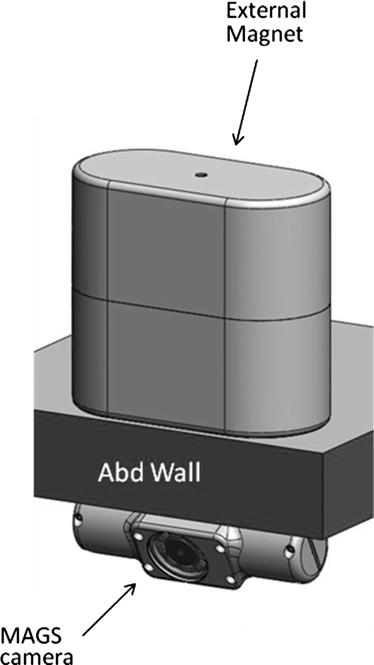
Cartoon drawing depicting the external magnetic handle coupled across the abdominal wall to the MAGS camera. Note the set 30° downward angle of the camera component. MAGS, magnetically anchored and guided systems.
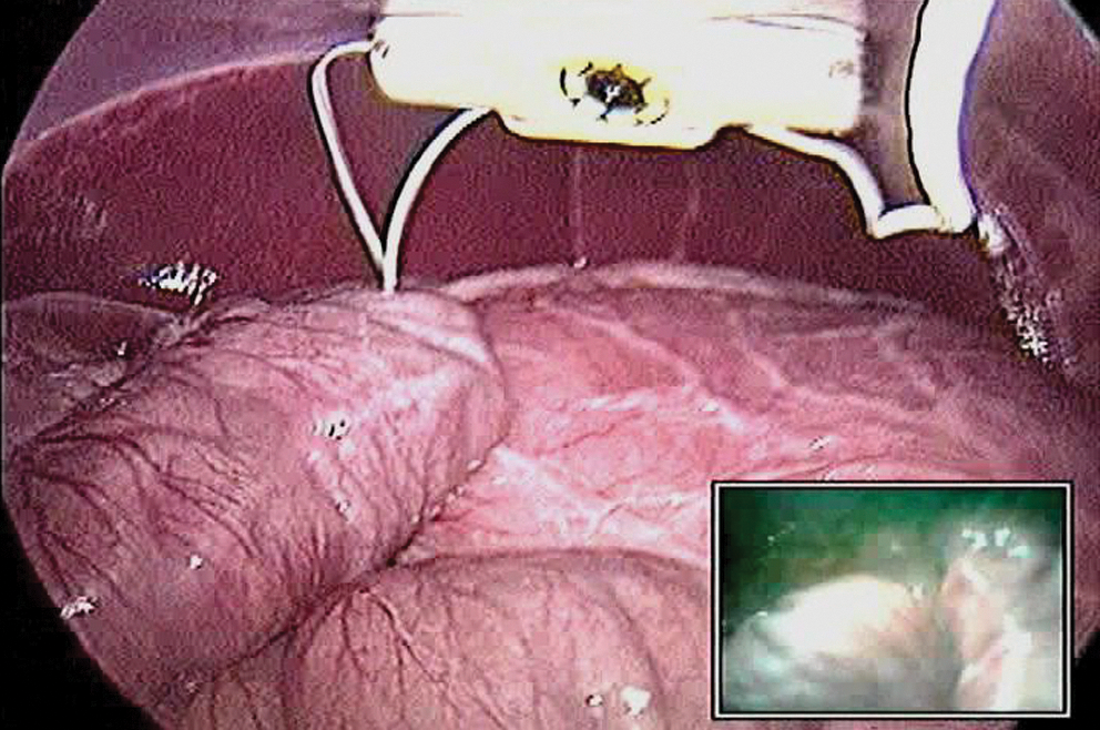
The novel MAGS camera in place inside the peritoneal cavity, suspended from the anterior abdominal wall. The picture-in-picture view is out of focus due to extraneous light generated by the standard laparoscope being used to take the photo.
The MAGS camera used in this investigation is an ∼2×6 cm cylinder with the imager/lens centered along the long axis (Figs. 1 and 2). The imaging device itself is made by Misumi Electronics Corporation (Jhonghe City, Taiwan) and consists of a ¼″ color CCIQ camera, 656×492 pixels, 400 TV lines contained in an 8 mm cube. The magnets in the device are positioned such that once coupled to the external magnetic handle, the camera sits at a 30° downward angle, mimicking the angled laparoscope traditionally used during conventional laparoscopic surgery. The device has its own on-board LED light array surrounding the central camera lens (Fig. 2).
After obtaining approval from our institutional animal care and use committee, three female farm pigs were utilized in this nonsurvival, pilot study. A 3 cm transperitoneal incision was made at the level of the umbilicus and the MAGS camera was then inserted into the peritoneal cavity through the incision. A single incision port device (Single Site Laparoscopy Access System; Ethicon Endosurgery, Inc., Cincinnati, OH), was then inserted alongside the camera's tether cable (providing transmission of video) and the abdomen was insufflated with carbon dioxide to 15 mmHg pressure (Fig. 3). A standard laparoscope was briefly inserted through the port to visualize the coupling between the camera and the external magnetic handle. This was accomplished by placing the handle on the abdominal wall and gently depressing it slightly until the camera is “pulled up” to the anterior peritoneal surface via magnetic attraction. The laparoscope was then removed and not used during the rest of the procedure. The MAGS camera was then manipulated into position by sliding the handheld magnet across the skin surface until the kidney could be visualized (Figs. 3 and 4).
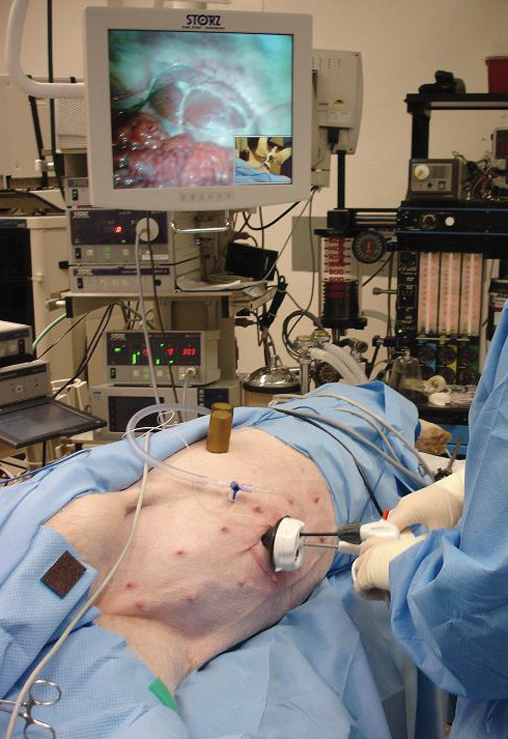
Intraoperative setup for solo surgeon laparo-endoscopic single site nephrectomy. The rust-colored object sitting on the pig's flank is the external magnet, holding the camera in place.

Image of the kidney generated by the MAGS camera.
LESS nephrectomy was performed using a 5 mm rigid straight Maryland grasper and an AutoSonix™ ultrasonic laparoscopic transducer (Covidien, Mansfield, MA) to incise the peritoneum and to dissect the renal vasculature such that it could be ligated with a 12 mm laparoscopic stapler. The kidney was freed from the perinephric attachments and the ureter transected. Since the camera was suspended from the anterior abdominal wall via magnetic forces, no camera-holding surgical assistant was required. Instead, the surgeon periodically used the extracorporeal magnetic handle to adjust the view if needed. Once the kidney was free and hemostasis was ensured, the kidney was grasped by the ureter and extracted through the surgical incision. The camera was removed from the peritoneal cavity via the tether after decoupling the external magnet from the skin surface.
Results
LESS nephrectomy was completed in all three animals without complication and without the use of a standard laparoscope beyond visualization of magnetic coupling, as described. Characteristics of the three operations are listed in Table 1. The image generated by the MAGS camera was excellent and sufficient to perform the operation (Fig. 4). Approximately once per procedure, the camera became fogged and was cleaned by steering the camera until it visualized the port, at which point a grasper holding a small piece of gauze was introduced, moved toward the camera, and used to clean the lens. Decoupling (inadvertent separation of the two magnetic components) did not occur during this investigation, but in our experience, recoupling is easily accomplished by gently depressing the external magnet to reduce the distance between the components such that recoupling occurs.
Due to decreased instrument conflict within the port (there was no laparoscope sharing the space), the surgeon was able to perform all procedures using only rigid, straight instruments. This resulted in a subjective sense of greater ease of the procedure compared with standard LESS nephrectomy with a 5 mm laparoscope and articulating instruments. There were no instrument collisions with the MAGS camera. Additionally, no camera-holding assistant was needed during the procedure since the MAGS camera was self-retaining and required only periodic adjustment by the surgeon. This improved the ergonomics at the bedside by reducing surgeon crowding.
Discussion
Recently, alternatives to conventional laparoscopy have emerged, namely NOTES and LESS surgery. These approaches attempt to minimize the invasiveness of surgery by decreasing the number of transabdominal ports used. Though the advantages of LESS are considered largely cosmetic at this point, some authors have demonstrated improvements in early postoperative convalescence as measured by duration of hospital stay or pain. 6 –8 Although other authors may not have found postoperative gains for LESS, they have demonstrated equivalence to standard laparoscopy in areas such as complication rates, 9 –11 operative time, 9 and duration of hospital stay. 7,9 –11 This demonstration of equivalence allows the consideration for cosmetic improvements to be weighted more heavily.
Despite these potential benefits, NOTES and LESS have not “taken off” in mainstream urologic practice. Indeed only one report of “pure” NOTES nephrectomy exists in the urologic literature, 12 though there are numerous papers on urologic LESS. The main reason for the failure of their widespread adoption is likely related to the significant technical challenges associated with these techniques. Passing all of the instruments including the camera through the same incision can lead to instrument collisions and “swordfighting,” and impaired tissue handling and retraction problems caused by the loss of instrument triangulation. Finally, especially with NOTES, unfamiliar viewing angles can significantly decrease surgeon comfort during the operation.
Several approaches have been used to attempt to address these challenges, including use of flexible endoscopes and articulating instruments that “bend away” from the midline, recreating a degree of triangulation. However, these tools themselves present their own technical challenges and are associated with a learning curve of their own. We have taken a different approach to the problem through the development of magnetic instrumentation termed MAGS. These tools consist of two parts: an internal magnetic instrument such as a camera, cautery dissector, or retractor that is inserted through a laparoscopic port site into the peritoneal cavity and then coupled across the abdominal wall to a second magnet, a handheld extracorporeal device that can be moved across the skin surface to move the internal device to any position within the peritoneal cavity. This technology essentially “decouples” the instruments from the incision site, meaning that the tools can be placed anywhere they are needed, regardless of where the port site is. We have previously reported using these instruments to perform LESS and NOTES porcine nephrectomies 3,5,13 and two human cases (appendectomy and simple nephrectomy). 2 Through their ability to be moved to a location remote from the incision site, these instruments can restore both a degree of triangulation and conventional viewing angles to LESS and NOTES. Additionally, they decrease clashing of the instruments that are still passed through the port site by eliminating one or more of the transabdominal devices.
The most recent innovation in this arena, the MAGS camera described in this report, maintains all of these advantages while offering additional benefits. The improved optics of this new generation camera provides a high quality image that is suitable for the fine dissection required for renal surgery. Additionally, the set 30° downward viewing angle, paired with its ability as a MAGS instrument to be positioned anywhere along the anterior peritoneal surface, allows the surgeon to work with the same viewing angle that would be achieved with conventional laparoscopy. As opposed to our first generation MAGS camera 2 where the imager and LEDs were at end of long axis of device, the imager in the current design is centered in the long axis (Figs. 1 and 2) with the internal magnets on either side. The downward viewing angle, in combination with the magnetic anchoring and the imager centered in the MAGS camera tool has an additional benefit of not requiring a camera-holding assistant. Since LESS is associated with internal (instrument) and external (surgeon) competition for limited space, this may provide a useful advantage.
Although this pilot study is small and limited to an animal model, our subjective experience is that the decreased instrument collisions and improved triangulation associated with the use of MAGS make LESS much easier to learn and perform. Recent investigations have demonstrated that these instruments do not cause tissue damage of the abdominal wall 14 and that the strength of the magnets is likely sufficient to span the distance of most human abdominal walls. 15 We are therefore hopeful that development of this technology may make widespread adoption of LESS and even NOTES more practical. Future studies will aim to objectively compare intra- and postoperative findings of both MAGS-assisted and conventional LESS surgeries.
Conclusion
The new generation MAGS camera provides excellent image quality and significantly improves the ergonomics of LESS by removing the camera from the transabdominal port space. Additionally, the unique self-anchoring capability of this novel technology may eliminate the need for an assistant during traditional laparoscopic and LESS operations.
Footnotes
Disclosure Statement
Jeffrey Cadeddu, Raul Fernandez, Richard Bergs, and Daniel Scott are consultants and investigators for Ethicon Endosurgery. This project was funded in part by Ethicon Endosurgery.