
Abstract
Background and Purpose:
Struvite in kidney stones is an important marker for infection. In kidney stone samples, struvite is known to be prone to chemical breakdown, but no data exist on the stability of samples stored in dry form. The objective of this study was to examine stability of struvite under increasingly poor conditions of storage.
Materials and Methods:
Samples of struvite kidney stones were broken to obtain 38 pieces averaging 67 mg in weight, and these were randomized into four storage conditions: Airtight containers stored in the dark, open containers in the dark, open containers in ambient light, and open containers at elevated temperature (40°C). Pieces were left for 6 months, and then analyzed for changes using micro CT and Fourier transform infrared spectroscopy (FT-IR).
Results:
Initial samples proved to be struvite, indicating no transformation in the large specimens that had been stored in airtight containers in the dark for more than 6 years before this study. Pieces of struvite taken from these large specimens appeared unchanged by micro CT and FT-IR after being stored in closed containers for 6 months, but 8 of 9 pieces in open containers showed the presence of newberyite in surface layers, as did 10 of 10 pieces in open containers out in ambient light. All pieces stored at 40°C showed transformation of struvite, with 60% of the pieces showing the presence of amorphous phosphates, indicating complete breakdown of struvite in the surface layers of the pieces.
Conclusion:
We conclude that struvite in dry kidney stone samples is stable when the specimens are stored in airtight containers at room temperature, even after several years.
Introduction
Thus, the assessment of the presence of struvite is an important part of normal stone analysis, and the indication of struvite should be of great consequence in guiding treatment of the patient. Nevertheless, there is evidence that the reporting of struvite in stone specimens can be inaccurate, either because of laboratory error or because the submitted specimen was nonrepresentative of the total stone composition. 7 In addition to the potential for error of struvite analysis, struvite also has the potential to disappear from stone samples; it has been shown that specimens of struvite, stored dry and at room temperature, can lose ammonia and change their mineral composition. 8 –11 One study found that loss of struvite can be detected over a period of only 6 days. 10
Retrospective studies in our laboratory sometimes necessitate the assessment of stone specimens that are several years old. We undertook the present study to find out if struvite is stable in stone specimens stored this long, and what conditions of storage would lead to loss of struvite from a stone.
Materials and Methods
Stone specimens were obtained as discards from a commercial stone laboratory (Beck Analytical Services, Indianapolis, IN). Although specimens were de-identified, they still carried the original analysis results, and three large specimens were found that had originally been analyzed as 100% struvite, at least 6 years previous to the present study. Two of these specimens had a very compact morphology by micro CT, 12 and the third consisted of struvite crystals ∼500 μm held together in a looser configuration. These stones were cleaved into pieces, which were again scanned by micro CT (Fig. 1), using the SkyScan 1172 system with voxel sizes ranging from 14 to 18 μm (using 50–60 kVp, 167–200 mA, and 0.5 mm Al filter). Pieces from each stone were verified as being similar by micro CT, and composition of a few pieces from each original stone were verified as struvite 13 –16 by Fourier transform infrared spectroscopy (FT-IR) using the KBr pellet method and a Bruker Alpha-T Spectrometer. This process resulted in 38 pieces of struvite that averaged 67 mg in weight (10 from one stone, 13 from another, and 15 from the third).

Typical pieces of struvite from the three stones used in this study.
The pieces were distributed randomly—using a spreadsheet “random” function—into four groups. In the first group, nine pieces were placed individually into airtight containers (snap-closed vials, internal diameter 36 mm, height 16 mm; from LA Packaging, Yorba Linda, CA), and stored in the dark at room temperature; this storage condition mimicked that of the stone analysis laboratory. The second group (nine pieces) were stored in the same dark cabinet as the first, but containers were left with lids open to the air. The third group (10 pieces) was in open containers and left out at room temperature in a west-facing window. The fourth group (10 pieces) was placed in open containers in an oven kept at ∼40°C. Thus, the four groups represented situations of increasingly poor storage conditions for the struvite specimens. At the end of 6 months, pieces were again scanned by micro CT and weighed; scrapings of the surfaces were taken from each piece and analyzed by FT-IR in a manner blinded to the storage site of the pieces.
Volumes of the pieces were measured on the micro CT image stacks, using ImageJ (
Statistics
Data were compared using analysis of variance, Tukey-Kramer Honestly Significant Difference test, contingency analysis with chi-square, or linear regression as appropriate. Statistical tests were performed using JMP 7 (SAS Institute, Inc, Cary, NC).
Results
Randomization of the struvite pieces into four groups was successful, with two-way analysis of variance showing no effects of stone source or initial storage site (P in all cases ≥0.5) on weight (0.067±0.038 g) or density (1.684±0.067 g/cc). The surface area to volume ratio averaged 2.40±1.02 μm2/μm3, and this did not differ among the four storage locations (P=0.4), but was significantly higher in pieces coming from the stone composed of struvite crystals loosely held together (as in panel C of Fig. 1) compared with pieces from the other two stones (3.78±0.98 μm2/μm3 for the 10 pieces from that source vs 1.85±0.38 and 1.95±0.39 for pieces from the other two stones; P<0.0001 by Tukey test).
After 6 months of storage, density was lower on average in pieces from all storage conditions; pieces stored in the dark/closed condition lost 5.4±4.6% of their density, in dark/open 3.6±4.7%, light/open 4.6±4.5%, but in pieces stored as 40°C/open the density loss was 16.4±8.8%, significantly greater than the other three groups (Tukey test, P<0.002). Density loss correlated with surface-area-to-volume ratio; that is, pieces with higher surface-area-to-volume tended to have higher rates of loss of density (P<0.004 for linear regression of all data), and this relationship was significant even when corrected for storage condition (two-way analysis of vairance, P<0.03).
Blinded observations of the micro CT images of the pieces before and after 6-month storage indicated visible changes only in the pieces stored at 40°C (Fig. 2), with all of those pieces showing a loss of x-ray attenuation in the regions nearest the surface.
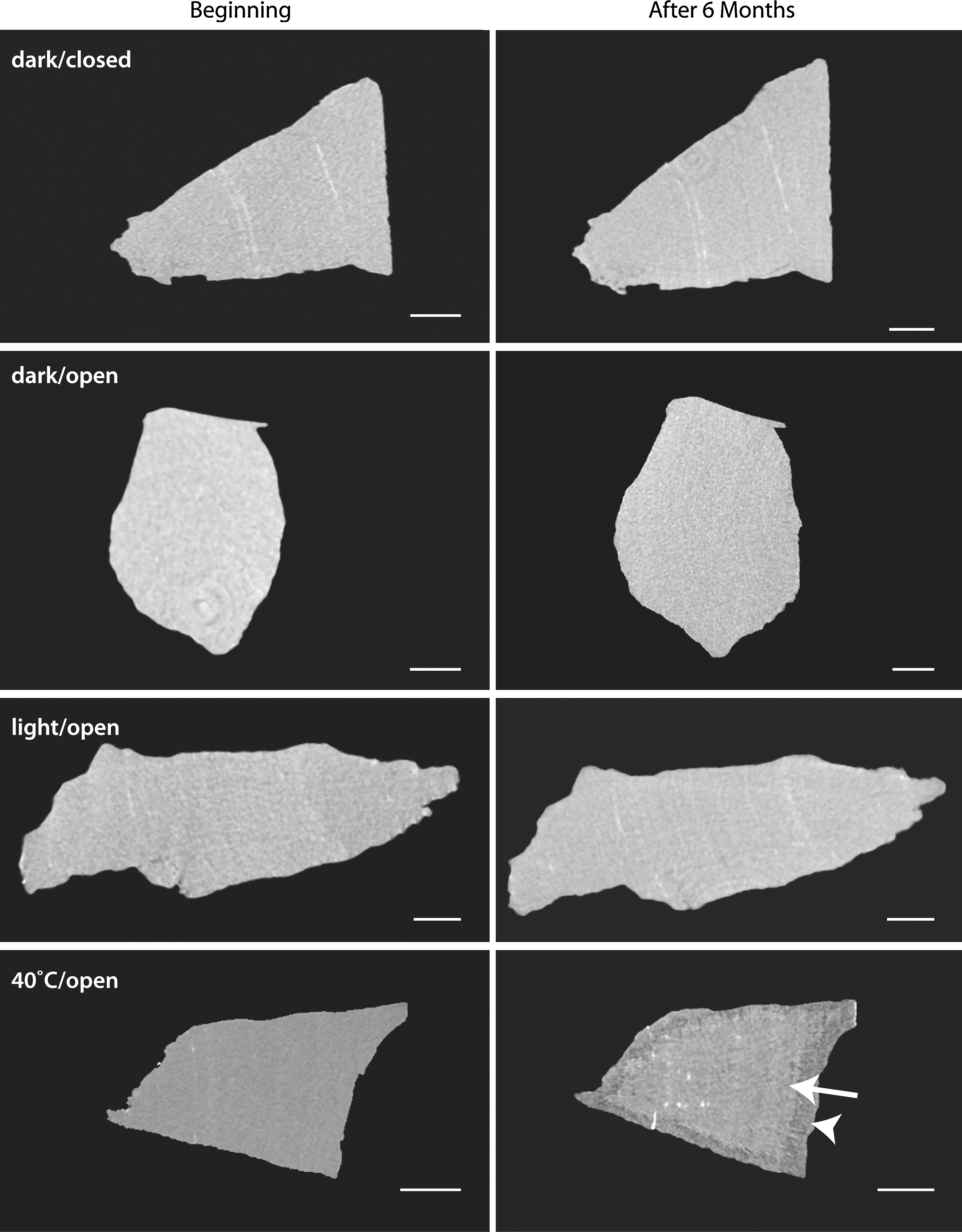
Representative micro CT slices of struvite stones used in this study. Scans show stones before and after 6 months of storage. Stones stored at 40°C developed distinct regions of changes in mineral content by Fourier transform infrared spectroscopy. Scans of these stones showed lower x-ray attenuation values around the outer edges of the stone (arrowhead) and attenuation values closer to the original values in the core of the stone (arrow). Bars indicate 1 mm.
FT-IR of scrapings from the surfaces of the pieces stored in closed containers in the dark showed them to be struvite (Figs. 3 and 4). In contrast, 8 of the 9 pieces stored in the dark in open containers showed the presence of newberyite in surface layers, and 10 of 10 of the pieces stored in open containers in the light showed similar transformation. For the pieces stored at 40°C, all showed transformation of struvite, and 6 of the 10 showed the presence of amorphous material, 18 which is the most dramatic evidence of transformation of struvite. 19 Samples from the interior of pieces, however, showed only struvite by FT-IR, regardless of storage condition.
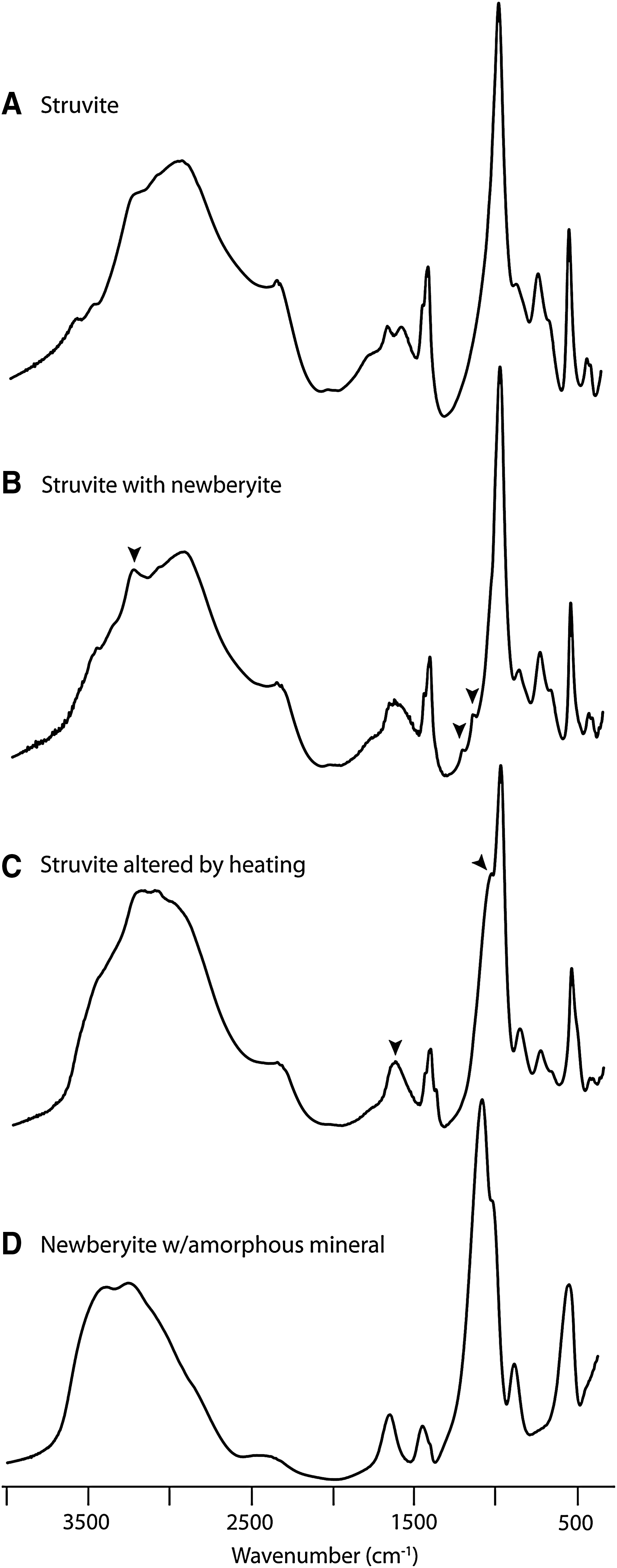
Representative Fourier transform infrared spectroscopy spectra for results in this study.
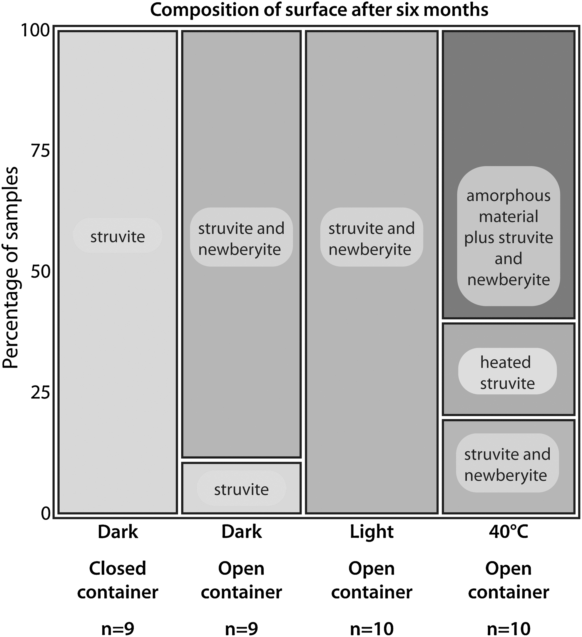
Results of infrared spectroscopy of material scraped from the surfaces of pieces of struvite stored for 6 months in various conditions. Contingency analysis (using the likelihood ratio) showed highly significant differences among the storage conditions, P<0.0001. Examples of spectra are shown in Figure 3: Struvite as in Figure 3, panel A; addition of newberyite as in panel B; heated struvite as in panel C; and spectra indicating amorphous mineral as in panel D.
Discussion
Evidence for loss of struvite from stone samples was first reported by Lonsdale and Sutor, 8 and shortly afterward, Whitaker 10 reported that struvite crystals in air at room temperature could show conversion to newberyite in as little as 6 days. The short time scales reported in the latter study suggest that loss of struvite content in kidney stone samples could lead to an underestimate of struvite content in subsequent analysis.
The evidence in the present study shows that transformation of struvite in kidney stone samples can indeed occur, but that this loss of struvite is prevented by proper specimen storage. The small pieces of struvite that were stored in sealed containers remained pure for the 6 months of the study, as is consistent with the results for the original (larger) specimens, which were originally analyzed as pure struvite more than 6 years before. Thus, kidney stone samples that are kept in airtight containers are unlikely to lose struvite, even in extended storage. A weakness in the present study was not testing specifically the effects of storing samples in closed containers, but in the light; the results in open containers, however, do not show much difference between specimens stored in daylight or in the dark, so it seems likely that the degradation of struvite is not especially accelerated by light conditions. We infer from this that samples stored in sealed containers but left out for a time in an office setting should have most struvite intact.
An unexpected finding was that all of the specimens—even those stored in sealed containers—lost density over time. These density measurements were based on weights and micro CT scans before and after the storage times, so that the density changes appear to be real. Thus, it appears that these small specimens, even in sealed containers, evaporated some material, although the total loss was less than one water per struvite molecule, and perhaps could have come from additional water held in the stone structure. This density loss did not result in any compositional changes visible by FT-IR, but it does point to the basic problem of mass loss that leads to composition change in this mineral. Presumably, in a sealed container, the gas surrounding the specimen eventually becomes saturated with the volatile materials so that the loss of struvite is slowed.
The present study does show that poor storage conditions—especially a high temperature, such as could be found in a warehouse in the summer—can result in loss of struvite from the specimen. In the milder examples of poor storage (such as in an unsealed container), however, the struvite is first replaced by newberyite, a mineral type that is readily recognized by FT-IR or x-ray diffraction.
It should be noted that newberyite—the primary mineral identified as appearing in poorly stored samples of struvite—can also appear in human stones and not as an artifact. 20,21 The rate of finding of newberyite in stones is very small (only 0.05% in the large study of Daudon et al. 20 ), however, so that its presence in a stone sample is most likely from improper storage. 5
A unique aspect of the present study is the use of micro CT, which allows the entirety of a stone specimen to be examined in a nondestructive manner. The sensitivity of micro CT can lead to the identification of heterogeneity within stones that is not detectable otherwise, 12,22,23 and such is the case in the present study. Figure 1 shows, for example, that by micro CT, all pieces of “pure” struvite showed regions of x-ray attenuation too high to be struvite 12 (Fig. 1). These nonstruvite regions always represented volumes <1% of the total, so they would not be detectable using FT-IR or x-ray diffraction analysis. 24 That is, such microscopic heterogeneity in stone composition is not readily detected by methods other than micro CT. These micro CT results are consistent with the statement by Reveillaud and Daudon 25 that “struvite stones from humans are never pure.” Apparently this is true even at the microscopic level.
Conclusion
Struvite in dry kidney stone samples is stable when the specimens are stored in airtight containers at room temperature in the dark, even after several years. Thus, there is no danger of struvite disappearing from a specimen of kidney stone if the stone is dried and kept in a sealed container; this is the standard for specimen storage in the commercial laboratories with which we are familiar. In addition, it should be possible to undertake retrospective study of older samples of stone from patients, even years after the specimens were collected, as long as the samples were stored in this manner.
Footnotes
Acknowledgments
This work was funded by NIH R01 DK059933. We thank Beck Analytical Services for providing stones for this work, and Dr. Amy Krambeck for helpful discussion.
Disclosure Statement
No competing financial interests exist.