
Abstract
Background and Purpose:
Drug-eluting stents proved to minimize neointimal hyperplasia in coronary vessels. Hyperplastic reaction is the most common unwelcome event related to the use of metal mesh stents in the ureter. We evaluated the effect of zotarolimus-eluting stent (ZES) Endeavor Resolute in the porcine and rabbit ureter.
Materials and Methods:
A ZES and a bare metal stent (BMS) were inserted in each ureter of 10 pigs and 6 rabbits. The insertion was performed by the retrograde approach. CT was used for the evaluation of porcine ureters while intraoperative intravenous urography (IVU) was used for rabbit ureters. The follow-up included CT or IVU every week for the following 4 weeks for pigs and 8 weeks for rabbits. Renal scintigraphies were performed before stent insertion and during the third week in all animals. Optical coherence tomography (OCT) has been used for the evaluation of the luminal and intraluminal condition of the ureters with stents. Histopathologic examination of the these ureters embedded in glycol-methacrylate was performed.
Results:
Hyperplastic reaction was present in both stent types. BMSs in seven porcine ureters were completely obstructed while porcine ureters with ZES stents had hyperplastic tissue that did not result in obstruction. Two rabbit ureters with BMS stents were occluded while no ZES was associated with ureteral obstruction. The function of the seven porcine renal units and the two rabbit units with obstructed ureters with stents was compromised. The OCT revealed increased hyperplastic reaction in the ureters with BMS stents in comparison with those with ZESs. Although, hyperplastic reaction was present in all cases, pathologic examination revealed significantly more hyperplastic reaction in BMSs.
Conclusion:
ZESs in the pig and rabbit ureter were not related to hyperplastic reaction resulting in stent occlusion. These stents were related to significantly lower hyperplastic reaction in comparison with BMSs while inflammation rates were similar for both stent types.
Introduction
Establishment of nephrostomy tubes is accompanied by the highest success rate of 98.7% regarding the relief of the obstruction while significant morbidity and impact to the quality of life of these patients have been reported. 4 BMSs represent a solution for the management of ureteral obstruction that initially had promising success rates. 5 Nevertheless, long-term efficacy of BMSs has been proven recently to be up to 62%. 6
The low success rates of retrograde ureteral stent plcement in cases of malignancy, significant morbidity of percutaneous nephrectomy, and the low long-term patency rates of BMSs have led to the proposal of novel ureteral stent designs, such as the full metal Double-J stent (Resonance, Cook Medical). 7,8 The Resonance has been evaluated in a series of 50 patients, and a high success rate for the management of malignant ureteral obstruction was observed. 9 Nevertheless, the stent was not equally efficient in alleviating obstruction by benign ureteral pathology while it necessitated exchange approximately every 12 months. 9
Drug-eluting metal mesh stents (DES) represent a significant development in the field of vascular interventional procedures. Endothelial hyperplasia frequently compromises vascular stent patency. The controlled release of pharmaceutical substances by the DESs reduces endothelial hyperplasia and improves patency rates. 10 Similar hyperplastic reaction has been observed with the use of MSs in the ureter. In fact, intraluminal hyperplasia represents the most common cause of stent obstruction observed during short-term as well as in long-term insertion in the ureter. 5,6
Thus, the implantation of DESs for the management of ureteral obstruction represents an intriguing perspective. We present an experimental evaluation of a zotarolimus-eluting MS in an animal model in an attempt to elucidate the efficacy of the latter stent in the reduction of ureteral hyperplastic reaction.
Materials and Methods
Animals
Ten domestic female pigs weighing between 20 and 25 kg and six female rabbits weighing 3 kg were included in the experiment. The protocol was approved by the Animal Care Committee of our institution.
Materials
Sixteen zotarolimus-eluting stents (ZES) (Endeavor Resolute, Medtronic Inc, Miami, FL) and 16 R-Stent BMS (Orbus Medical Technologies, Hoevelaken Netherlands) were used. One ZES was implanted in one ureter of each animal while a BMS was inserted in the contralateral ureter as a control. The Endeavor is composed of a cobalt alloy metal mesh platform coated by phosphorylcholine polymer that provides a special surface mimicking red blood cell surface and is considered to prevent red blood cell aggregation while providing controlled release of the zotarolimus. The latter is a semisynthetic derivative of the immunosuppressive rapamycin and represents the active substance of the stent.
Anesthesia
At least 72 hours of recovery from the stress of transportation was allowed to all animals. Food was withheld for 12 hours before anesthesia. The induction to anesthesia included a combination of ketamine, xylazine, and atropine sulfate. All pigs were intubated and ventilated. Intravenous propofol 5% was administered to maintain the anesthesia during the procedure. Postoperative analgesia, when indicated, was achieved by intramuscular administration of morphine sulfate. Prophylactic, perioperative, and postoperative antibiotics were administered to all animals. Specifically, all animals received an appropriate for their weight dose of a second-generation cephalosporin intravenously during the anesthesia and intramuscular injection of the same medication for the following 5 days.
Intervention
Stent insertions were performed in retrograde fashion under fluoroscopic guidance (C-arm equipment) in a standard operating room. In short, each pig or rabbit was placed in the supine position and a 0.035-inch hydrophillic guidewire was placed bilaterally in each ureter via cystoscopy. Then, 4F angiographic access sheaths were inserted over each guidewires, and retrograde pyelographies were performed for the evaluation of the urinary tracts of the rabbits. The hydrophilic guidewire was exchanged for the appropriate stent guidewire, and the stents was inserted over the latter guidewire.
All stents were inserted on the upper ureteral segment near the ureteropelvic junction in an attempt to avoid stent migration. Stents inserted in pig ureters were 30 mm in length and 5 mm in diameter while the respective figures for rabbit ureters were 12–15 mm and 3–4 mm.
Measurements
Imaging evaluation of the urinary tract included the performance of CT for the pigs while the rabbits underwent retrograde pyelography during stent insertion and intravenous urography (IVU) during the follow-up period. The selection of two animal models for the current study was based on a practical issue: Pigs represent an excellent experimental ureteral model. Nevertheless, they have a rapid growth resulting in significant difficulty to manipulate these animals during the follow-up period. Thus, an extended follow-up for several weeks was possible with the use of rabbits. Follow-up period was 4 weeks for pigs and 8 weeks for rabbits.
The CT protocol for the pigs included abdominal CT before stent insertion in an attempt to recognize any urinary tract abnormalities and to obtain control images. On the first day after stent insertion, a CT scan was performed to ensure correct stent placement. CT scans were also performed weekly afterward. Ultrasonographic investigations were performed between CT evaluations to elucidate any signs of significant obstruction. The rabbits underwent IVUs on the first day after the implantation and weekly afterward to delineate the patency of the inserted stents. The selection of different imaging methods for the evaluation of the two animal kinds was based on the excellent imaging of the rabbit ureter by the IVU while avoiding the additional cost of a CT scan.
The severity of the hyperplastic reaction as observed by enhanced images in CT scans and IVUs was graded as “0=patent, no stenosis,” “1=minor stenosis, <50% stenosis,” or “2=stenosed, >50% stenosis” and “3=occluded, complete obstruction.” A similar scoring system has been used previously. 11 Enhanced images of consecutive CT scans and IVPs were used for the comparative evaluation of the ureters with stents.
Renal scintigraphies were performed before stent insertion and during the third week in all animals in an attempt to depict the effect of the patency of the stent to renal function. Diuretic renography was performed with the standard protocol used for human renal studies. The anesthetized animal was placed supine on the imaging table. Serial images (10 sec per image) of the abdomen in the posterior projection were acquired for 20 minutes, starting immediately after the injection of 150 MBq of technetium-99m-mercaptoacetyltriglycine. Furosemide was then administered intravenously at a dose of 1 mg/kg, and additional images were obtained dynamically for another 20 minutes. Data processing included the creation of background-subtracted renographic curves. The differential renal function was calculated by the integral method, during the 60 to 120 second interval.
Optical coherence tomography (OCT) has been used for the evaluation of the luminal and intraluminal condition of ureters with stents. OCT generates ultrahigh resolution cross-sectional images of tissue layers using back-reflected light with a bandwidth in the near-infrared spectrum. Near-infrared light is used because it is less absorbed and scattered than visible light; it results in improved image quality and renders OCT superior for visualization of microscopic structures such as vessels and ureters with stents. 12,13
The optical beam scans radially the lumen of the ureter in a sequential manner similar to radar, and optical reflections from the vessel wall are measured. Information during the movement of the optic fiber from the reflective boundaries and the intensity of backscattering light are collected (time domain OCT). The speed of light prevents the direct measurement of the optical echoes; hence, the reflected light from the tissue is measured by comparing it with a light of a known reference path, which allows precise measurement of distance (low-coherence interferometry).
Current OCT systems use a central wavelength of approximately 1300 nm, which results in tissue penetration limited to 1 to 3 mm while the axial resolution (related to the light wavelength) ranges from 15 to 10 μm. 13 The above high resolution properties result in images reaching histology-level capacity for detection and quantification of even small layers of newly formed tissue over ZES struts. The layers of the ureter were evaluated by the OCT according to the description used by Mueller-Lisse and associates 14 that was successfully correlated to histologic results. The OCT examinations were scheduled to be performed in all stents before sacrificing the animals. Obstructed stents observed in the CT or IVU were not included in the OCT evaluation.
Pathologic examination
The animals were sacrificed after the 4-week follow-up period for the pigs and 8-week period for the rabbits. High doses of anesthesia were administered for the euthanasia. Resection of both ureters followed, and the specimens were sent for histologic examination. The histology specimen was fixed in formalin and underwent embedding process in 2-hydroxyethyl-methacrylate (glycol-methacrylate, GMA) resin (SPI-Chem “low acid” GMA for light miscroscopy, SPI supplies/Structure Prob Inc, West Chester, PA). The embedding of the stent-containing specimens in resins such as methyl-methacrylate or GMA is considered as one of the best methods for histologic evaluation of vascular stents because the processing of the tissue for the pathologic evaluation does not affect the combined structure of the tissue and stent. 15
For the embedding process, the instructions of the manufacturer were followed. The embedding kit consisted of GMA g monomer (low acid), a polymerization initiator or catalyst (benzoyl peroxide), and a plasticizer or filler (polyethylene glycol). The first step of the embedding was the infiltration process that was performed by using a mixture of GMA monomer and benzoyl peroxide (infiltration mixture). The latter mixture is diluted by water to different concentrations mentioned below. The fixed specimen was rinsed from formalin by water and placed in small baths containing increasing concentration of the infiltration mixture. The consecutive concentrations of the infiltration mixture were 85%, 97%, and 100% GMA. The specimen was left in each of first two concentrations for approximately 2 hours. Then, the specimen was left to the final stage of the infiltration process overnight. The infiltrated specimen was transferred to custom-made plastic vessels containing prepolymerized GMA mixture. The latter mixture was composed of GMA monomer, benzoyl peroxide and polyethylene glycol.
The embedding process took place under natural ultraviolet light condition and took approximately 1 week for the GMA material to complete the polymerization process. The embedded specimens were then cut in sections of 4 μm by a tungsten-carbide knife placed on a standard microtome. The sections were stained with hematoxylin/eosin by a previously described staining process for resins 16 and examined under light microscopy. The inflammation of the ureteral wall was graded according to Nakada and colleagues. 17 Normal appearance in light microscopy of the ureteral wall was graded as 0 while severe inflammation was graded as 3. 17
The SPSS v.16 statistical software package was used for all data analysis and statistical calculations.
Results
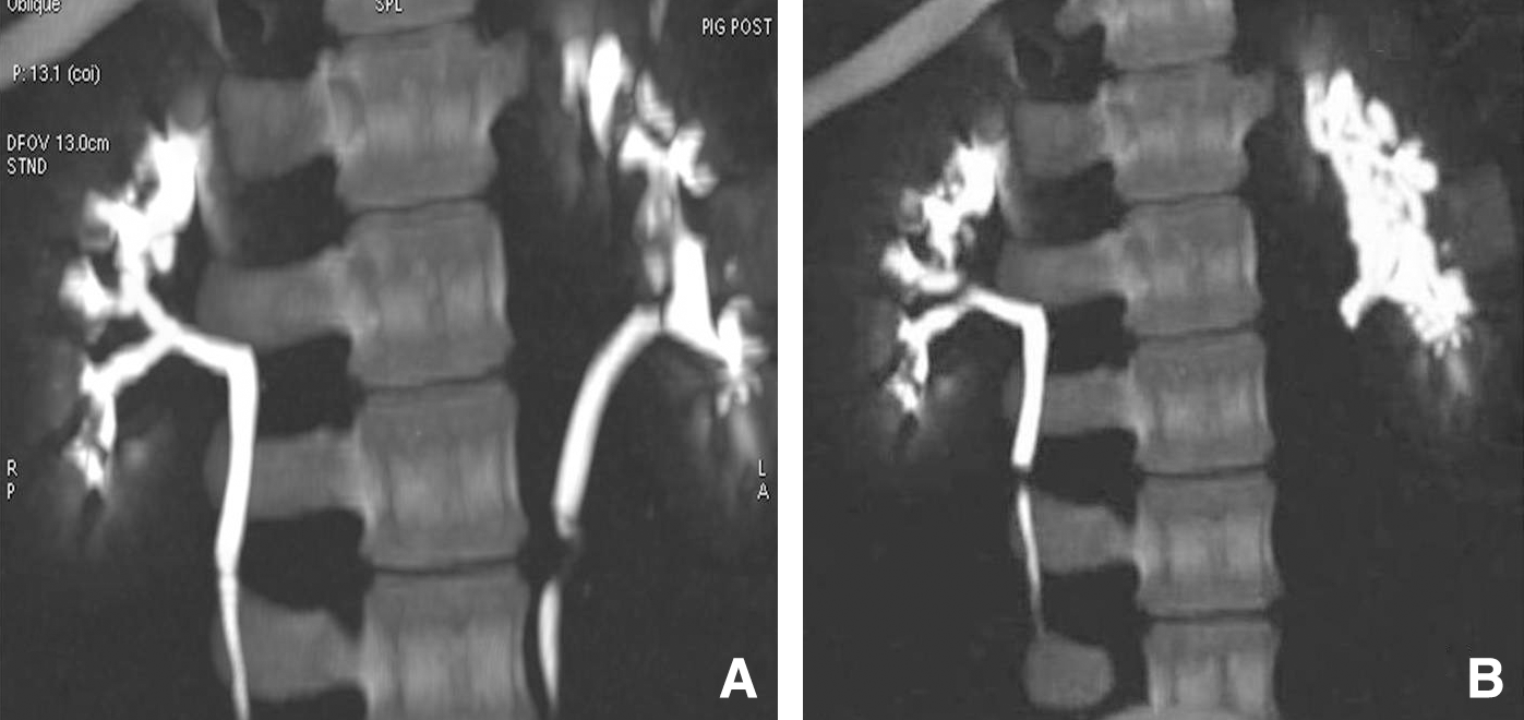
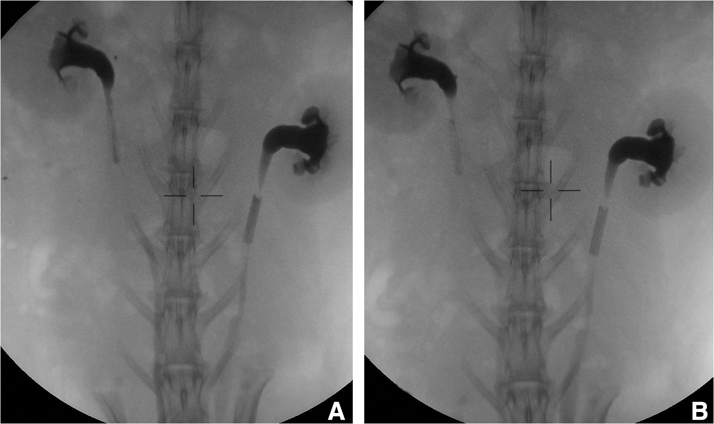
Stent implantation and follow-up were successfully completed in all animals. The CT scans revealed that hyperplastic tissue was present in both stent types. BMSs in seven porcine ureters were completely obstructed while ZESs had hyperplastic tissue that did not result in obstruction (Fig. 1). The rabbit ureters with a BMS were occluded in two cases while no ZES was associated with ureteral obstruction (Fig. 2). No obstruction remission was observed during the follow-up periods either in pigs or in rabbits. In fact, four of seven obstructions of the BMSs in pigs were evident on the first week follow-up CT evaluations while the remaining were seen on the second week follow-up. In rabbits, the obstructions were observed on the second week follow-up IVU examination. ZESs were associated with renal dilatation in two pig ureters while rabbits did have any marked dilatation. Obstructed ureters with BMSs were associated with significant dilatation in the obstructed renal units in pigs and rabbits. Moreover, dilatation of the pelvicaliceal system was observed in two rabbit renal units with the ureter containing a BMS.
The function of the seven porcine renal units and the one rabbit unit with obstructed ureters with stents was significantly compromised (Fig. 3). Two pig ureters with ZES and two rabbit renal units with ureters with BMSs were observed to have delays to achieve the peak of renographic curve, which was not influenced by the intravenous administration of furosemide. Thus, renal excretion was compromised without significant effect to the renal function.
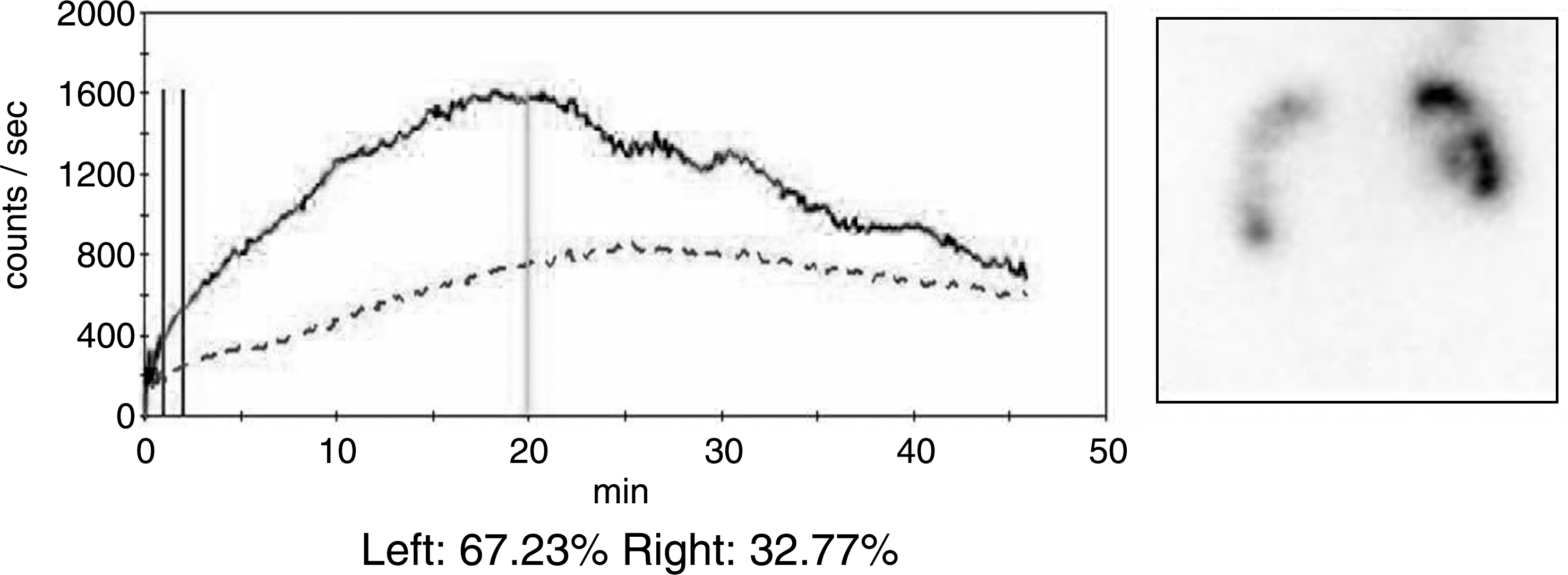
Renal scintigram of a pig on the third week follow-up. There is significant reduction in the function of the right kidney in comparison with the left kidney. The right ureter was stented by a BMS. The solid line represents the left and the broken line the right kidney.
Before the sacrifice of the animals, patent ureters underwent evaluation with OCT. Only patent ureters were evaluated, because it was impossible to insert the optic fiber through these stents. As a result, 23 ureters with stents were evaluated. The OCT revealed increased hyperplastic reaction in the ureters with BMSs in comparison with ZESs. In fact, evaluation of the ureteral wall was possible, because stent struts were observed as shadows in the serial images provided by the OCT and marked the border between the urothelium and muscular layer of the ureter for either pigs or rabbits. Moreover, the texture of the tissue representing the urothelium is markedly different in comparison with the underlining muscular layer (Fig. 4).

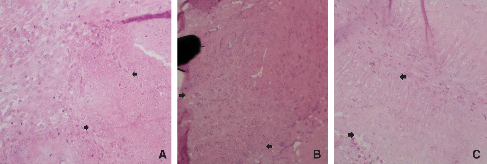
Hyperplastic reaction was present in all cases, and pathologic evaluation revealed significant more hyperplastic reaction in BMSs in comparison with ZESs (Figs. 5 and 6). On the contrary, mild to moderate inflammation was present in all ureters without any significant difference among stent types (Table 1). Table 1 summarizes pathology results.

Characteristic presentation of a ureter with an implanted BMS. The tissue between a strut of the stent and the ureteral lumen is presented. The stent has been covered by muscular (black arrows) and urothelial cells (white arrows). The extensive hyperplastic reaction of the urothelium extends inside the lumen of the ureter and compromises patency.
BMS=bare metal stent; ZES=zotarolimus-eluting stent; SD=standard deviation.
Discussion
BMSs have been proposed for the management of both benign and malignant ureteral obstruction in an attempt to address challenging urologic cases with a minimally invasive method. Benign cases, such as ureteropelvic junction obstruction or ureteroileal obstruction after urinary diversion, have been successfully managed by BMSs. 5 Extrinsic obstructions from the involvement of the ureter by malignant disease have been addressed by BMSs. 5 In the latter cases, the retrograde insertion of polymeric stents is associated with high failure rates to alleviate the obstruction, and the use of nephrostomy tubes results in significant deterioration of the quality of life of the patient. 1 –5 Thus, BMSs provide an alternative that remains in limited clinical application, because the use of these stents has been related to complications such as encrustation, migration, and intraluminal hyperplasia. The latter complication is the most common cause of obstruction of BMSs inserted in the ureter. 4,5
Ureteral patency can be compromised by the development of hyperplastic tissue protruding through the stent struts. The hyperplastic tissue is responsible for restenosis of the ureter with a stent. An experimental study in the porcine model by Desgrandchamps and coworkers 18 revealed that only one of eight stents was patent 35 days after insertion. Hyperplasia seems to be prevented by avoiding overextension of the strictured area of the ureter followed by careful stent implantation. Urothelial hyperplasia regresses within 4 to 6 weeks after the BMS insertion, resulting in ureteral lumen narrowing. 18 –23 Flueckiger and colleagues 24 suggested that the initial effect (first 1 to 2 weeks) of the inserted BMS in the ureter is reactive swelling of the ureter and not hyperplasia of the urothelium leading to constriction of the ureteral lumen. A characteristic trumpet-like morphologic configuration of the ureter located on the upper extremity of the stent has been described. The latter configuration has not been related to ureteral patency risk. 5,23,25
Neointimal hyperplasia represents a similar hyperplastic reaction that has been observed in vascular MSs. The latter phenomenon results in stent occlusion by the formation of blood clot and eventually thrombosis of the stent, which could be life-threating for the patient. In an attempt to reduce or minimize the intraluminal hyperplastic reaction, DESs were introduced. DESs have been proven to provide higher patency rates than BMSs and have been established as the standard of care in interventional cardiology for several years. 10
Urologic stent research on DESs has been limited mostly on polymeric DESs with promising success. 26 –28 Nevertheless, drug-eluting metal mesh stents have been evaluated in only one experimental study reported by our group. In the latter study, a paclitaxel-eluting metal stent was evaluated in the ureters of 10 pigs. The majority of the DESs (8 of 10) remained patent while 5 of 10 BMSs were occluded. Pathologic evaluation revealed that DESs had lower inflammation and hyperplasia in comparison with BMSs. 11
The current study aimed to evaluate a ZES in an attempt to confirm the efficacy of ZES in the ureter. Moreover, various methods used in the evaluation of the vascular stents were used for the assessment of the ureteral stents for the first time.
The currently presented evidence confirmed the results of our previous study that showed that BMSs are associated with high occlusion rates in the pig ureter while ZESs provide more reliable patency. Rabbits were also used in an attempt to extend the time of follow-up, because pigs are animals with rapid growth, which results in significant difficulties for long periods of observation. A follow-up period of 8 weeks was possible for the rabbits. The extended follow-up did not reveal any changes regarding the patency of the stents. The possible regression of the stenosis after a period of 4 to 6 weeks was not confirmed in the rabbit ureters. 18 –24 The pathology outcome of the current study did not reveal severe inflammation, and all cases were associated with mild or moderate inflammation. The latter observation does not concur with our previous report on DESs. 11
Zotarolimus in implanted stents were investigated by consecutive CT scans or IVUs for patency control. These methods provided the base for further evaluation of the stents. Renal and ureteral dilatation as well as the stent luminal patency were reliably depicted by the above methods, and these results could be directly compared with newly introduced intraureteral stent patency assessment methods. OCT was used for the assessment of the luminal condition as well as the depiction of the ureteral wall layers. In fact, the method provided information regarding the thickness of the hyperplastic urothelium. Other methods for intraluminal depiction such as ureteroscopy or virtual endoscopy provide only information of the intraluminal condition. 11 Nevertheless, the accurate correlation of the OCT and the pathologic cross-sectional images obtained by the aforementioned embedding process need further investigation.
The use of GMA for the embedding of the ureters with stents provided a significant advantage because the stent is not removed from the tissue for the pathologic examination. Thus, the intergration of the stent in the ureter and the stent-ureter structure could be observed in an intact condition under light microscopy. GMA and other resins have been used for the examination of vascular specimens containing stents and are considered as excellent material for the above purpose. 15
The main limitation of the study was that the evaluation model was based on the comparison of the ZESs with control BMSs. Consequently, the effect of these stents in cases of ureteral pathology was not evaluated and remains questionable. Nevertheless, the assessment of the efficacy of ZES in different pathologies of the ureter was not the purpose of the current investigation.
An additional limitation is lack of thorough statistical analysis with appropriate sample calculation and direct comparison of patency and inflammation results as presented in Table 1. The presented statistical evaluation was performed in an attempt to additionally validate the observation regarding the stent patency and inflammation. The results of the current analysis concur with the observations, and the extension of the current sample (16 animals, 32 stents) may not provide more information because the current study documents the differences among stent types by the use of several methods resulting in solid conclusions for the animal model.
Conclusion
ZESs in the pig and rabbit ureter were not related to hyperplastic reaction resulting in stent occlusion. These stents were related to significantly lower hyperplastic reaction in comparison with BMSs while inflammation rates were similar for both stent types.
Footnotes
Disclosure Statement
No competing financial interests exist.