
Abstract
Background and Purpose:
Cone beam CT (CBCT) is a novel imaging modality that combines the versatility of conventional C-arm imaging with the functionality of cross-sectional imaging. This is a pilot study to evaluate the capabilities of this new technology to obtain percutaneous access and for the immediate postoperative evaluation of residual fragments in percutaneous nephrolithotomy (PCNL).
Materials and Methods:
A retrospective analysis of all PCNL cases performed between April 2007 and November 2007 was performed. One urologist (NSS) and one radiologist (JFA) reviewed the studies postoperatively. Preoperative films were evaluated to see if CBCT influenced or improved percutaneous access. Postoperative films were evaluated that compared CBCT with conventional noncontrast CT to determine efficacy in finding postoperative stone fragments. Parameters of stone size, location, and quantity of fragments were compared.
Results:
For preoperative access, CBCT was used in 52 cases of PCNL between April 2007 and November 2007. In eight of these cases, CBCT altered the percutaneous access. In postoperative evaluation, 26 cases had both CBCT and conventional CT for comparison. In 11 cases with residual stones, conventional CT identified a greater number of fragments, but these were less than 2 mm. The postoperative recommendation for a secondary procedure concurred in 22 of 26 studies.
Conclusions:
CBCT may provide advantages of improved preoperative imaging, which may result in better percutaneous access, and improved postoperative imaging, which allows surgeons to have “real-time” access to CT quality images. The intraoperative availability of these high quality tomographic images may obviate the need for other postoperative imaging and subsequent adjunctive procedures for residual fragments.
Introduction
Cone beam CT (CBCT) is a novel imaging modality that uses an imaging head similar to that of a conventional C-arm to provide high-resolution, three-dimensional, CT-like images (Fig. 1). These images can be presented axially, coronally, and sagitally to show calcified or air-filled areas, and to differentiate soft tissue from fat. CBCT has been used for intraoperative image guidance in neurosurgical procedures. 1 CBCT provides near CT-quality imaging in accessing small abdominal structures without subjecting the patient to travel between the interventional radiology suite and the CT scanner. 2 It should be noted that CBCT does not use real-time imaging like CT fluoroscopy, but allows the surgeon to obtain CT quality images using a machine that provides full function fluoroscopy as well. Fluoroscopic images are equivalent to those using conventional fluoroscopic equipment. It is has a large footprint and requires a fixed location in a dedicated room. Estimated costs of the system can range from $200,000 for the main imaging head and software component to up to $1,000,000 for a de novo complete system.
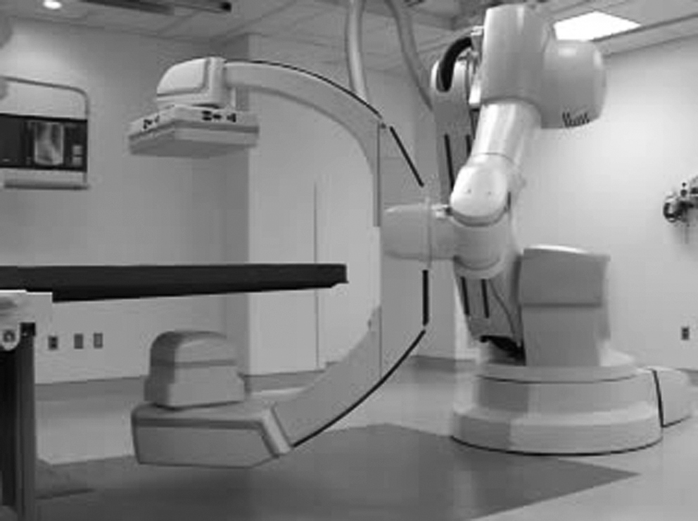
Operating room setup of DynaCT system. Imaging arm, similar to conventional C-arm, can be placed on either side of the operating table, opposite to the percutaneous access instruments. The head of the patient and anesthesia equipment would be on the far left of the table in this image.
Complications of percutaneous access have been well described. Other investigators have shown that rates of pleural injury, colon injury, and anterior or incorrect access may be improved with better intraoperative imaging for detection 3 and guidance for needle placement. Matlaga and colleagues 4 suggested CT-guided access to the renal collecting system may be as safe and effective as conventional fluoroscopy, and it may be preferable in nondilated systems. With this in mind, we postulated that the improved resolution of CBCT may offer the advantage of improved safety and efficacy over conventional fluoroscopy in percutaneous access for PCNL.
Current practice of assessing postoperative stone burden in PCNL is either a stone protocol CT (noncontrast spiral CT) or antegrade nephrostography. Although stone protocol CT provides improved sensitivity and characterization of residual stones, 5 it must be performed outside the PCNL operating suite. CBCT may offer equivalent resolution for improved sensitivity and characterization of residual stone burden with the advantage of allowing the acquisition of these images either intraoperatively or immediately postoperatively within the PCNL suite.
In this pilot study, we examined the ability of this new technology, CBCT, to aid in percutaneous access during PCNL and evaluate residual stone burden by comparing with conventional stone protocol CT.
Materials and Methods
In a protocol approved by the Institutional Review Board, we retrospectively studied all PCNL cases using CBCT between April 2007 and November 2007 at a single institution (University of Virginia) and found all cases with both postoperative CBCT images and conventional stone protocol CT images.
Imaging equipment
The CBCT we used for this study is the Syngo Dyna CT (Siemens). Standard slice thickness is 0.4 mm. This size can be adjusted upward. Most body imaging for this CBCT is optimized at 3 mm slice thickness to improve resolution-to-sharpness ratio. Field of view (FoV) dimensions are standard at 18 cm height and 24 cm diameter. In cases necessitating a larger FoV, diameter can be increased to 47 cm.
For our conventional, standard stone protocol CT, we used the 64-slice GE Litespeed–VCT. Slice thickness for standard stone protocol is 3.75 mm. FOV is 70 cm in maximum diameter.
Preoperative analysis.
Most patients (48 of 52) have preoperative imaging using a conventional noncontrast CT. Other imaging, such as contrast-enhanced CT or excretory urography may be performed at the discretion of the attending surgeon. A preoperative CBCT is obtained while the patient is in the prone position. Access to the collecting system is then obtained in the conventional fashion, using a fine-gauge needle to opacify the collecting system, then obtaining caliceal access and placing a wire down the ureter using fluoroscopy. In selected cases with unusual anatomy or difficult access, CBCT may be performed after access is obtained to confirm position. One urologist and one radiologist retrospectively evaluated all of the above-mentioned cases for the number of times CBCT facilitated a change in percutaneous access from traditional fluoroscopy.
Postoperative analysis.
CBCT is obtained at the end of the case after the patient is cleared of stones with the endoscope. If significant residual fragments are detected, the endoscope may be reintroduced. Postoperatively, all patients undergo a standard noncontrast CT on the evening of surgery to again assess for residual stones for purposes of comparison in this study. One faculty radiologist and one faculty urologist then independently compared the postoperative CBCT with the stone protocol CT for each case to assess residual stone burden, including quantity, size, and location of residual stones. In addition, the faculty evaluator made a judgment regarding need for a secondary procedure. This decision was compared between the two imaging modalities (CBCT vs conventional CT).
Results
Percutaneous access
CBCT was used in 52 cases of PCNL between April 2007 and November 2007. In eight of these cases, CBCT altered the percutaneous access. In six of these cases, CBCT was able to identify an anterior caliceal or infundibular access before dilation that was not recognized on conventional fluoroscopy. In these cases, after second puncture was performed, CBCT was repeated to demonstrate more favorable access. In one case, preaccess CBCT demonstrated a posterior-lying colon, not appreciated on standard supine preoperative CT, which altered the initial access site (Fig. 2). In one other case, CBCT identified a low-lying pleura, which altered the initial access site. CBCT was repeated after access was obtained in both of these cases to confirm favorable access.
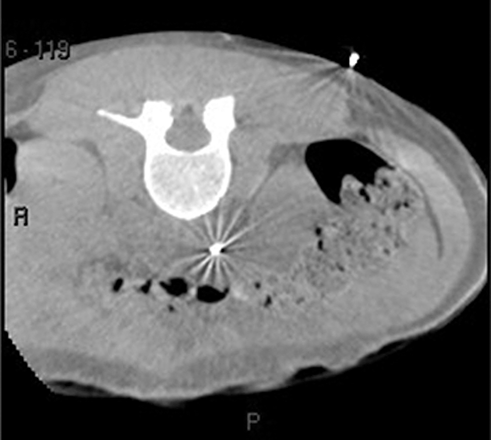
Cone beam CT demonstrates that the colon is posterior to the lower pole. The access site was moved superiorly and medially.
Residual stone burden
Twenty-six cases were found that had conventional CT images to compare with perioperative CBCT images. Overall image quality between CBCT and stone protocol CT is similar (Fig. 3). CBCT is able to identify most stones and soft tissue structures seen on stone protocol CT.

Comparison of conventional and cone beam CT images in the same patient demonstrating similar quality in identifying calcified structures and soft tissues.
Stone quantity
Of 11 cases with significant residual stone burden, stone protocol CT showed more residual stones than CBCT (Table 1). All of these additional stones, however, were 3 mm or less in size. Overall stone-free rates were 75% on stone protocol CT and 81% on CBCT. In three cases, CBCT showed additional stones, although these were also small (3 mm or less). These stones may have passed through the ureter in the interval between the CBCT and the stone protocol CT. Three showed no difference in quantity between the two imaging modalities.
Does not include cases where cone beam computed tomography was of poor quality
CT=computed tomography.
Using conventional stone protocol CT as the gold standard, CBCT demonstrated sensitivity of 64%, specificity of 72%, positive predictive value of 72%, and negative predictive value of 64%.
Stone location
Only three of seven cases showed a difference in location of significant (>2 mm) residual stones. One 15-mm stone was seen at the ureteropelvic on stone protocol CT but not on CBCT, because of contrast seen on the CBCT that obscured the stone. One 8-mm upper pole stone was seen on CBCT that was not present on stone protocol CT. One case had significant steinstrasse in the distal ureter. Because the current CBCT scanning protocol does not scan the distal ureter, this was missed by CBCT. Overall, stone protocol CT detected more stones in the lower pole, but these were not clinically significant (2 mm or less).
Stone size
There was no significant difference in stone size for stones identified by both CBCT and stone protocol CT.
Secondary procedure
In 22 of 26 cases, CBCT and stone protocol CT resulted in the same recommendation from each evaluator as to the need for a secondary procedure to treat residual stone burden. In one case, CBCT did not visualize a steinstrasse that needed treatment. In three cases, CBCT interpretation warranted additional procedures where stone protocol CT would not. In all three of these cases, CBCT misinterpreted contrast injected during the procedure for residual stone burden.
Discussion
Fluoroscopy is the most common method used to obtain percutaneous access. When opacified with contrast, it provides an accurate image of the renal collecting system and can give multiple views by rotating the C-arm. It does not provide helpful data of the soft tissues surrounding the kidney, however, and can only render two-dimensional views. In addition, increased intrapelvic pressure from injection of contrast may lead to extravasation. Conventional CT provides excellent soft-tissue detail and complete three-dimensional viewing without using injectable contrast. It does not, however, provide any intraoperative use because of its large size, circumferential enclosure, and location outside the PCNL suite.
CT-fluoroscopy offers the advantage of the high soft-tissue resolution of CT at much faster speeds, 6 but it lacks integrated conventional fluoroscopy with real-time large FoV, which is essential for PCNL. The improved soft-tissue resolution and three-dimensional images obtained using CT axial images may prove superior to conventional fluoroscopy in obtaining percutaneous access to the renal collecting system. 7 This can be even more pronounced when there is no hydronephrosis, as is often the case in PCNL.
In our study, CBCT has similar soft-tissue resolution to conventional noncontrast CT for purposes of identifying stones. In addition, CBCT has the ability to optimize percutaneous access, identifying anterior caliceal or infundibular access when conventional fluoroscopy did not. Our study demonstrated that in at least one case, CBCT could be helpful in avoiding colonic injury, which is a rare, but significant complication affecting 0.3% of patients undergoing PCNL. 8 This may be because of changes in the position of the colon in the prone position as opposed to that seen in the preoperative supine stone protocol CT. 9
In assessing for residual stone burden, CBCT has excellent sensitivity for stones when compared with stone protocol CT. In fact, there was no significant difference in the detection of clinically significant stone fragments (>2 mm) between the two modalities. In our current imaging technique with CBCT, the distal ureter is not imaged, but this is easily rectified by moving the CBCT imaging head more caudad during imaging.
The single greatest advantage of CBCT for imaging residual stones is its location in the PCNL operating suite. Therefore, if significant residual stone burden remains, the urologist can simply reinsert the nephroscope and lithotrite without ever having to move the patient or replace the access sheath. This may have significant positive implications by reducing the number of second-look PCNLs or secondary shockwave lithotripsy for residual stones. Currently, the average number of procedures for patients with staghorn calculus undergoing primary PCNL is 1.9. 10 The costs, inconvenience, and morbidity associated with these secondary procedures are a burden to the patient and healthcare system.
Two limitations apply to CBCT when assessing for residual stone burden. Active respiratory movement causes significant motion artifact that can make the images uninterpretable. We now routinely have the anesthesiologist suspend respirations (the patients are always intubated for PCNL at this institution) during the accrual of images. It takes approximately 2 minutes to set up and then acquire the images during which breath-holding is required for 8 seconds. After this, it takes 3 minutes to perform the software reconstructions to provide the axial, sagittal, and coronal images.
The other limitation is the use of contrast. Because CBCT is performed intraoperatively, residual contrast used during the procedure may persist at the end of the procedure and may mimic a residual fragment. Our routine practice now consists of minimizing intraoperative use of contrast, elimination of the standard nephrostography at the conclusion of the procedure, and thorough irrigation of the collecting system before performing CBCT.
Exposure to radiation energy for CBCT is comparable to that of conventional imaging for stone disease. CBCT uses the same radiation energy, X-ray, as conventional CT and fluoroscopy. Stone protocol CT exposes the patient to the effective dose of 8.5 mSv. 11 CBCT images for an average PCNL patient in our study were acquired during two 8-second runs exposing the patient to an average of 9.9 mSv of radiation. Typically, this is only 30% of the entire effective dose during PCNL, with the remaining 70% attributed to conventional fluoroscopy. Therefore, using CBCT does not appear to expose the patient to significant additional radiation risk relative to conventional methods of imaging.
Conclusions
CBCT is an investigational modality already used in other surgical specialties for image-guided surgery. This is a pilot study that shows potential advantages to current imaging modalities for obtaining percutaneous access in PCNL. CBCT may also have equivalent ability to stone protocol CT in detection of residual stone burden with the advantage of being performed while the patient is still in the operating suite.
Footnotes
Acknowledgments
We wish to thank Allen Goode, Department of Medical Physics, Radiology, University of Virginia Health System.
Disclosure Statement
No competing financial interests exist.