
Abstract
We describe our experience using an iodinated contrast solution to hydrodissect adjacent structures before percutaneous renal cryoablation. Hydrodissection was performed before cryoablation with placement of a 20-gauge, 15-cm introducer needle into the retroperitoneum under CT or ultrasonographic guidance followed by infusion of 5% dextrose in water and 2% iodinated contrast between the kidney and the adjacent organ. Ten patients underwent hydrodissection with an iodinated contrast solution at our institution. The mean tumor size was 3.1±1.2 cm. The organs displaced included colon (n=7), small bowel (n=1), pancreas (n=1), and in one case, both the colon and ureter were displaced. The average displacement of all organs from the kidney was 2.8 cm (range 2.2–3.5 cm). There were no complications and no injuries to any adjacent structures. The injection of iodinated contrast allows for safe mobilization and differentiation of adjacent structures from the renal tumor and parenchyma leading to potentially safer cryoablation.
Introduction
The anatomy of the retroperitoneum dictates that other structures, such as the colon, duodenum, ureter, psoas muscle, and pancreas, may be immediately adjacent to the renal tumor. Given that the ice ball must exceed the tumor margin by ≥3 mm to cause complete cell death, adjacent organs may need to be displaced to decrease the risk of injury to adjacent structures. 6 –9
Hydrodissection using 5% dextrose in water (D5W), saline, or sterile water has been described as a method to displace structures at risk of injury during thermal ablation. 10,11 When used in combination with CT as a guidance and monitoring technique, however, these solutions may be difficult to differentiate from surrounding organs and muscles. To increase the visibility of injected fluids at CT, we combine D5W with iodinated contrast material. We describe our experience using D5W doped with iodinated contrast to hydrodissect colon, pancreas, and ureter during percutaneous renal cryoablation.
Technique
This study was approved by our Institutional Review Board, and the need for informed consent was waived. We performed a retrospective review of all percutaneous renal cryoablation cases performed at our institution from 2003 to 2010 for patients undergoing hydrodissection as a protective technique. Patient medical records and imaging studies were reviewed, and demographic data, tumor size, and details regarding hydrodissection of adjacent organs on CT were extracted.
Percutaneous cryoablation procedures were performed by the same team of radiologists and urologists working together. 12 Patients were placed in either the prone or decubitus position in the CT gantry after intubation and induction of general anesthesia. Hydrodissection was performed before cryoablation by placement of a 20-gauge, 15-cm introducer needle into the retroperitoneum and the infusion of D5W and 2% iodinated contrast between the kidney and the adjacent organ. The introducer needle was placed under CT guidance, and displacement of the adjacent organ was verified via CT before cryoablation (Figs. 1 and 2). Because of migration of fluid in the retroperitoneum or peritoneal cavity, different volumes of fluid were infused to maintain adequate displacement of adjacent structures away from the renal tumor.

Hydrodissection of the colon from the right kidney (patient in prone position).
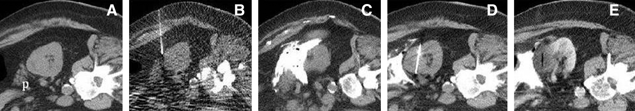
Hydrodissection of the pancreas from the right kidney (patient in decubitus position).
All patients were treated with 1.7-mm cryoprobes (CryoCare, Endocare). Real-time ultrasonography was also performed for targeting and placement of cryoprobes. A combination of ultrasonography and unenhanced CT was used to monitor ice ball formation. Our standard treatment regimen consists of two 10-minute freeze cycles with an intervening 5-minute passive thaw. The ice ball size was closely monitored on ultrasonography and CT, and the percentage of argon flow was modified to obtain a 5- to 10-mm margin. After probe removal, a contrast-enhanced CT with delayed images was performed to evaluate for hematoma or acute bleed and assess the renal collecting system in cases of endophytic tumors.
Measurement of organ displacement before and after instillation of the hydrodissection fluid was performed on CT fluoroscopic images. Region of interest measurements were performed of the renal tumor and adjacent hydrodissection fluid using a picture archiving and communication systems workstation (McKesson, Inc, San Francisco, CA). Follow-up imaging consisted of a contrast-enhanced MRI within 6 months of the ablation, and ablation success was defined as adequate coverage of the entire tumor by the ablation zone, and a lack of tumor enhancement. Statistical analysis was performed using paired Student t test; P value < 0.05 was considered significant.
Of the 101 percutaneous renal cryoablations performed at our institution in the study period, 10 patients underwent hydrodissection using D5W and iodinated contrast. The mean patient age was 64 years. The mean tumor size was 3.1±1.2 cm.
Fluid was injected into the retroperitoneum to displace several different organs, including colon (n=7), small bowel (n=1), pancreas (n=1), and in one case, both the colon and ureter were displaced. The mean distance between the renal tumor and the adjacent organ before hydrodissection was 0.15 cm (range 0.01–0.80 cm). A mean of 389 mL of fluid (range 100–1000 mL) was injected for displacement resulting in a mean postinjection displacement of 2.7 cm (range 2.2–3.5 cm) away from the target tumor. The mean attenuation of the hydrodissection fluid was 305 Hounsfield units (HU) (range 73–883 HU) and statistically different compared with 48 HU (range 16–75 HU) for the kidney (P=0.01). There were no intraprocedure or postprocedure complications, and no known injuries to any adjacent structure. No recurrences were noted on follow-up imaging with a mean follow-up of 5.4 months.
Discussion
Cryoinjury to adjacent structures during percutaneous cryoablation has been described and can have devastating consequences. 4 The anatomy of the retroperitoneum in reference to the renal tumor and the need for the ice ball to extend at least 3 mm beyond the outer tumor margin to result in complete cell death are important factors that may lead to injury of adjacent structures. 6,7 Damage to hollow thin-walled structures (bowel, ureter, gallbladder) is more severe than freezing adjacent solid organs (liver, spleen) because of the risk of mural necrosis and secondary abscess or stricture. 13,14 Large blood vessels such as the inferior vena cava tend to be at lower risk for cryoinjury because of the heat sink effect of blood flow. 15,16
The location of the renal tumor frequently dictates the need for hydrodissection. The colon is the most commonly displaced structure and is generally located anterior and inferior to the kidneys. Lower pole or anterior renal tumors may thus be more likely to need hydrodissection. Lower pole tumors may also need hydrodissection and/or stent placement because of the close proximity of the ureter. 11 In the past, we approached many of these lesions under laparoscopic assistance because of concerns regarding adjacent organ injury without previous mobilization. Whenever feasible, however, we favor a percutaneous approach because of the decreased morbidity and costs when compared with laparoscopic cryoablation, and the use of hydrodissection techniques has increased the number of cases that can be performed percutaneously. 12
Hydrodissection with the use of sterile water or D5W has been described. 10 Both of these fluids, however, are similar in attenuation to the renal tumor, normal renal parenchyma, and adjacent structures (including bowel, which is often fluid filled) making accurate targeting and monitoring difficult. A recent retrospective study by Bodily and associates 11 described hydrodissection in 50 cases of percutaneous renal cryoablation. The structures displaced included the colon (n=41), body wall (n=3), duodenum (n=2), jejunum and ileum (n=2), ureter (n=1), and psoas muscle (n=1). Hydrodissection was successful in 50 of 52 cases, displacing structures a mean distance of 1.6 cm. The two failed cases occurred early in the series and were because of insufficient displacement of the colon from the index tumor. There was one complication of hemorrhage resulting from injury to an intercostal artery branch that necessitated termination of the procedure before fluid infusion and endovascular intervention to embolize the injured vessel.
DeBenedectis and colleagues 17 described using a mixture of D5W and iodinated contrast medium to aid hydrodissection for radiofrequency ablation. They performed hydrodissection using an iodinated contrast medium in 21 patients who were undergoing percutaneous radiofrequency ablation. Organs displaced included colon (n=19), small bowel (n=4), pancreas (n=3), and spleen (n=1), with a mean displacement distance of 2.6 cm. Ablation was performed successfully in all 21 cases with no injury to adjacent structures. All patients also underwent a follow-up CT scan an average of 3 weeks after the procedure, and no residual hydrodissection fluid was noted.
The choice of hydrodissection fluid varies with ablation modality. For radiofrequency ablation, tissue heating is created by ionic agitation as current is conducted between electrodes and ground pads. Thus, maximum protection of vulnerable structures is gained by the choice of nonionic, poorly conducting fluids, such as sterile water or dextrose in water. Saline is an ionic fluid that conducts electricity and is thus poorly suited for this purpose when used during radiofrequency ablation. 10,18
For cryoablation, the choice of fluid type is less important because the object of hydrodissection is solely to increase displacement between the ice ball and vulnerable structure. We used a 2% iodinated contrast solution based on the results of Roen and coworkers, 19 which demonstrated that the ideal concentration of 300 mg/mL of iohexol in physiologic saline and D5W is between 0.5% and 2%. At these concentrations, there is sufficient contrast between the infused fluid and abdominal organs without producing beam hardening artifacts. 19 We found that as the hydrodissection fluid dissipates in the abdomen, there will be some variability in the HU attenuation of the fluid.
In our study, the amount of displacement of adjacent structures (2.8 cm) was similar to that reported by other studies (range 1.6–2.6 cm). 10,11,18 We show that the use of a D5W with 2% iodinated contrast allows for a clear differentiation between the renal tumor (mean attenuation of 48 HU) and the hydrodissection fluid (mean attenuation of 305 HU). The ability to differentiate between the hydrodissection fluid and the renal tumor and adjacent structures is helpful to prevent inadvertent freezing of nontargeted structures and assure adequate coverage of the tumor by the ablation zone.
Limitations of our study include a small number of patients and the retrospective nature of this study. Another potential limitation is the subjectivity of the improved visualization with an iodinated contrast solution vs D5W, although a significant difference in HU attenuation was noted between the tumor and hydrodissection fluid.
Conclusion
The injection of D5W 5 mixed with 2% iodinated contrast allows for safe mobilization and differentiation of adjacent organs from the renal tumor and parenchyma during percutaneous cryoablation.
Footnotes
Disclosure Statement
Dr. Henshaw is a stockholder and on the Board of Medical Advisors for Neuwave Medical. For all other authors, no competing financial interests exist.