
Abstract
Purpose:
To report on integrated endourology suites (IES), remote monitoring and supervision (RMS) of urology residents and to evaluate patients' opinions, acceptance, and satisfaction level with IES and RMS.
Patients and Methods:
Patients undergoing flexible cystoscopy in the IES with RMS were surveyed using a questionnaire. All procedures were performed by junior urology residents (UR-1 level) using RMS. Patients were studied using a nine-question survey to evaluate their comfort level, acceptance, and level of satisfaction with RMS. Six questions used a scale of 1 to 10 (1=strongly disagree; 10=strongly agree), and the remaining three questions solicited a “yes” or “no” response.
Results:
100 patients were studied (59% Caucasians, 40% African Americans, and 1% Hispanic). Median age was 63 years. The highest level of education was middle school in 2% of patients, high school in 55%, undergraduate in 33%, and postgraduate in 10%. Patients scored a mean of 9.50/10 (highly satisfactory) regarding their comfort with RMS; 96% scored ≥7, 4% scored 5 to 6, and none scored <5. Patients were satisfied having a urology resident perform the procedure (9.48/10), other residents and medical students watch the procedure (9.41/10), a video camera in the room (9.40/10), and two-way sound communication (9.40/10). None perceived compromise to their privacy or quality of care.
Conclusions:
RMS in IES is highly acceptable to patients undergoing endoscopic procedures. RMS has the potential to positively impact residency training, efficiency, regulatory compliance, safety, and productivity.
Introduction
The increase in clinical and financial productivity is often in direct conflict with the mission of residency training. This consumes time and resources and tends to challenge efficiency and safety. For example, operative times increase and efficiency decreases as a result of resident participation. 2 With urology residency requiring training in multiple types of surgical techniques and a maximum 80-hour work week, both the teacher and student are under a time constraint. 3 Residents must learn with great efficiency while performing their duties to achieve the necessary training to fulfill the requirements for board certification. Finding ways to train residents efficiently and facilitate their learning without negatively impacting safety and cost is not only desirable but also key to the future success of urology residency training in the United States.
Technologic advances such as electronic medical records (EMR) 4 –7 and telecommunication 8,9 have provided significant benefits in clinical practice. EMR has had a positive impact on efficiency, quality of care, safety, and cost. 5 –7 In one report, the use of EMR resulted in an estimated net savings between $8400 and $140,100 per physician. 4 Such technologic advances can be similarly used to enhance resident training, improve efficiency, and decrease cost without compromising safety.
In our practice, we have implemented remote monitoring and supervision (RMS) in residency training through advanced telecommunication in integrated endourology suites (IES). The objective of the study is to report on IES and RMS and to evaluate patients' opinions, acceptance, and level of satisfaction with the system. This is the first reported survey of patients' opinions and satisfaction regarding RMS in the operating room.
Patients and Methods
The study was approved by the Institutional Review Board at Emory University and by the Research and Development Committee at the Atlanta Veterans Affairs (VA) Medical Center. This was a prospective study to evaluate patients' opinions and acceptance of RMS of urology residents during flexible diagnostic cystoscopy.
Two new state-of-the-art IES were used for the study. Each IES was equipped with live visual, audio, and telestration communication that allowed for RMS of urology residents by urology faculty members from a control room (CR). Visual signals were displayed in the IES on six color monitors, the main two being 24-inch high definition (1080 p) ceiling mount monitors (Fig. 1). Audio signals were communicated to the IES through a ceiling mounted microphone and speakers, which allowed all personnel in the IES (residents, nurses, anesthesiologists, and the patient) to hear all conversation. Wireless audio communication was available but was not used because it limited audio access to the urology team and prevented the patient from listening to the conversation.
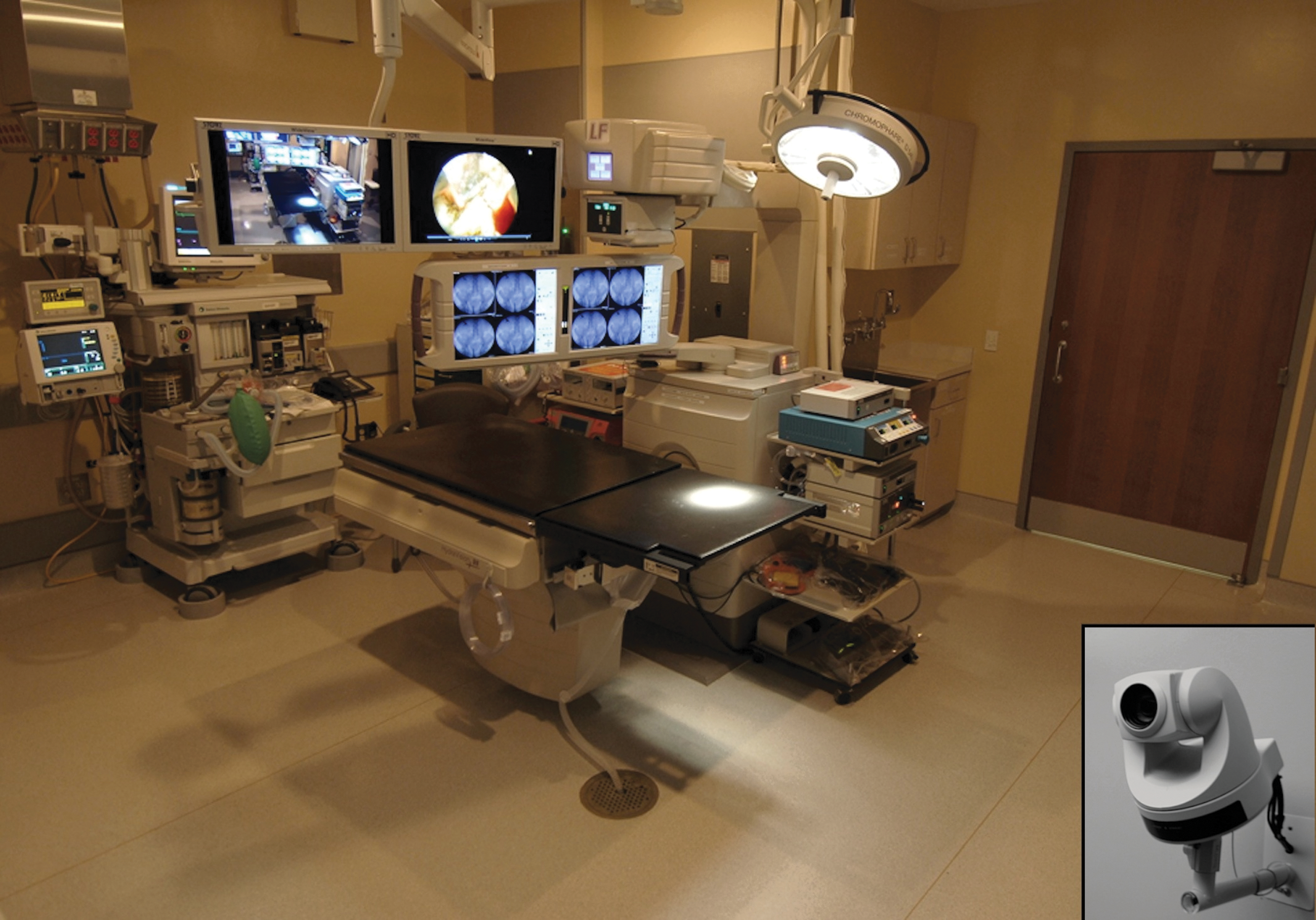
Integrated endourology suite. Insert shows the wall-mounted room camera.
All communications were routed and managed centrally through the designated CR located adjacent but separate from the IES. The CR was equipped with two 46-inch high definition (1080 p) flat screen monitors, two touch screen monitors that provided all the functionality to manage and control the IES, desk space, and multiple dual monitor computers (Fig. 2). The CR provided the supervising urologist full capability to remotely control and manage all equipment in the IES and the communication between the IES and CR. This included control of sound, volume, lights, dimmers, input to display monitors, room camera (motion, zoom, focus, and aperture), endoscopic video camera, fluoroscopy, plain radiography, picture capture, printing, video recording/streaming, display of electronic medical records (notes, labs, radiographs, etc.), presettings, and other auxiliary equipment and signals. The same CR capabilities were also available at a secondary remote site located in the academic office of the chief of urology on the same floor as the IES. This allowed the urology team to consult with the chief of urology at any time when faced with unique and difficult cases/situations. All communication signals are routed and integrated through three network towers in a designated hub located in a storage site outside the IES. Access to all communication signals was secured and limited to the operating team and the supervising urology in compliance with Health Insurance Portability and Accountability Act regulations. The supervising urologists were present and available on site during the entire procedures.

Control room.
Inclusion criteria for the study consisted of all patients undergoing diagnostic flexible cystoscopy who were willing to participate in the survey, a minimum education level of sixth grade, and an appropriate level of comprehension and cooperation. Patients with significant cognitive disorders, active chemical dependence, and psychiatric illness were excluded from participating in the study. Before cystoscopy, the resident reviewed the medical records to determine patient suitability to participate in the study.
Informed consent was obtained from all patients. All procedures were performed by junior urology residents (Postgraduate Year 2; Urology 1), while being monitored and supervised remotely by a faculty urologist in the CR. Topical intraurethral anesthesia (20 mL of 2% lidocaine jelly) was the primary method of anesthesia. Conscious sedation was not used to allow full patient awareness of the visual and audio monitoring during the procedure.
After the procedure, patients completed a survey regarding RMS of the procedure by completing a brief, nine-question survey. The first six questions evaluated patients' opinions and comfort with regard to: (1) Audible conversation between IES and CR on the speakers; (2) use of a video camera in the IES; (3) having a trainee (urology resident) perform the procedure; (4) remote viewing of the procedure by other members of the urology team; (5) overall comfort with privacy; and (6) overall comfort with RMS. These six questions were evaluated on a visual liner scale of 1 to 10 (1=strongly disagree; 10=strongly agree) (Fig. 3). The remaining three questions addressed patient satisfaction with the way the procedure was performed and whether the patient would undergo the procedure again in the same way or recommend it to friend or a family member if needed in the future. These three questions solicited a “yes” or “no” response.

Visual linear scale (1–10).
One of the authors (NJT), a specialist in behavioral instrument design and reading level, supervised the design of the survey instrument. The questions were written at a sixth grade reading level and designed to minimize response bias. Patients were informed that their participation in the study was completely voluntary and that their treatment would not be affected by their decision to participate in the study or by any of their answers. In addition, none of the Personal Health Record was used in the study. Data were obtained regarding age, sex, race, education level, literacy level, number of previous cystoscopy procedures, complications, diagnosis, and other potential demographic and clinical variables such as history of psychiatric condition or chemical dependency. Because the hospital was undergoing reconstruction at the time of the study, data were also collected regarding any potential aggravating factors related to the patient's trip, check-in, wait time, or any other hospital-related factor on the day of the procedure. Documentation of any potential aggravation was deemed relevant because of its potential to impact survey results.
Statistical analysis was performed using the Student t test. Statistical significance was declared by a P value of<0.05.
Results
One hundred patients completed the survey. The vast majority of patients (99%) were men, which is likely related to the predominantly male population at the VA. Median age was 63 years (range 37–92 years). Caucasians constituted 59% of the patients, followed by African Americans (AA) (40%) and Hispanics (1%). The highest level of education was middle school in 2% of patients, high school in 55%, undergraduate in 33%, and postgraduate in 10%.
Of the 100 patients, 48% underwent cystoscopy for the first time. The remaining 52% had previously undergone cystoscopy with 24% having had five or more past procedures. Eleven percent of patients had additional manipulation during cystoscopy, such as dilation of an incidentally discovered urethral stricture or bladder irrigation to enhance visualization. The examination revealed abnormal urothelial lesions consistent with bladder neoplasm in 11% of patients. The remaining 91% had either negative results on examination or findings consistent with benign conditions such as enlarged prostate and bladder trabeculation.
Four patients had low visual acuity, which challenged their reading ability. For those patients, the questions were read to them. Thirty percent of the patients had a documented history of psychiatric illness and/or chemical dependency. These comorbidities were well controlled and did not interfere with their ability to participate in the study. Nineteen percent of patients were considered aggravated before the cystoscopy because of a transportation issue, heavy traffic, and/or difficulty finding a parking spot at the medical center.
Patients scored a mean of 9.50/10 (highly satisfactory) regarding their comfort with having the procedure performed under RMS. The vast majority (96%) of patients scored ≥7, 4% scored 5 to 6, and none scored <5. Patients felt similarly comfortable and satisfied with hearing the conversation between the supervising physician and the resident over the speakers (9.40/10; range 5–10), having a urology resident perform the procedure (9.48/10; range 5–10), a video camera in the room (9.40/10; range 4–10), having other residents and medical students watch the procedure (9.41/10; range 5–10), and with their privacy during the procedure (9.40/10; range 4–10). All patients (100%) were satisfied with their experience, and all would choose to have the procedure performed the same way on themselves, family members, or friends if needed in the future.
There was no significant difference in the satisfaction levels of patients with a history of cystoscopy (P=0.19), psychiatric comorbidities (P=0.69), and aggravating factors related to traffic and parking on the day of cystoscopy (P=0.09). Similarly, satisfaction levels were not adversely impacted by race (AA vs non-AA, P=0.85), college degree (n=53, P=0.92), additional manipulation during cystoscopy (n=11, P=0.66), or the diagnosis of new bladder tumor (n=11, P=0.46).
Discussion
This study documents patients' acceptance and high level of satisfaction with the use of telecommunication advances to provide RMS in the daily practice of urology. Patients had a high level of satisfaction with the presence of a video camera in the cystoscopy suite, hearing the conversation on the speakers, having urology residents perform the procedures, or having other team members watch the procedures from a remote site. Further, the vast majority of patients did not feel that their privacy was compromised. Overall, patients were comfortable and highly satisfied having their procedures performed under RMS. While anecdotal, the staff received additional feedback and comments from patients confirming a high level of satisfaction. Many thought they had a more thorough and careful examination because of the oversight provided by more experienced supervising surgeons. A few patients compared the situation to that of an air traffic controller communicating with a flight pilot to ensure safe landing.
The current regulatory environment for residency training continues to mandate more scrutiny, monitoring, supervision, and documentation of residents' activities. Compliance with these mandates consumes time and resources, resulting in significantly higher costs for residency training. In one study, $35,000 was spent per year to train each resident in internal medicine. 10 In another study, the daily cost to train a resident was $200 to $300 and $100 to $200 per day for each medical student on internal medicine clerkship. 11 These figures do not address the impact of resident training on clinical productivity. When productivity was examined, Johnson and associates 12 reported that Rush University lost $163,949 in productivity during the 2004 to 2005 academic year to support internal medicine residency training. The use of RMS in IES provides an efficient approach to training residents, ensures compliance with regulatory mandates, and improves safety and productivity. Our study shows that it can be successfully implemented in urology practice with high acceptance and satisfaction rates.
RMS offers residents autonomy with oversight. The resident has complete access to the supervising urologist for any questions or clarifications throughout the procedure. The supervising urologist can advise and instruct the resident at any time through voice communication and/or telestration. Both parties work in harmony to achieve the clinical and training objectives. The success of RMS is dependent on clear and precise communication. As such, RMS forces the supervising urologist to communicate with clarity using words instead of hand signals, gestures, or mumbles, which are often confusing to residents. For example, if a resident cannot find the right ureteral orifice during flexible cystoscopy, the supervising urologist can instruct him/her to “take thumb off, pull back the scope to the bladder neck, and then advance the scope slowly with right wrist rotation and slight thumb up.” Residents convey that such clear communication provides a more comprehensible instruction and more time to process and learn. The same learning experience can be extended to other residents and medical students in the CR without interfering with the procedure or the patient. Video recording of the procedures can also be used to highlight technique and findings, and provide constructive feedback. While not studied in this project, we have found that residents tend to achieve training and learning milestones at a faster rate using RMS.
For endoscopic procedures necessitating general anesthesia, RMS serves as an efficient tool for the urologist to check and ensure that all perioperative steps are executed properly, such as timeouts, correct site surgery check, prophylactic antibiotics, sequential compression devices, patient positioning, and skin preparation. In addition, the urologist can visually verify through the remotely controlled room camera that all instruments and disposables are available in the room befoe the start of the case. These steps improve safety and minimize delays and interruptions.
RMS also reduces secondary conversations among members of the nursing and anesthesia teams because the room has to be kept relatively quiet so not to interfere with the communication among urologists. Awareness of RMS by nursing and anesthesia and room turnover teams has positively impacted the dynamics in the operating room at our institute. Employees work more efficiently with less frequent personal conversations, phone calls, texting, and/or web browsing. Overall, RMS has promoted higher levels of professionalism and accountability.
To date, the use of advanced telecommunication and high fidelity has involved interactive teleconferencing, telesurgery, bench models, and simulators. 9,13 –18 The first report of high fidelity communication in the operating rooms for teaching purposes involved simulation on mannequins. 19 The study concluded that simulation was an effective tool for resident training. None of these studies surveyed patients' opinion and satisfaction. As noted, our study is the first reported survey of patients' opinions and satisfaction regarding RMS in the operating room. RMS provides an excellent tool for residency training. The results of the study highlight patients' acceptance and satisfaction. In our opinion, residency programs would benefit tremendously from implementing RMS in the operating room.
Conclusions
The study confirms patients' acceptance of and high level of satisfaction with the use of RMS in IES during endoscopic procedures. RMS has the potential to positively impact residency training, efficiency, regulatory compliance, safety, and productivity.
Footnotes
Acknowledgment
Isabella M. Issa provided editorial assistance.
Disclosure Statement
Dr. Ritenour is an investigator for Coloplast Corporation and a consultant/advisor for GlaxoSmithKline. For the remaining authors, no competing financial interests exist.