
Abstract
Background and Purpose:
Ureteroscope breakage is commonly related to laser fiber damage. Often, the damage is mechanical and not energy related. As such, we evaluated a novel laser fiber and sheath system in preventing mechanical ureteroscope damage during fiber insertion.
Materials and Methods:
We assessed 200-μm/272/3-μm laser fibers with the laser sheath in a flexible ureteroscope. Diminishment of active deflection and irrigation flow rates were compared with a standard laser fiber alone. Eight nonassembled working channel components were tested in a 0-degree/90-degree/210-degree deflection model. After insertion cycles, external and endolumenal damage to the working channel were classified. We also tested the sheath system in a 0-degree/90-degree/210-degree deflection model for fiber failure and laser damage.
Results:
In all test trials with the sheath and for standard laser fibers in the 0-degree model, there were no channel perforations or damage. With standard laser fibers, in the 210-degree model, superficial scratches and demarcated abrasions were visible after 10 and 60 to 70 insertions for the 273-μm laser fiber and after 30 insertions (superficial scratches) for the 200-μm laser fiber. In the 90-degree model, superficial scratches occurred after 20 insertions for the 273-μm fibers and after 40 insertions for the 200-μm laser fibers. No demarcated abrasions were seen after 100 insertions. In the 210-degree model, there was one perforation with the 272-μm fiber, but none with 200-μm fiber. There were no fiber failures with sheath use; however, the sheath did not prevent laser energy damage. The laser sheath resulted in a 4.7-degree/3.8-degree (1.2%/1.5%) diminishment in deflection (up/down) for the 200 μm and a 3.5-degree/4.3-degree (1.8%/1.5%) diminishment for 272-μm laser fiber compared with standard 200/272-μm laser fiber. Irrigation flow was diminished with the sheath on both the 200-μm and 272-μm laser fiber by 28.7% and 32.6%, respectively.
Conclusion:
The Scope Guardian Sheath prevented mechanical working channel damage with minimal diminishment of deflection and irrigation flow.
Introduction
The increasing role of ureteroscopy in urologic practice and well-documented issues associated with ureteroscope durability and the cost of repairs are increasingly salient issues, especially in high volume centers. 1,2 Commensurate with continued ureteroscope miniaturization has been increased instrument fragility, time in which the equipment is not available, and repair and maintenance costs. 3
The most common application of the ureteroscope is for urolithiasis. For endoscopic stone treatment, the holmium:yttrium-aluminum-garnet (Ho:YAG) and neodymium:yttrium-aluminum-garnet laser has become an indispensable adjunct for many urologists. Landman and coworkers 3 documented that 70% of the ureteroscope failures are the result of the incorrect use of holmium laser energy. While endoscope damage from laser energy is well recognized, mechanical damage to the working channel because of repeated insertions of laser fibers is a less recognized and appreciated mechanism for ureteroscope damage via working channel perforation.
As such, the current study evaluates laser fiber ureteroscope channel perforation with and without a laser fiber sheath. Metrics evaluated include ureteroscope performance and working channel damage with newly developed ScopeSafe laser fibers (SSF), the Scope Guardian Sheath (SGS), and conventional 200-μm/272-μm laser fibers.
Materials and Methods
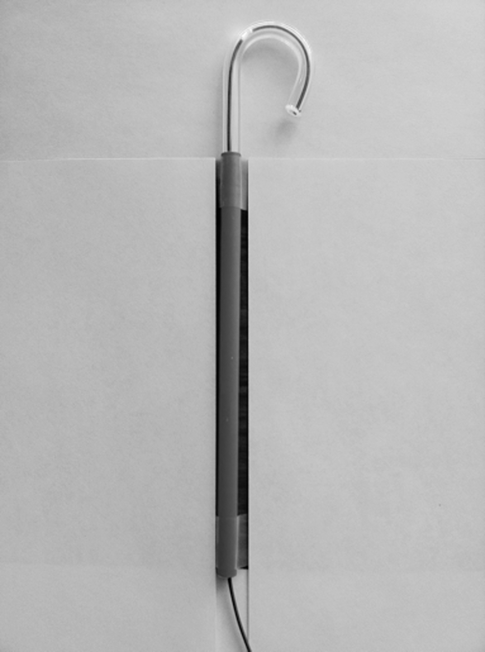
A new 200-μm/272-μm SSF with SGS (Optical Integrity Inc., Panama City Beach, FL) was objectively assessed in a new 7.5F flexible distal sensor chip ureteroscope (Storz Flex- XC, Karl Storz, Tuttlingen, Germany) (Fig. 1). Measured data included active upward and downward deflection (up and down) and irrigation flow rates compared with a standard 200-μm/273-μm laser fiber (Cook Urological Inc., Spencer, IN).

ScopeSafe Fiber with Scope Guardian Sheath.
Each ureteroscope was evaluated for flow and deflection with an empty working channel and with a 200-μm and 273-μm OptiLite single use holmium laser fiber (Cook Urological Inc., Spencer, IN) and a 200-μm/272-μm SSF with SGS (Optical Integrity Inc., Panama City Beach, FL). Before insertion of the laser fibers, the fibers were prepared as described in the manufacturer's instructions.
Deflection
Measuring of upward and downward deflection was made by photocopying the ureteroscope completely deflected and taking measurements using a protractor as described by Parkin and colleagues. 4 Measurements for each laser fiber were taken three times. The intersection angle between the tangents to the active deflection segment and deflected tip was considered the deflection angle.
Irrigation flow
Irrigation flow was measured by connecting the working channel inlet of the ureteroscope to an irrigation system set to a pressure of 100 mm Hg. After a 5-minute equilibration time, three flow measurement for each condition were made and recorded. 5
Force
In the completly defected Flex XC, we assessed the force that is needed to pass with the SSF and the SGS (200 μm and 272 μm) compared with standard laser fibers (200 μm and 273 μm) by using a tensiometer (Digital Force Gauge DS-2, Imada Inc, Northbrook, IL). Each tensiometry measurement was repeated five times and the mean value was calculated.
Working channel model
Ten nonassembled working channel elements from the Storz Flex-XC ureteroscope were tested in a 0-degree, 90-degree, and 210-degree deflection model as shown in Figure 2. The 200-μm/272-μm SSF with SGS and 200-μm/273-μm standard laser fibers were inserted into an irrigated working channel (0.9% NaCl) in cycles of 10 insertions. After 40 insertions, the insertion cycle was reduced to five insertions. After air drying the working channel with pressured air, each test cycle was followed by an external visual inspection for damage and an endolumenal video examination of the working channel using a 2.4F flexible Storz fiberscope (Karl Storz, Tuttlingen, Germany) that was passed into the working channel elements (Fig. 3). Damage to the working channel was classified as superficial scratches, demarcated abrasions, or perforations.
The 210-degree deflection model.
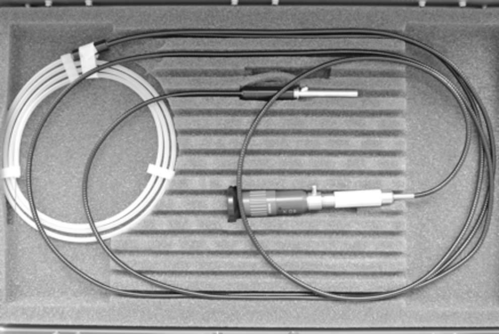
The 2.4F flexible Storz fiberscope.
Laser model
Evaluation of the SSF with SGS consisted of different deflected configurations (0-degrees, 90-degrees, and 210-degrees) in a water bath, with the fiber in the sheath. We fired a 20 W Ho:YAG laser (Calculase II, Karl Storz, Tuttlingen, Germany) with the fiber tip beyond the end of the sheath for 30 seconds at 1.2 J and 10 Hz or until fiber failure. All fibers were bent to 90-degrees and to 210-degrees with a radius of 3 cm. The radius was then decreased to 1.5 cm. Afterward, the bend radius was decreased by 0.5 cm steps until a radius of 0.5 cm was reached. Six trials per fiber were completed.
In addition, the SSF and SGS were also deflected (0-degrees, 90-degrees, and 210-degrees) as shown in part 1 in a water bath, with the fiber in the sheath. The tip of the fiber was aimed directly at the point of maximal deflection of the sheath at a radius of 3, 1.5 and 0.5 cm. The laser was fired for 30 seconds at 1.2 J and 10 Hz with the goal to characterize whether the sheath provides any protection against worst case stray energy and was performed in triplicate.
Statistical analysis
Statistical comparison for continuous variables were analyzed using a Student t test. All analyses were performed with SPSS version 19 (IBM Corporation, Armonk, NY).
Results
Deflection
The maximal deflection (up/down) for the Storz XC with an empty working channel was 296.8 degrees/292.7 degrees. The standard Cook laser fibers (200-μm/273-μm) diminished the deflection (up/down) by 7.66 degrees/6.50 degrees and 40.0 degrees/42.5 degrees, respectively. By using the new SSF with SGS, we measured a diminishment in deflection by 11.16 degrees/10.84 degrees and 44.66 degrees/46.34 degrees, respectively. Consequently, the use of the SGS with the SSF resulted in a 4.7-degree/3.8 degree (1.2%/1.5%, P=0.0001/P=0.0001) diminishment in deflection (up/down) for the 200 μm and a 3.5 degree/4.3 degree (1.8%/1.5%, P=0.0001/P=0.0005) diminishment for the 272-μm laser fiber compared with the standard 200/272-μm laser fiber (Table 1).
SD=standard deviation.
Irrigation flow
The irrigation flow for the ureteroscope with an empty working channel was 62.38 mL/min (SD 0.57 mL/min). The use of an 200-μm/273-μm Cook laser fiber resulted in a 30.33 mL/min/34.83mL/min (51.7%/53.4%) reduction in flow (P<0.0005). Irrigation flow rate was diminished with the use of the SSF with SGS on both the 200-μm and 272-μm laser fiber by 39.7 mL/min (60.1%) and 43.96 mL/min (70%), respectively (P<0.0005) (Table 2).
SD=standard deviation.
Force
For the 200-μm SSF with SGS, the force was 0.32N and for the 272-μm, 0.5N, respectively. Both 200-μm and 273-μm standard laser fibers could be barely inserted. The force of insertion reached maximal resistance; therefore, no measurements could be performed.
Working channel model
There were no channel perforations or damage with 100 laser fiber insertions with the 200-μm/272-μm SSF with SGS in the 0-degree, 90-degree model as well as in the 210-degree model.
With the standard laser fiber, in the 210-degree model, superficial scratches and demarcated abrasions (Fig. 4) were visible after 10 and 60–70 insertions for the 273-μm laser fiber and after 30 insertions (superficial scratches) for the 200-μm laser fiber (Table 3).
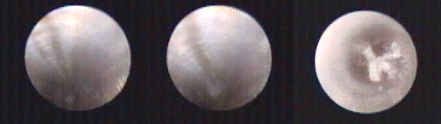
Working channel–scratches– abrasion–perforation.
In the 0-degree model, there was no note of either superficial scratches, abrasions, or perforations, whereas in the 90-degree model, superficial scratches occurred after 20 insertions for the 272-μm fibers and after 40 insertions for the 200-μm laser fibers. No demarcated abrasions were seen after 100 insertions. In the 210-degree model, we saw one perforation (Fig. 4) after 110 insertions with the 273-μm fiber, but none with the 200-μm fiber.
Laser model
No fiber failures were identified for the different laser fibers in the 0-degree, 90-degree, and 210-degree models for the different radiuses from 3 cm to 0.5 cm after six trials (3 min).
In the 0-degree and 90-degree models (radius 3 cm), the sheath and cladding for both laser fibers (200 μm and 272 μm) were damaged after a mean of 7 seconds, but the laser energy did not break the fiber.
In the 210-degree model (radius 1.5 cm) for the 200-μm fiber, the laser energy caused damage to the sheath and the cladding, but did not break the fiber during the application. Unfortunately, the fiber did break during the removal. In the 210-degree model with a radius of 0.5 cm, the fiber broke after a mean of 6.6 seconds.
In the 210-degree model (radius 1.5 cm) for the 272-μm fiber, the laser energy broke the fiber after a mean of 20 seconds (two fibers) and caused damage only to the sheath for one fiber. In the 210-degree model (radius 0.5 cm), the fiber broke after a mean of 8 seconds (Fig. 5).
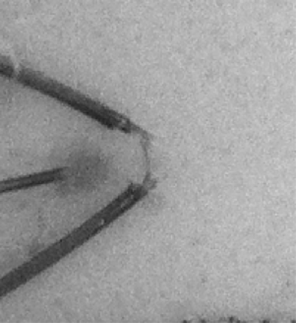
Laser model 210 degrees (radius 0.5 cm).
Discussion
The use of Ho:YAG laser lithotripsy in ureteroscopy is a well established technique for treating patients with ureteral and renal caliculi. Scope damage from laser fibers, however, remains poorly characterized. Several studies identified risk factors for working channel damage such as lower pole access, multiple passes of the laser fiber per operation, and potentially the ureteroscope processing technique. 6,7 As such, these endoscopes need repair after an average of 6 to 15 operative cases with a higher failure rate for lower calix cases. 1
Seto and coworkers 8 reported the sharp-edged distal tip of the laser fiber as the origin for the mechanical damage to the working channel. The working channel injury may be avoided by using a 2F protection tube for the laser fiber with no diminishment in deflection, but decreased flow as reported by Hollenbeck and associates. 9
The SGS was introduced to minimize the mechanical damage to the working channel during the insertion of the laser fiber in a deflected and nondeflected ureteroscope. The clinical setup for the SSF/SGS includes gentle insertion into the working channel of the ureteroscope. Once the tip of the laser fiber is visible at the tip of the ureteroscope, the SGS is retracted 1 cm and fixed in this position with a Tuohy-Borst Adapter (Cook Medical, Bloomington, IN) (Fig. 1).
The outer diameter of the 200-μm SSF plus SGS is 520 μm compared with 375 μm for the SSF alone, leading to a measurable diminishment in flow by 28.7%. The flow did decrease with low pressure irrigation. While we did not test various pressures, increased pressure may mitigate this decrease in flow because we only used a pressure of 100 cm of H20. In addition, this device has not been tested in vivo, so it is unknown how much this would affect visualization. Further studies in these areas are indicated.
Both the 200-μm and 272-μm SSF with SGS caused diminishment in deflection (up and down), but the total percent of deflection affected was minimal at 1.2%/1.5% and by 1.8%/1.5%, respectively (Table 1). These findings were supported by a study of Hollenbeck and colleagues 9 who showed a 16% to 25% decrease in deflection by using a 2F working channel catheter with a 200-μm laser fiber.
In addition, the force that is needed to insert both the 200-μm and the 272-μm SSF and SGS in a fully (270-degree) deflected scope is significantly less compared with the standard 200-μm /272-μm laser fiber. Similar findings were reported by Durak and coworkers, 10 who showed a reduced insertion force for a sheathed laser fiber in a deflected ureteroscope. We recognize that it is commonly suggested that the urologist not push a laser fiber through a deflected ureteroscope. In clinical practice, however, the advancement of a laser fiber through a deflected ureteroscope is common practice and does result in ureteroscope channel damage. The diminished force of passing the fiber and sheath through a ureteroscope and the documented channel preservation in the in vitro deflection model are strong evidence that the use of the SGS may diminish ureteroscope damage. The SGS, however, does not prevent damage from laser energy itself, as shown in our laser test. Fibers typically break at the point of the maximum deflection, and in this case, the SGS will not prevent working channel and ureteroscope damage.
Besides the purchase price, the cost of ownership—meaning maintenance and repair costs—plays a vital role in performing ureteroscopy in high-volume centers. Landman and colleagues 3 projected costs for ureteroscopy and demonstrated that ureteroscope repair is the most significant factor for the cost of ureteroscopy. Regarding repair costs, each of the four major ureteroscope manufactures (Storz, Olympus, ACMI, Wolf) stated that more than 70% of all ureteroscope failures are the result of damage of laser fibers either by the laser fibers alone or the incorrect use of holmium laser energy. 3
The new SSF with SGS has an official sales price of $1125 ($956.25 local price) for the single use 200-μm and 272-μm laser fiber compared with a sales price of $369.75 (local price) for the single use standard Cook laser fiber. Afane and associates 1,3 stated in their study that all the tested flexible ureteroscopes needed major repairs after 6 to 15 procedures with repair costs between $3500 and $5900. If we assess the cost of ownership with ureteroscope use in 50 cases a year (without included warranty), we deal with costs between $31,005 and $36,512 for the major ureteroscope manufactures. 3 Reports from these major ureteroscope manufacturers (Wolf, Storz, ACMI, Olympus) state that more of 70% of the ureteroscope failures are the result of working channel damage by incorrect use of laser fibers. 3 The additional costs of 12 procedures by using the SSF with SGS compared with a standard laser fiber amount to: 12 procedures×$586.50 (sales price difference)=$7038 compared with repair costs of $7000 to $11,800 (2×$3500 – 2×$5900). Therefore, the SSF with SGS needs at least double the number of procedures performed for a major repair to occur to be cost efficient.
The current article supports that the use of SSF and SGS prevents physical working channel damage resulting in prolonged maintenance and repair intervals and, hence, a prolonged utilizability. In addition, the application of the SGS may potentially decrease the cost of ownership and increase lifespan/durability, but clinical evaluations have to prove this statement. Clinical evaluation will be needed to confirm these in vitro results.
Conclusion
The SSF with prevents working channel damage with limited diminishment of deflection and irrigation flow. By preventing working channel damage, the SSF with SGS may decrease repair and maintenance costs and consequently expand the lifespan of flexible ureteroscopes. Clinical evaluation will be needed to confirm these in vitro results.
Footnotes
Disclosure Statement
No competing financial interests exist.