
Abstract
Purpose:
This experimental study was designed to explore the protective effect of apocynin, the NADPH-oxidase inhibitor, on kidney damage induced by ischemia/reperfusion (I/R) in a rat model.
Methods:
Thirty-two rats were randomly divided into a control group and three I/R groups (1-hour ischemia followed by 23-hour reperfusion). Three I/R groups were treated by apocynin (20 mg/kg, i.p.) at two different time points (before ischemia and during ischemia). The histopathological findings, including apoptotic changes, and also tissue malondialdehyde (MDA), superoxide dismutase (SOD), catalase (CAT), glutathion peroxidase (GPX), reduced glutathione (GSH), myeloperoxidase (MPO), blood urea nitrogen (BUN), and serum creatinine (Cr) levels, were determined.
Results:
Kidney tissue MDA and MPO, and serum BUN and Cr levels were found to be significantly higher in the I/R group, but there was no statistically significant difference in the levels of SOD, CAT, GPX, and GSH between the I/R and the control groups. Although apocynin significantly reduced MDA and MPO in group 3 and increased GPX in both treatment groups when compared to the I/R group, the elevated BUN and Cr levels were significantly reduced in treatment groups. Renal I/R injury also induced extensive tubular necrosis, glomerular damage, and apoptosis in the histological evaluation. Apocynin, especially when used during ischemia, ameliorated these histological damages in different amounts in treatment groups.
Conclusion:
The beneficial effects of apocynin on renal I/R injury were evaluated for the first time.
Introduction
Many studies have shown that production and releasing of ROS by inflammatory cells, endothelial cells, platelets, injured cells, and cell debris, via many enzymatic mechanisms 7 is one of the most important reasons for reperfusion injury. 8 –10 ROS, which also take part in host defense mechanism, show their effect on cell by changing the structure of DNA, proteins, and lipids. 7,11,12 To prevent or decrease the kidney damage that can be due to I/R injury, several antioxidants and anti-inflammatory agents, which have effects on production of ROS and inflammatory reactions, have been used in experimental studies. 13 –16 However, there have been no studies concerning the protective effect of apocynin against renal I/R injury.
NADPH-oxidase (NOX) is a major enzyme that uses NADPH to generate superoxide, initial ROS molecule, from oxygen. Apocynin (4-hydroxy-3methoxy-acetophenone), naturally occurring methoxy-substituted catechol, is an inhibitor of NOX and extracted from the roots of Apocynum cannabinum (Canadian hemp) and Picrorhiza kurroa (Scrophulariaceae). 17 It was used to treat dropsy and heart troubles in India and currently seems to be an encouraging therapy for hypertension in the light of many experimental studies. 18,19
Here we explore the protective effect of apocynin on kidney damage induced by I/R in an in vivo rat model. To examine this, we evaluated histopathological findings and biochemical analyses (including apoptotic changes, and tissue malondialdehyde [MDA], superoxide dismutase [SOD], catalase [CAT], glutathione peroxidase [GPX], reduced glutathione [GSH], myeloperoxidase [MPO], blood urea nitrogen [BUN], and serum creatinine [Cr] levels).
Materials and Methods
Animals and experimental protocol
The experimental protocol in the current study was approved by the Ethics Committee on Animal Research of Inonu University, and the Guidelines for Animal Research from the National Institutes of Health publication were followed in the all experimental procedures. Thirty-two female, postpubertal (10–12 weeks) Wistar Albino rats weighing 180 to 220 g were obtained from the Inonu University Laboratory Animals Research Centre and maintained in a light (12-hour light and 12-hour dark cycle), temperature-controlled (21°C±2°C), and humidity-controlled (60%±5%) room, on a standard commercial pellet diet and water ad libitum.
The rats were randomly divided into four groups (n=8), as follows: group 1, control, only underwent right nephrectomy; group 2, I/R; group 3, 20 mg/kg apocynin (Sigma-Aldrich) given i.p. 30 minutes before the ischemia; group 4, 20 mg/kg apocynin given i.p. 30 minutes after the beginning of the ischemia. The experimental model in this study was designed in accordance with our previous studies. 20,21 Also, the dosage of apocynin was chosen according to the previous dose–response study of Connell et al. 22
Surgical procedure
Ketamine (70 mg/kg) and xylazine (8 mg/kg) were used as anesthetics before the operation. The surgeons were blinded to the treatment groups. Each animal's lumbar area was shaved with electric clippers, and then prepared with povidone-iodine (Poviiodeks®; Kim-Pa Corporation). During the surgical procedure, a local sterile environment was provided for aseptic conditions. Following preparation of the surgical area, a midline laparotomy was carried out and right nephrectomy was performed on the rats in all the groups. After the right nephrectomy, no further surgical procedure was performed for 30 minutes, to enable for circulatory adjustment. Then, the left renal artery and vein were occluded together by an atraumatic clamp for 60 minutes. Following 60 minutes of ischemia, a reperfusion period began by removing the clamp. Left kidney reperfusion was maintained for 24 h by removing the clamp; re-initiation of pulsation through the vessel was verified visually. After controlling the bleeding, the skin and skin textures were sutured again. At the end of 24 hours, all animals were euthanized with high doses of the anesthesia mixture, and the kidneys were quickly removed, decapsulated, and divided equally into two longitudinal sections. One section was placed in a formaldehyde solution for routine histopathological examination by light microscopy. The other half of the kidney was placed in liquid nitrogen and stored at −70°C until assayed for MDA, SOD, CAT, GPX, GSH, and MPO. Trunk blood was extracted to evaluate serum levels of BUN and Cr, using an Olympus Autoanalyzer (Olympus Instruments).
Biochemical analyses
Determination of MDA production
The MDA contents of the homogenates were determined spectrophotometrically by measuring the presence of thiobarbituric acid reactive substances (TBARS). 23 Three milliliters of 1% phosphoric acid and 1 mL 0.6% thiobarbituric acid solution were added to 0.5 mL of homogenate pipetted into a tube. The mixture was heated in boiling water for 45 minutes. After the mixture cooled, the colored part was extracted into 4 mL of n-butanol. The absorbance was measured by a spectrophotometer (UV-1601; Shimadzu) at 532 and 520 nm. The amount of lipid peroxides was calculated as TBARS of lipid peroxidation. The results were given in nmol/g tissue, according to a prepared standard graph.
Determination of SOD activity
The total SOD activity was measured according to the method of Sun et al. 24 The principle of the method is the inhibition of nitroblue tetrazolium (NBT) reduction by the xanthine–xanthine oxidase system as a superoxide generator. One unit of SOD was defined as the enzyme amount causing 50% inhibition in the NBT reduction rate. The SOD activity was calculated as U/g protein.
Determination of CAT activity
The CAT activity was determined according to Aebi's method. 25 The principle of the assay is based on the determination of the rate constant (k, s−1) or the H2O2 decomposition rate at 240 nm. The results were given as k/g protein.
Determination of GPX activity
The GPX activity was measured by the method of Paglia and Valentine. 26 An enzymatic reaction in a tube containing NADPH, GSH, sodium azide, and glutathione reductase was initiated by adding H2O2, and the change in the absorbance at 340 nm was observed by a spectrophotometer. The GPX activity was calculated as U/mg protein.
Determination of GSH
The GSH concentration in homogenate was measured spectrophotometrically according to the method of Ellman. 27 Each homogenate sample was mixed with 10 mM 5,5′-dithiobis(2-nitrobenzoic acid) in 100 mM potassium phosphate buffer (pH 7.5) and 17.5 M ethylenediaminetetraacetic acid (EDTA). The reaction was started by the addition of 0.5 U of GSH reductase and 0.4 mM NADPH. The absorbance was measured at 410 nm after 5 minutes. The concentration of GSH was calculated against a standard curve. The results were given in μmol/g tissue.
Determination of MPO activity
The MPO (EC 1.11.1.7) activity was determined by using a 4-aminoantipyrine/phenol solution as the substrate for MPO-mediated oxidation by H2O2 and change in the absorbance at 510 nm was recorded. 28 One unit of the MPO activity was defined as the amount causing degradation of 1 μmol H2O2/min at 25°C. The results were given in U/g protein.
Histological analyses
The kidney tissue was fixed in 10% formalin, and then embedded in paraffin. The 5-μ-cut sections were stained with hematoxylin and eosin. The sections were evaluated for the presence of the tubular cell necrosis, desquamation, interstitial congestion, glomerular shrinkage, and enlargement of the Bowman's space (Fig. 1). For immunohistochemical analysis, thick sections were taken onto polylysine-coated slides. After rehydrating, the samples were transferred to a citrate buffer (pH 7.6) and heated in a microwave oven at 65°C for 20 minutes. After cooling for 20 minutes at room temperature, the sections were washed with phosphate-buffered saline (PBS). The sections were kept in 0.3% H2O2 for 7 minutes, and then washed with PBS, after which they were incubated with primary rabbit-polyclonal cysteine aspartate-specific proteinase (caspase-3) (Neomarker). 29 The sections then were rinsed in PBS and incubated with biotinylated goat anti-polyvalent for 10 minutes and streptavidin peroxidase for 10 minutes at room temperature. The staining procedure was completed with chromogen+substrate for 15 minutes, and the slides were counter-stained with Mayer's hematoxylin for 1 minute. Caspase-3 was used according to the manufacturer's instructions, with a minor revision. Stained tubules with caspase-3 were measured in ten different fields, using a Leica Q Win Image Analysis System (Leica Micros Imaging Solution Ltd.) (X20). The histological slides of the kidney tissue were evaluated for semiquantitative analysis and graded as follows: 0, normal; I, areas of tubular epithelial cell necrosis and desquamation involving <25% of cortical tubules; II, similar changes involving 25%–50% of cortical tubules; III, similar changes involving 50%–75% of cortical tubules; IV, similar changes involving >75% of cortical tubules (Table 3). The sections were examined with a Leica DFC 280 light microscope by an experienced observer unaware of the animal treatment groups.

Histological photographs of kidney tissues from groups:
Statistical analysis
For detecting even minor effects, the required sample sizes used in this experiment were identified by using statistical power analysis. The sample sizes necessary for a power of 0.80 were estimated by using NCSS software. Data were analyzed using the SPSS software program for Windows, version 15.0, (SPSS, Inc.). The normality of the distribution was confirmed through a Kolmogorov–Smirnov test. According to the results obtained from the normality test, one-way analysis of variance (ANOVA) and a Kruskal–Wallis H test were used for the statistical analysis, as appropriate. Multiple comparisons were carried out by the Tamhane's test after the ANOVA. The results are expressed as mean±standard deviation. After a significant Kruskal–Wallis H test, a Conover test was also carried out for SOD, GSH, BUN, and renal tissue weight. p<0.05 was considered statistically significant. The values were given as median (min–max).
Results
Body and kidney weight
No animals died during the experiment period. There were no differences between the body weights before and after the experiments among the groups (data not shown). The kidney weights of rats of the I/R group were significantly higher than those of the control group (1.02 [0.97–1.40] vs. 0.76 [0.67–0.98], respectively), whereas apocynin did not reduce the kidney weight to the control levels (Table 1).
p<0.05 versus group 1; b p<0.05 versus group 2.
BUN=blood urea nitrogen; Cr=creatinine; SD=standard deviation.
Effect of apocynin on serum parameters
As shown in Table 1, I/R caused a significant increase in serum levels of BUN and Cr (334.8 [225.4–378.0] and 2.34±0.501 mg/dL, respectively), when compared to the control group (56.9 [49.5–80.0] and 0.45±0.046 mg/dL, respectively). In treatment groups (group 3 and 4), the elevated BUN and Cr levels were significantly reduced when compared to the I/R group.
Effect of apocynin on renal tissue enzymes and lipid peroxides
As shown in Table 2, kidney tissue MDA and MPO levels were found to be significantly higher in the I/R group (24.88±6.15 nmol/g tissue and 25.75±5.24 U/g prot, respectively), when compared to the control group (11.21±3.01 nmol/g tissue and 10.96±3.64 U/g prot, respectively). However, there was no statistically significant difference in the levels of SOD, CAT, GPX, and GSH between the I/R and the control groups.
p<0.05 versus group 1; b p<0.05 versus group 2.
MDA=malondialdehyde; SOD=superoxide dismutase; CAT=catalase; GPX=glutathione peroxidase; GSH=reduced glutathione; MPO=myeloperoxidase.
In the treatment groups, the elevated MDA levels were reduced to nearly control levels and this reduction was statistically significant only in group 3 (apocynin given 30 minutes before the ischemia) when compared to the I/R group. In addition, this ameliorating effect was found to be parallel to the results of GPX, which reached statistically meaningful levels compared to the I/R group, in the treatment groups. Although apocynin reduced the levels of MPO, the reduction was statistically significant in group 3 when compared to the I/R group (16.77±3.75 U/g prot vs 25.75±5.24 U/g prot, respectively). On the other hand, there was no significant change in the levels of SOD, CAT, and GSH in the treatment groups.
Histological results
The slight histological alterations, such as epithelial desquamation, were seen in the control group (Fig. 1A). On the other hand, group 2 exhibited extensive cortical damages. Moreover, widespread tubular desquamation, tubular necrosis, and interstitial congestion were observed. There was also shrinkage of nearly all the glomeruli with enlargement of the Bowman's space in the same group (Fig. 1B). The tubular necrosis and desquamations like patchy areas were revealed in the renal cortex in group 3 by light microscopy. The damaged glomeruli were still present in group 3 (Fig. 1C). Although mild tubular changes, such as tubular atrophy and desquamation, were present, the glomeruli were intact in group 4 (Fig. 1D). The results of the semiquantitative histological analyses with ratios are shown in Table 3.
The caspase-3 immunostaining was not seen in the tubules of the control group (Fig. 2A). On the other hand, in group 2, caspase-3-positive cells were found to be significantly increased when compared with the control group (Fig. 2B). Although apocynin given before I/R did not prevent apoptosis (group 3), its administration during I/R significantly decreased the expression of apoptotic cells (group 4). The coloring density and the number of caspase-3-stained tubular cells were lower in group 4 than in group 3 (Fig. 2C, D). Caspase-3 immunostaining was not observed in glomeruli of any groups. The results of staining with caspase-3 are shown in Table 4.
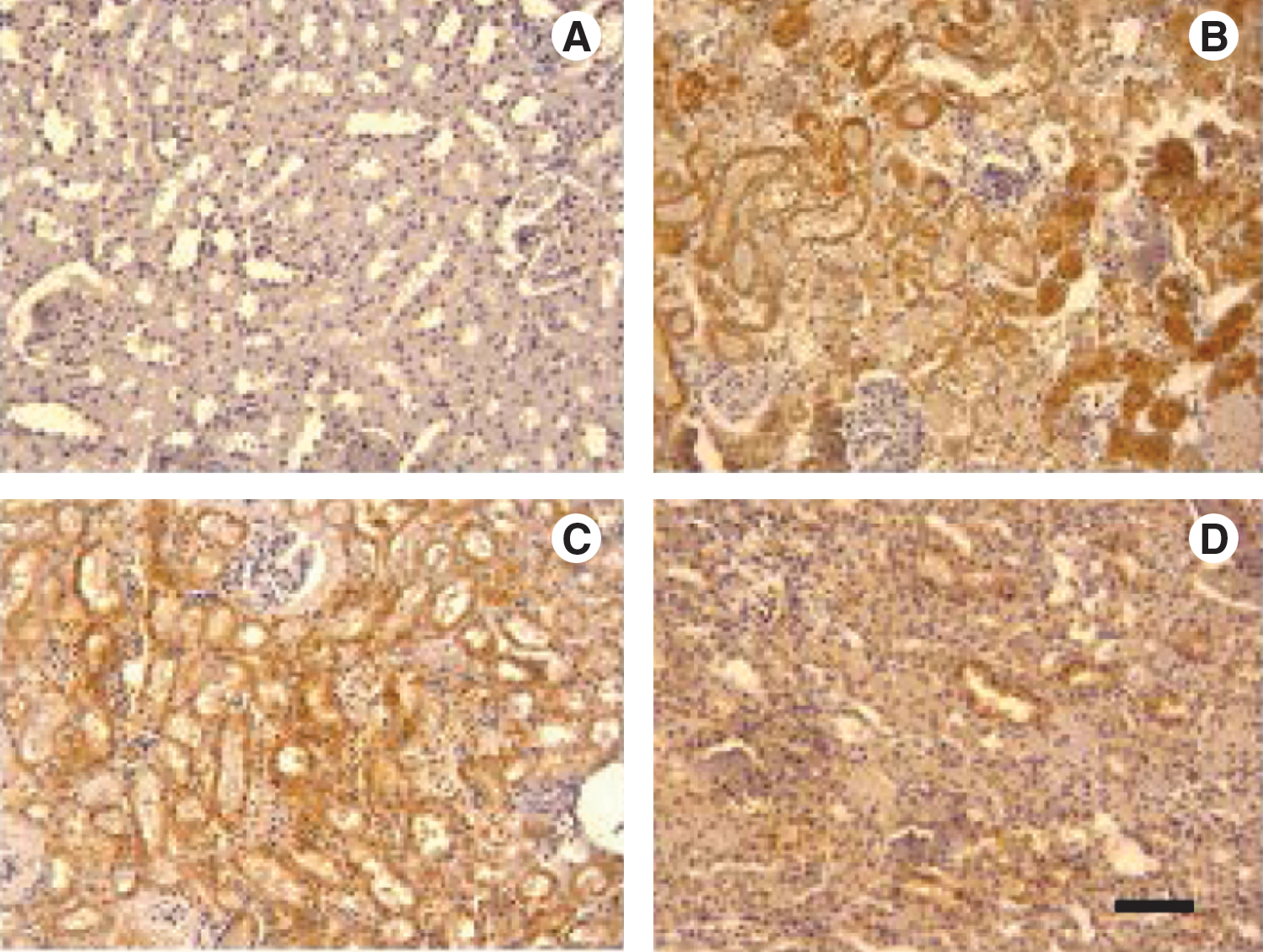
Histological photographs of kidney tissues after caspase-3 immunostaining:
Significantly increased when compared with group 1, p<0.0001.
Significantly decreased when compared with group 2, p<0.0001.
Discussion
Renal I/R injury is a major reason for renal dysfunction 30 that occurs via intracellular injury and inflammatory reactions, which are related to the each other. Renal ischemia, which can be due to sepsis, shock, hypotension, 1 some renal surgeries, including renal stone surgery, partial nephrectomy, renal transplantation, and renal artery revascularization, or cardiothoracic surgery, 2 –5,31 is the beginning of the injury process and starts with a reduction of energy production in the mitochondria. Hence, cellular ion imbalance, increase of activity of proteases, and phospholipases exist, which are all causes of an increase in cell membrane permeability. The duration of the ischemic injury is the main factor for viability of the cell. 7 Eventually, a necrotic type of cell death can occur due to a long ischemic period.
The intracellular pathologic changes happened in the ischemic period result in further tissue damage during the reperfusion period following ischemia. 7,32 The damaged enzymes, which have the role in electron transferring in the mitochondria, cause electrons transfer to oxygen, in turn resulting in the generation of superoxide, that is, the initial ROS molecule. The overproduction of ROS, which is a part of host defense mechanism, gives rise to oxidative stress that has a major role in the formation of I/R injury, 31,33,34 thus causing membrane lipid peroxidation, oxidation of cell proteins, damage to the DNA helix, and cell death. 7,11,12 Therewithal, the reperfusion period initiates an inflammatory response cascade that can take a long time and causes irreversible tissue damage. 7,11,35 Many drugs that block the oxidative stress and inflammatory response cascade have been used to prevent or decrease renal I/R injury in several experimental studies. 15,16,20,36 –39 Nowadays, ice-slush, mannitol and furosemide have been used to protect the renal tissue, as much as possible, during some renal surgical procedures, such as partial nephrectomy. 40,41 However, the new experimental studies will help us to find the most appropriate feasible treatment.
Apocynin, naturally occurring methoxy-substituted catechol oxidized by peroxides to a more potent dimer in the cell, 42 is a selective NOX inhibitor, which generates superoxide. The superoxide anion is a central and initial ROS molecule and converts to more reactive and toxic-free radicals, such as hydrogen peroxide, hydroxyl radical, or peroxynitrite, under conditions of oxidative stress. 43 Although the process how apocynin inhibits the NOX activity is poorly understood, it is thought to act by preventing the translocation of its cytosolic components, p47phox, to gp91phox. 44 –46 In addition to NOX inhibition, many studies have shown that apocynin may inhibit cytochrome P450, thromboxane synthase, and COX-2 expression. 18,47,48 Apocynin, which has low toxicity and specificity, may be of promising potential therapy for asthma, arthritis, neurological, and cardiovascular diseases 18,22,49 –52 via antioxidant and anti-inflammatory effects. Also, it has been used in many experimental studies related to I/R injury. In these studies, apocynin was shown to be effective in protecting the tissue against the harmful events induced by I/R injury. 22,53,54
In our study, the MDA levels significantly increased by I/R procedure, whereas these high levels reduced to nearly control levels in the treatment groups. However, the reduction of MDA levels was statistically meaningful only in group 3, in which apocynin used 30 minutes before the ischemia, when compared to the I/R group. MDA is a major indicator of oxidative stress, and it increases due to lipid peroxidation, which is one of the harmful consequences of I/R injury. 55 I/R rapidly causes cellular injury associated with lipid peroxidation. Whereby lipid peroxidation is the main pathway of oxidative stress, regardless of the source of free radicals, blocking this pathway may be an effective strategy to prevent ROS-mediated kidney damage. 13
MPO is a particular oxidase in polymorphonuclear leukocyte (PMN) and the MPO activity in tissue is used to estimate the PMN chemotaxis and infiltration. 56 PMN infiltration during the reperfusion period may cause generation and releasing of additional large amount of oxidants that exacerbates this harmful cascade. Apocynin, which is also activated by MPO, 57 reduces the generation of inflammatory mediators by inhibiting NOX. However, it does not impress the defensive property of PMN. 17 In the current study, I/R injury caused a significant elevation in tissue MPO activity when compared to the control group. Apocynin usage ameliorated the MPO activity in the treatment groups and this improvement was significant when apocynin used 30 minutes before the ischemia.
In addition to many properties within the cell cycle, GSH and GPX are two important components of the protective mechanism of the cell against lipid peroxidation and oxidative stress, which occur during I/R injury. 58,59 GSH and GPX convert hydrogen peroxide to water; thus, they prevent the formation of more reactive-free radicals. Some studies showed that GSH can be reduced by apocynin, contrary to expectation, and oxidized by apocynin-free radicals, which are formed via activation by MPO, in alveolar cells. 46,60 In the current study, although apocynin did not cause considerable change in GSH levels, it significantly increased the GPX activity in the treatment groups when compared to the I/R group.
The levels of SOD and CAT did not reach a statistically meaningful level in the groups. SOD and CAT are antioxidant enzymes that are components of the defense mechanism against ROS activities. The levels of these enzymes within host increase to protect the tissues during I/R injury. 61,62 Any agent used to increase the level of these enzymes can prevent the possible damages that occur during oxidative stress. However, apocynin, a significant inhibitor of NOX and uses the NOX pathway to suppress oxidative stress, did not show its effect by increasing these enzyme levels in the present study. On the other hand, according to our knowledge, there are some other pathways, using xanthine oxidase or GSH, that continue to process during I/R injury.
Also, herein renal I/R caused an elevation in BUN and Cr levels in the serum specimens when compared to the control group. One of the other important findings of our study was an improvement of serum levels of BUN and Cr with apocynin. Although BUN and Cr levels did not reach to the control levels, there was significant amelioration in the treatment groups when compared to the I/R group. Elevation of serum levels of BUN and Cr is a sign of impairment in glomerular function. 14 Recently, it has been reported that for humans, serum Cr, in association with certain other clinical characteristics, may be a more definite assessment of the glomerular filtration rate than Cr clearance. The impairment in glomerular function was accompanied by an increase in BUN. In the earlier phases of kidney disease, the serum Cr level is more meaningful than the BUN level. However, BUN begins to elevate only after a prominent renal parenchymal injury. These findings are coherent with ischemic injury related to kidney damage. 63
In the current study, apocynin prevented particular effects of renal I/R injury, such as the decreased level of GPX and the increased levels of MDA, MPO, BUN, and Cr. The kidney weights of the I/R rats were significantly higher than those of the control group. I/R injury also caused both an increase in the kidney weight and extensive tubular atrophy and necrosis, marked tubular cell apoptosis, and glomerular damage as seen in the histological evaluation. However, apocynin improved these histological damages in different amounts in the treatment groups (Figs. 1 and 2). There are many studies that have shown that renal I/R injury induces renal blood flow changes via microvascular injury and impaired renal vascular reactivity, and as a result, infiltration of inflammatory cells, tubular epithelial cell injury, glomerular damage, apoptosis, and fibrosis occur. 64 –70 In our study, apocynin ameliorated tubular necrosis and glomerular alterations; however, the improvement was more obvious when it was used during the ischemic period, in the treatment groups. However, as we know, apocynin may induce apoptotic cell death as a different strategy to protect the tissue from the additional harmful effect of necrotic cell death occurred during I/R injury. 22,71 Parallel to the knowledge mentioned above, we found that apocynin induced an increase in apoptosis when it was used before I/R injury, whereas it, when used during I/R injury, reduced apoptosis in tubules. Also, Connell and colleagues reported same effect of apocynin in cerebral I/R injury. 22 Although apocynin caused alterations in the histological damages, it did not decrease the kidney weights in our study.
Conclusion
In this study, the beneficial effects of apocynin on renal I/R injury were evaluated for the first time. Apocynin improved the kidney damage, which occurred after I/R injury, in different amounts in the treatment groups. Although improvement in the histological appearance was more clear when apocynin was used during the ischemia, levels of MDA and MPO, which have been used as the general biochemical determinants of I/R injury, were significantly reduced when apocynin was used before the ischemia.
In sum, apocynin, a NOX inhibitor, is considered safe, but less selective. According to our results, apocynin could be used to prevent the kidney damage induced by I/R injury occurring in many ways, after further clinical and experimental trials, including different treatment dosages and timing for inductions.
Footnotes
Disclosure Statement
No competing financial interests exist.