
Abstract
Background and Purpose:
The recently described Galdakao-modified supine Valdivia position for percutaneous nephrolithotomy (PCNL) has become increasingly popular. We have made further modifications to this and describe our recent experience compared with our previous prone cases.
Patients and Methods:
From April 2011, all patients undergoing PCNL have been placed in the modified supine position. A suction beanbag is used to secure the patient and improve renal access. Data on patient age, comorbidities, stone size, operative time, radiation exposure, complications, stone clearance, and length of stay was collected, analyzed, and compared with data from our previous year's prone surgery.
Results:
Thirty-six patients in each group underwent 41 PCNLs. The groups were well matched for age, sex, and comorbidity. The supine patients tended to have a higher body mass index. Stones in the supine group were larger (32.6 vs 25.7 mm, P=0.0402), and the operative time was shorter (86.2 vs 116.6 min, P=0.003). Radiation time was similar in the two groups, but the dose was higher in the supine group. Stone clearance rates, length of stay (2.5 days), and complications were similar. Nineteen (46%) patients underwent simultaneous lower urinary tract procedures including 4 (10%) with complete staghorn calculi for which ureterorenoscopy was used to fragment ureteral and upper renal pole stones.
Conclusions:
The modified supine position for PCNL has a number of advantages for the patient and staff compared with the prone position. Despite a more obese study group with larger stones, we have maintained stone clearance rates and significantly reduced operative time with no increase in complications. The technique has been easy to learn and teach. A major advantage has been simultaneous access to the lower urinary tract for ureteroscopy and stent placement, and this has helped with complete stone clearance.
Introduction
Patients and Methods
From April 2011, all patients with large or complex urinary tract stones who were planned to undergo PCNL were placed in the modified supine position as described by Ibarluzea and colleagues. 9 One of the issues we encountered was a paucity of good descriptions for the position, with most articless merely referring to pictures. 10,11 As we developed our experience, we made further modifications. We currently ask the patient to lie on a C-shaped vacuum beanbag on the operating table, with the concave area of the beanbag under the operation side. We rotate the lower cushion of the operating table so that the cutout is below the operating site, which allows a greater range of movement for instruments when in the kidney.
Once anesthetized, the patient is pulled down the table and the legs placed in the lithotomy position. The ipsilateral hip is flexed with a flexed knee, and the contralateral leg is abducted and supported in an extended position. The beanbag is rolled under the hips and shoulders to tilt and support the torso at approximately 20 to 30 degrees. The ipsilateral arm is supported with a flexed elbow over the chest with the contralateral arm tucked next to the torso with an extended elbow. Suction is applied to the beanbag, thus supporting the patient in the tilted position (Fig. 1). A standard cystoscopy drape and PCNL drape are positioned after both the genitalia and flank have been prepped.

Modified supine position for left-sided percutaneous nephrolithotomy. Posterior axillary line is marked. Patient is propped up and supported by a vacuum beanbag.
Cystoscopy is performed, and a 6F retrograde ureteral catheter (Cook Urological, Spencer, IN) is placed in the renal pelvis and fixed to a 16F urethral catheter. Percutaneous access is achieved simultaneously by a second surgeon with a 19-gauge tetrafluoroethylene three-piece needle (Cook Urological) through the posterior axillary line. The image intensifier is tilted to 20 to 30 degrees so that an anteroposterior view of the renal tract is maintained (Fig. 2). Depth of the tract is assessed by returning the intensifier to 0 degrees.
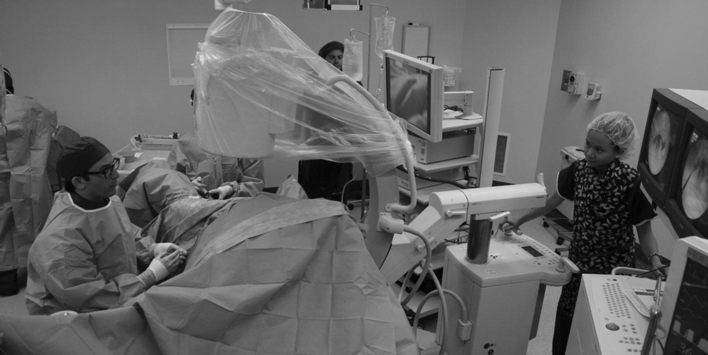
Simultaneous retrograde and left-sided caliceal puncture. Positioning of monitors and image intensifier is demonstrated.
After insertion of guide and safety wires using a 7F Safety Wire Guide Introducer (Cook Urological) the tract is dilated to allow either a 24F or 30F Amplatz tube using balloon dilators (Boston Scientific, Natick, MA or Cook Urological). Nephroscopy is performed using either 17F or 24F Storz rigid nephroscopes with final checks using an Olympus 16F flexible scope. When needed, ureteroscopy is performed simultaneously by the second surgeon using 7.5F Wolf semirigid ureteroscopes or ureterorenoscopy through an access sheath (Boston Scientific) using an 8F Olympus flexible ureterorenoscope.
Stone fragmentation is achieved with our standard selection of lithotripters–Swiss LithoClast Master (EMS Medical, Munich, Germany) and holmium:yttrium-aluminum-garnet laser (Verapulse PowerSuite Holmium 100W, Lumenis, Yokneam, Israel). In the majority of cases, a 24F silicone tube is left as a nephrostomy and fixed to the skin with a 2-0 absorbable suture, although no tube is left after short cases with no stone fragments or tract bleeding.
Patient data collected prospectively included age, sex, comorbidity, and body mass index (BMI), stone size (defined as the maximum length of stone either on CT or plain radiography of the kidneys, ureters, and bladder [KUB]) and position, operative time (defined as start to end of procedure, not including anesthetic, positioning, or recovery time), radiation time and dose, complications, length of inpatient stay, and stone clearance (defined as no stone fragments on review on plain radiography or CT of the KUB). The results over the first year were compared with similar data from the immediately preceding procedures in an equivalent number of patients performed by the same surgeon with the patients in the prone position.
Statistical analysis
Data were prospectively collected on a Microsoft Excel spreadsheet and analyzed with the Analyse-It (Leeds, UK) statistical package. Parametric data were compared using the Student t test. Correlation was calculated with the Pearson coefficient. Results were considered significant if P<0.05.
Ethics
The study was approved by the Southern Health Human Research Ethics Committee (Number 12336Q).
Results
Thirty-six patients (23 male, 13 female) underwent 41 PCNLs in the modified supine position. One patient underwent consecutive procedures for bilateral stones, and four others had two procedures each for staghorn or complex stones. The position was also used in an additional eight patients who may have needed PCNL, but in one case, a nephrostomy was left in place when pus was encountered on puncturing, and in the others, the stone was considered more appropriate for a retrograde approach. The procedure of one patient (number 5 in the series) was converted to the prone position because access to the collecting system from the supine position proved difficult.
Fourteen (34%) procedures included simultaneous ureteroscopy or ureterorenoscopy for either actual or suspected ureteral stones or to assist in fragmentation of large/staghorn renal stones (four cases, 10%). An additional four (10%) procedures involved retrograde stent insertion, and one (2.4%) case involved simultaneous litholapaxy.
The majority of punctures were lower pole (25, 61%) with 9 (22%) midpolar punctures, 6 (14.6%) upper pole punctures, and one (2.4%) direct puncture onto a caliceal diverticulum. The majority of punctures were performed by the lead author, although seven trainees also successfully accessed the collecting system.
The results from these patients were compared with those of the previous 41 PCNLs performed by the same surgeon with the patient in the conventional prone position. Again, 36 patients were involved, with five patients undergoing second procedures for staghorn or complex stones. The comparative results are shown in Table 1.
Results are mean (standard deviation). Number of patients in each group, 36; number of procedures, 41.
There was no significant difference in the age or sex distribution of the patients. The stones in the supine group were significantly larger, and while the BMI was not significantly different, there was a trend to larger patients in the supine group. The median BMI in this group was 30.3 (range 14.7–69.9), with only nine (25%) patients considered normal or underweight. Eleven (30.5%) patients were obese (BMI>30) and five (14%) morbidly obese (BMI>40). Overall operative time was significantly shorter by an average of 30 minutes. Radiation time was similar in the two groups, although the radiation dose was higher in the supine group. When this was analyzed separately using all measured variables, the only correlation was with BMI (r=0.66, P<0.0001). Length of stay and complications were similar in the two groups. Two (5%) patients in each group needed blood transfusions and had angiography performed. No patients needed embolization. There was no significant difference in total stone clearance, although there was a trend toward better rates in the supine group (71%–65%).
Discussion
The prone position has been preferred for PCNL ever since the first descriptions, 3 mostly for historic reasons. Valdivia and associates, who first reported the supine position, for many years described excellent results with little uptake elsewhere. 6,7 With a number of other centers taking up the supine position and making further modifications, there has been a recent debate about whether prone is still the best position. 11 –14
We initially positioned patients as described by Ibarluzea and colleagues 9 who used 3 L fluid bags to prop the patient as suggested by Valdivia and coworkers. 6 We found that the patient was not well supported and that there was some movement of the torso while attempting renal access. The addition of the vacuum beanbag has prevented this movement and also kept the patient more securely placed on the operating table. We also slightly modified the leg positions, because flexing the knee on the abducted ipsilateral side gave more room for the operating surgeon and draping proved easier with the extended contralateral leg.
We have found that the modified supine approach reduces operative time for all stones with an average time saving of 30 minutes. This has been reported previously, 15 –17 although some articless have suggested increased time for larger stones. 7 The majority of the time saving is from not having to reposition the patient from the lithotomy to the prone position with the consequent reprepping, redraping, and staff rescrubbing and regowning. There is also some time saved with the ability to perform simultaneous retrograde and caliceal puncture using two surgeons, and we have noted less requirement for repeated nephroscope insertion because of previously reported gravity-induced stone clearance. 17,18
The supposed advantages of the prone position are easier access to the renal upper pole calices, better clearance of staghorn calculi, a more distended collecting system because of the effects of gravity on the irrigating fluid, and an unrestricted range of movement for the nephroscope. 11,12 We previously performed all our PCNL with the patient in the prone position and, in comparison with the modified supine position, found no difference in accessing the most appropriate calix; have shown no change and possibly better clearance of staghorn calculi; have not noticed any observable difference in renal views; and, if anything, have found a better range of movement. We had always found that large buttocks impinge on accessing the renal pelvis and upper pole calices from lower pole punctures. Our described cutout in the theater cushions and the beanbag allow for a vast range of movement.
In particular, we have been impressed with the procedures for the 10% of patients with large/staghorn calculi. Having two surgeons attacking the stones, one with the ureterorenoscope and laser and the other through the renal tract with a nephroscope sped up the procedure and reduced trauma to the patient. In the prone position, these cases often necessitate creation of additional tracts. 19 Fragmentation with the laser does not have to be as complete as during conventional retrograde procedures, because the larger stones can be passed down to the nephroscope operator and removed that way. We have also found no problems with small ureteral fragments, because ureteroscopy or ureterorenoscopy can easily be performed simultaneously. We can therefore almost guarantee complete stone clearance using this position.
Despite ureteral access being easier in this position, even compared with standard lithotomy, we did find that the additional ureteral instrumentation resulted in more postoperative colicky pain and now routinely insert ureteral stents in these circumstances. This is easily achieved with either a retrograde or antegrade approach, and stent position can be confirmed at both ends.
As described previously, 11 the theater can be quite crowded during these procedures, particularly when complete stone clearance is attempted with combined antegrade and retrograde approaches. This necessitates two operating monitors, two scrub teams, and both ultrasonic/pneumatic lithotripters and a laser, in addition to the image intensifier and anesthetic team and kit. Modern monitors allow picture-in-picture enabling image intensifier pictures and camera views on a single screen, thus saving some space, but careful preoperative planning of equipment placement is needed. Our standard theater setup is displayed in Figure 2.
The prone position has a number of potential concerns. 11 There are a number of cardiovascular changes, including a decrease in the cardiac index, obstruction of the inferior vena cava, and potential thrombosis. There can be an increase in sympathetic activity. Turning the patient from the supine to the prone position can cause central and peripheral nerve injuries and affect flow in the carotid and vertebral arteries. Anesthetic difficulties include ensuring appropriate placement of a reinforced endotracheal tube, the need for an increased depth of anesthesia, and ensuring free movement of the abdomen, which often necessitates expensive additional mattresses. 20 Eye problems can include corneal abrasion if the eyes are incorrectly protected and blindness from ischemia, globe damage, and decreased perfusion reported as a rare complication. 20 Direct pressure injuries are also well reported with a probable underreported liver damage related to prolonged procedures. 20
In contrast, there are few reported concerns with the supine position. There may be more mobility of the kidney when attempting access, 9 although we have not noticed that since moving to the vacuum beanbag. Others claim to have similar results using rolled up towels as opposed to 3 L fluid bags. 21 One of the major concerns seems to have been a possible increased risk of perforating the colon and major vessels, and some authors suggest this is the major reason for the supine approaches not being widely used. 22 It seems that the risk of colonic perforation is actually reduced in the supine position, 6,8,11,14,17,22 with CT studies showing the colon is pushed closer to the kidney in the prone position.
It has been suggested that there is an extended learning curve in moving from the prone to the supine position, 23 although the figures quoted compare favorably with the learning curve for prone PCNL. 24 In our study, confidence in achieving access was gained by the lead author inside 10 cases. The main difference was that needle movements were opposite to those learned with the patient in the prone position. The one patient whose procedure was converted to the prone position revealed that the initial attempt at making the tract was too medial compared with the normal technique, and this lesson made subsequent punctures more successful. The majority of the trainees had minimal experience with prone punctures and found the supine technique very easy to learn.
Previous reports have suggested a reduction in operator radiation exposure. 16 We have shown no difference in overall radiation time, although an increased generated radiation dose, most likely associated with the increased patient size. An additional benefit was markedly reduced finger irradiation for the operator, especially the trainees. Tilting the patient and the image intensifier takes the surgeon's hands almost completely out of the radiation field.
Increasing patient obesity is one of the scourges of modern medicine, and there is evidence that obesity increases the risk of stones. 25 Previous reports have shown that PCNL is safe even in the morbidly obese. 25,26 There has been some debate about whether obese patients are best served by the prone or a supine approach. 12,27 We have found no difficulty in renal access and stone removal in our predominantly obese population. After the success of this initial series, our anesthetists are now insisting that any obese patients are only listed for PCNL in the modified supine position, and other centers have suggested major benefits for this group. 20 Theater staff have been impressed with the reduced patient handling, particularly of the large patients. The tilted position helps with skin rolls falling away from the operating side, and access has been relatively straightforward, although extra long needles, Amplatz sheaths, and balloons may have to be used.
Conclusions
Our experience with both positions favors complete stone clearance in the modified supine position. It is essentially the same operation with less patient handling and operator radiation exposure. Despite more complex stones and more obese patients, the operative time has been significantly reduced, and overall results are equivalent with no increase in complications.
Footnotes
Acknowledgments
The ongoing developments in the position have been a team effort, and thanks go to Indra Jolayemi and her team in Casey Hospital theaters. The registrars and Fellows Dan Spernat, Renu Eapen, and David Connelly have all made helpful suggestions.
Disclosure Statement
No competing financial interests exist.