
Abstract
Purpose:
To investigate the feasibility of the all-seeing needle for safe entry and creation of pneumoperitoneum in pediatric urologic laparoscopy.
Patients and Methods:
A total of 14 children underwent various transperitoneal urologic laparoscopic procedures. The all-seeing needle, which is 4.85F in diameter, was used for safe entry into the abdominal cavity at the site of the umblicus in all cases. The microoptic was integrated with the light system and connected via a zoom ocular enabling direct visualization of the layers between the skin and the peritoneal cavity. Once the intraperitoneal access was obtained, CO2 pneumoperitoneum was created from one port of the three-way connector attached to the proximal part of the needle. Then the laparoscopic trocars were placed under vision of the microoptical system.
Results:
Mean age of the children was 4.5±2.9 years. In all children, the all-seeing needle was safely introduced into the abdominal cavity under direct vision. Then, CO2 pneumoperitoneum was succesfully performed. The mean time for optical puncture was calculated as 1.1±0.8 minutes. No complication was encountered during the introduction of the needle, creation of the pneumoperitoneum, and placement of the trocars.
Conclusions:
The all-seeing needle appears to be beneficial in safe entry and for creating pneumoperitoneum in laparoscopic pediatric urology cases. It eliminates the disadvantages of the Veress needle, which is blunt insertion, and may possibly prevent complications.
Introduction
The technique for creating pneumoperitoneum and the first trocar insertion is a critical step during laparoscopic procedures. The two main and worldwide accepted approaches for intra-abdominal entry are the open insertion of the trocar with Hasson technique and blind insertion with the Veress needle. 1,2 Both techniques have unique advantages, depending on the age and characteristics of the patients, in terms of safety, but neither can completely prevent iatrogenic injuries. Nevertheless, the debate has still continued for the best available option for this first step of laparoscopic procedures, even during the last decade. 3 –7
The all-seeing needle has recently been introduced for confirming renal access during percutaneous nephrolithotomy. 8 It has been stated that this instrument increases the safety for creation of the percutaneous renal tract and may decrease the risk of tract-related complications, such as bleeding and bowel injury. This very thin (1.6 mm) instrument, however, has not been applied in laparoscopy yet.
In this study, we aimed to investigate the feasibility and safety of the all-seeing needle (microoptical system) for safe entry and creation of pneumoperitoneum in pediatric urologic laparoscopy. The main advantages of this technique are performance under direct vision and its smaller size. It eliminates the most important disadvantage of the Veress needle, which is the blind insertion.
Patients and Methods
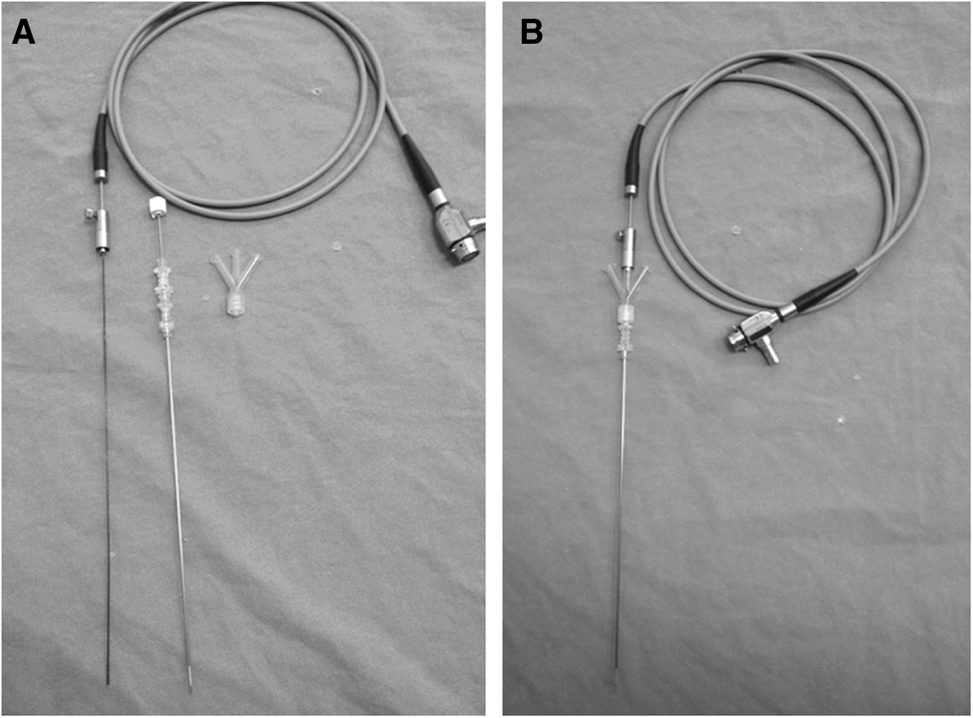
The all-seeing needle is a novel instrument consisting of three main pieces (Fig. 1A). The first one is the 4.85F (1.6 mm, 16 gauge) needle that has an inner stylet. The diameter of the inner stylet is 3.9F (1.3 mm). The second piece of this system is the Y-shaped three-way connector. The third piece is the highly flexible 2.7F (0.9 mm) microfiber optics with resolution of 10,000 pixels (PolyDiagnost, Pfaffenhofen, Germany). Those three pieces are connected with each other in the following order:
1. The inner stylet of the 16-gauge needle is removed.
2. Three-way connector is attached to the proximal end of the needle.
3. Microfiber optic is inserted inside the middle port of the three-way connector.
4. The length of the optical system is adjusted with the aid of an adapter that is attached on the working sheath.
After completion of these steps, the all-seeing needle or the microoptical system is ready for use (Fig. 1B). The microoptic is integrated with the light system and connected via a zoom ocular. Those parts (ocular, camera, light cables) are located outside the sterile area and are stabilized with a special arm, which helps the surgeon for comfortable use. The pressure pump system for temporary irrigation is attached to another port of the three-way connector and is controlled by the surgeon with a foot pedal. Water dissection gives the possibility of better visualization whenever the vision is blurred. Physiologic saline (0.9% NaCl) is used as the irrigant. Once intraperitoneal access is obtained, CO2 pneumoperitoneum is created via the other port of the connector with high-flow insufflation under direct vision.
Operative technique
The all-seeing needle was used instead of a Veress needle for various treansperitoneal laparoscopic urologica procedures in children (Figs. 2A, 2B). All procedures were performed under general anesthesia. The prone position and the 60-degree left or right lateral decubitus positions were used according to the type and the site of the procedure. In all cases, the puncture was performed at the edges of the umblicus, and the skin was retracted by two Langenbeck retractors. Introduction of the needle was visualized from the video monitor in real time, making it possible to identify the subcutaneous tissue, fascial layer, the peritoneum, and the abdominal cavity (Figs. 3A–D).

Intraoperative appearance of the layers between the skin and the peritoneal cavity. Note that the puncture is performed at the edge of the umblicus.
After successful entrance of the all-seeing needle and the creation of penumoperitoneum, the trocars were inserted under direct vision of the optical system. The location of the trocars is decided according to the type and the site of the procedure.
Patient demographics including age, sex, and type of operation were recorded. The duration of the optical puncture was also documented for each patient. In addition, operative time and complications were noted.
Results
A total of 14 children underwent various transperitoneal laparoscopic procedures between November 2011 and December 2012 (Table 1). The mean age of the study population was 4.5±2.9 years. The majority of the children were placed in the supine position (64.2%) for laparoscopic intra-abdominal testis exploration, whereas the 60-degree lateral decubitus position was chosen for the rest of the patients. In all cases, the all-seeing needle was safely introduced into the abdominal cavity. CO2 pneumoperitoneum was successfully performed under direct vision in all children. The mean time for optical puncture was calculated as 1.1±0.8 minutes. No complication was encountered during the introduction of the needle, creation of the pneumoperitoneum, and placement of the trocars.
Patient Demographics
SD=standard deviation.
Discussion
Regardless of the recent technical advances in minimally invasive surgery and the development of safer instruments, the best option for laparoscopic access still remains a hot spot in the literature. Many severe complications, including death, have been reported related to Veress needle insertion in several series. 5 –7 The occurence of those complications has been attributed to the blind needle/trocar insertion. Although an open technique was suggested to be safer than the Veress needle insertion in terms of bowel and major vascular injuries, the meta-analyses reveal that there is no evidence that the open technique is superior to other available techniques. 9,10
As a consequence, optical access trocars were developed and met with entusiasm. 11,12 The main purpose of the invention of the optical trocars was the visualization of the layers between the skin and the peritoneal cavity to reduce the risk of complications. It has been suggested that those trocars are good alternatives to the other available techniques, especially in obese patients and in patients with previous abdominal surgeries. It has been reported that even with the optical access trocars, however, serious complications, such as bowel injury, may occur. 13 Moreover, evidence-based studies do not suggest the superiority of the optical trocars over other trocar insertion techniques, at least at present. 9 When assessing the current information all together, none of the techniques of laparoscopic access may completely prevent complications and none is considered the gold standard.
The safety of the Veress needle to establish penumoperitoneum in pediatric urology is reported in different series. In a study that assesses the complication rates of American pediatric urologists, the Veress needle technique was associated with significantly higher complication rates in contrast to the open technique. 14 In another study that reported a consecutive 257 pediatric urologic laparoscopy experience, however, the only reported complication was preperitoneal insufflation in 7% of all children. 15 No major complication, such as vascular or bowel injury, was detected. There is still a lack of evidence, however, regarding the optimal access technique of laparoscopy in pediatric populations.
The all-seeing needle was introduced into the urologic terminology by Bader and associates 16 in 2010. It is a very thin instrument with the diameter of only 4.85F and is composed of three main pieces. The three-way connector is attached to the proximal part of the needle, and the microoptic is inserted through the needle. The microoptic is so highly flexible that it can be bent over itself. Then the system is integrated to the camera and the light cable for visualization. The pressure pump system controlled by a foot pedal allows water dissection and helps for better visualization during the access whenever necessary. The main purpose of this instrument is to facilitate the access into the renal collecting system and to reduce the tract-related complications during the management of renal stone diseases.
The working mechanism of this instrument is similar to optical trocars, and it was used to allow visualization of the whole tract during percutaneous access to reduce injury to the major vessels and the bowels. The feasibility study of this instrument in 15 cases with renal stones revealed the safety and the applicability of this novel instrument. 8 Subsequently, many investigators used this instrument not only for safe access but also for the management of the kidney stones. 17 –20 They have inserted a holmium laser from one port of the three-way connector and fragmented the stones through this needle. The system is now accepted as the smallest available percutaneous renal tract in the literature and is called the microperc (micropercutaneous nephrolithotomy).
On the other hand, this instrument have not been applied in laparoscopy yet. This is the first study investigating the feasibility of the all-seeing needle in laparoscopic procedures. We have used the all-seeing needle in 14 children instead of the Veress needle, and our initial results demonstrated that this instrument is safe and effective in intra-abdominal entry and for creation of pneumoperitoneum in the pediatric population. The optical puncture took only about 1 minute, and no complication was encountered in any of our patients.
The relatively poor quality of the visualization of the microoptical system can be considered as a disadvantage of this instrument. We were able, however, to introduce the all-seeing needle into the peritoneal cavity in all cases without difficulty. The visualization was fair enough to introduce the needle and to identify the layers between the skin and the peritoneum (Fig. 2). We believe that our case series are proof of the safety and the efficacy of this instrument for entering into the abdominal cavity in pediatric patients. On the other hand, we also acknowledge that this novel technology should technically be improved to enhance its use in different cases. The field of view of this instrument is 120 degrees in the air, which is sufficient for diagnostic laparoscopy. 8 In addition, if the lightening of this system can be strengthened and the resolution can be improved, this device can also be used for diagnostic laparoscopy not only in urology but also in several other disciplines.
Another limitation of the instrument is that the all-seeing needle (20 cm) is longer than the Veress needle (15 cm). Although this situation did not cause any significant difficulty during introduction, balancing the instrument is important. For this purpose, we have used Langenbeck retractors to facilitate the penetration of the needle by creating a counterpower.
In our patients (n=9) with impalpable testicles, we evaluated whether we were able to identify the ductus deferens, testicular vessels, internal inguinal ring, and the testis itself by using this microoptical system. Although in five patients we could be able to diagnose that the testicle was entering into the inguinal channel, in the other four patients, we could not discriminate the diagnosis of testicular abscence and intra-abdominal testis. Therefore, at least currently, we experienced that this system is not sufficient for diagnostic laparoscopy in impalpable testis. If the technology can be improved, however, we believe that the all-seeing needle can be a standard of care for these kinds of purposes in the near future.
Another drawback of our study is the small numbers of the cases presented. This is a pilot study investigating the safety and the feasibility of the microoptical system in various laparoscopic pediatric urology cases. Unless prospective, randomized, and comparative studies between the all-seeing and the Veress needle in larger series are conducted, our technique should be considered experimental. Despite the mentioned limitations, however, if further series are published, our study can be an inspiration for the future aspect of diagnostic laparoscopy cases in every field of surgery.
Conclusions
The all-seeing needle appears to be beneficial in safe entry and for creating pneumoperitoneum in pediatric urologic laparoscopy. It eliminates the disadvantage of the Veress needle, which is blunt insertion, and may possibly prevent complications. Our preliminary results suggest that the all-seeing needle may replace the Veress needle, if it can be technically improved and if further studies in larger populations are conducted.
Footnotes
Disclosure Statement
No competing financial interests exist.