
Abstract
A comparative in vitro research of the efficiency of nanosecond electropulse (Urolit-105M) and Holmium laser (Auriga) lithotripters is presented in this work. Four sizes of BegoStone cement stones of various densities were fabricated for these tests. A comparison of the efficiency of the lithotripters was performed in the experiments on pairs of probes corresponding to a predetermined stone size. The probes and stones sizes that were used simulate an actual clinical situation to some extent. During the execution of the tests, stones of the specified size were placed on a stainless steel grid with the 2×2 mm cells, immersed in a liquid. The distal part of each probe type was placed in contact perpendicularly with regard to the horizontal surface of a stone. The experiment was discontinued when the destroyed particles did not remain on the grid's surface (i.e., when the sample had been shattered into fragments of less than 2 mm). It was ascertained that, for all of the stone samples used in the given experiments the nanosecond electropulse lithotripter demands significantly less cumulative energy and less time for destruction of the stones than the laser lithotripter, that is, according to physical parameters, it is more effective. With that, various dependences from pulse energy and from stones properties at their disintegration for two examined methods of contact lithotripsy are confirmed experimentally. Operation of the compared lithotripters differs according to the mechanism by which the stones are destroyed, accounting for the variable influence of sample density on the received results.
Introduction
Principal methods currently being employed in the area of intracorporeal lithotripsy are ultrasonic-, pneumatic-, electrokinetic-, laser-, and electrohydraulic lithotripsy. Each lithotripsy method has its advantages and disadvantages. 3,4 Ultrasonic lithotripsy uses only rigid probes and rigid endoscopes. Its application is currently limited, mainly due to kidney and bladder stones. The impact of lithotripsy (pneumatic or electro-kinetic methods) is considered one of the most effective and safest ways among the contact methods for the destruction of stones. However, the usage of these lithotripters is also limited to rigid endoscopes, and the retrograde propulsion of stones during transurethral uretero-lithotripsy is considered a disadvantage of these methods. Electrohydraulic lithotripsy (EHL) and laser lithotripsy (LL) techniques can be used as effective stone fragmentation methods via both rigid and flexible endoscopes, which significantly expand their range of use in modern urology. However, EHL causes more complications as compared with other methods, because the shock wave produced by the spark discharge results in tissue damage when the discharge is too close to the walls of the urinary tract. LL is safer, but its fragmentation rate is low compared with other contact lithotripters. In addition, the laser lithotripter is relatively expensive equipment. Frequent damage of flexible ureterorenoscopes due to laser fiber breakage in the deflected endoscope section is a big disadvantage of LL. 3 –7
In order to overcome the disadvantages of current intracorporeal lithotripters, Lithotech Medical Ltd. has developed a new lithotripter 8 that allows safe direct contact destruction of stones in all parts of the patient's urinary tract with probes of different diameters, and which is compatible with both rigid and flexible endoscopes. This novel lithotripsy method is differentiated by its principle of operation from existing lithotripsy methods in that it uses electrical pulses to break stones at a substantially higher voltage and at a very fast discharge time (nanosecond range). 9 –12
This nanosecond electropulse lithotripter (NEPL) employs a new urinary calculus fragmentation technology 8 based on the following principle: The dielectric strength within a solid object (in our case a urinary stone) becomes lower than the dielectric strength within a liquid when using very fast high-voltage pulses that produce an electrical breakdown of various objects. Thus, electrical breakdown at direct contact occurs through the urinary stone as the solid object and not through the surrounding liquid, that is, the electric current flows through the plasma channel formed in the stone. This phenomenon was discovered in the 1960s 13 when it was demonstrated that, under the influence of a high-voltage pulse of microsecond duration in a solid dielectric substance such as geological materials placed in an insulating liquid (including water), penetration of an electrical discharge channel occurs through the solid state. This leads to tensile thermomechanical stresses in the stone, resulting in its fragmentation. 9 The conceptual basis for the electropulse method of material destruction is described in 14 –18 and is illustrated by experimental data showing its significant potential. Presentation of the results of investigations of the physical basis of the electropulse method is given in, 15 where the physical principles of electrical breakdown of solid dielectrics are considered.
In conventional EHL with the probe situated a small distance from the stone, the discharge always occurs through the liquid, which generates a shock wave due to much longer duration of used electric pulse and its lower voltage as compared with NEPL. The EHL-induced shockwave can do serious damage to tissue and is the reason that EHL has been widely abandoned for endoscopic lithotripsy. As compared with EHL, NEPL is more energy efficient in urinary stone fragmentation, because it acts directly on the stone and not via a shock wave generated in the liquid.
Figure 1 schematically illustrates the electrical breakdown for a solid material and a liquid medium at the same discharge gap depending on time variables. At the intersection point of the volt-second characteristics Ac, an equal probability exists of the electrical breakdown through either the solid object or the liquid. When exposed to a pulse voltage for less than 200–300 nanoseconds (to the left of point Ac), the dielectric strength in the solid object drops below the dielectric strength in the liquid, so that the electrical breakdown occurs in the solid state.
The principle of nanosecond electropulse lithotripsy (NEPL); comparison of volt-second characteristics of solid state and liquid media. Ac, the point where probability of a breakdown through liquid or solid is equal; U(t), pulse voltage in the absence of breakdown; Us(t), pulse voltage under breakdown of a solid dielectric.
Figure 2 schematically illustrates the working principle of NEPL that uses the inversion of the dielectric strength between a liquid and a solid body at very fast (nanosecond range) discharges rates.

Pulse voltage U(t), having parameters corresponding to the left side of the chart from the point Ac, is applied to the electrodes mounted on a solid-state surface (Fig. 2a). The breakdown within the solid state occurs in the gap but not along the shortest path on the surface of the solid (Fig. 2b). The phenomenon shown in Figure 2b is known as “discharge penetration into the solid state.” 9,15 The penetration stage of the process is characterized by the flow of the pulse current I(t) in the discharge channel and the release of energy. If the release of energy in the discharge channel is fast enough, a micro-explosion will occur in the solid, resulting in the formation of a crater and the separation of a portion from the solid material (Fig. 2c).
The newly developed NEPL utilizes the described method of fragmentation of solids by electrical breakdown in order to fragment urinary stones. 8 NEPL is in clinical use for several years in dozens of Russian clinics and has established itself as an effective and safe lithotripter. 10 –12
At the same time, we should note that the subject of tissues safety with the use of NEPL has been previously established. When the NEPL probe is not in direct contact with the stone and is located solely in a liquid, the discharge can occur through the liquid medium and produce a pressure wave. Safety studies have, therefore, been conducted in order to determine tissue safety in such situation. In these studies, the NEPL probe tip was positioned close to or in direct contact with tissue when the pulses were released. The low-trauma profile of NEPL was demonstrated in vivo on ureters and urinary bladders of sexually mature dogs. 19 NEPL safety was further demonstrated ex vivo on human tissue samples harvested after nephrectomy, ureterectomy, and cystectomy procedures where a direct electropulse exposure for approximately 1.0 J was shown to be safe for kidney, ureter, and bladder 20 urothelium.
The basic characteristics of the NEPL device are as follows: pulse rise time <50 nanoseconds; pulse duration 250–500 nanoseconds; discharge voltage for approximately 10 kV; and pulse energy range of 0.3 to 1.0 J.
The high voltage nanosecond pulse is transmitted to the stone through a special flexible coaxial cable in order to avoid transmission losses and signal distortion. The cable has different diameters in order to connect with the various French sizes of NEPL probes. Coaxial cable is inserted into a polyimide sheath with which the probe's flexibility can be controlled. A special part is assembled on the distal end of the probe that creates the nanosecond discharge on the probe tip.
Processes observed in urinary stones destroyed by NEPL as presented in Figure 3 support the effects described earlier: Fragmentation process of an artificial “urinary stone” sample by using the nanosecond electric pulse lithotripter: 1. The stone surface located between the electrodes is destroyed under the influence of an electric arc during breakdown and penetration of the discharge into the solid, which creates the effect of a micro explosion (Fig. 3b); 2. An accumulation of micro damages also occurs in the body of the stone, due to the spreading of thermomechanical tension caused by electrical breakdown; 3. Damages create a main crack that connects with the initial hole-fracture zone between the electrodes which, subsequently, results in splitting of the stone. (Fig. 3c).
The stone fragmentation mechanism employed in LL is also extensively described in the literature. 21 –25 Currently, LL is generally synonymous with Holmium laser treatment. 21 Stone fragmentation with Holmium lasers is associated with a combination of three basic phenomena: photothermal, photomechanical, and the cavitation bubble effect. Photothermal and photomechanical effects apply to direct absorption of laser energy by a stone. Molecules of a liquid contained in a stone absorb a specific length of laser light, which lead to their evaporation. In addition, the high temperature caused by the laser's pulse energy has the effect of destabilizing the stone's chemical structure such that the laser creates a crater by “stone meltdown” at the stone surface. In addition, the laser beam generates spherical cavitation bubbles, which then produce a shock wave by their collapse, and this shock wave produces the photoacoustic effect of stone fragmentation similar to phenomena observed in EHL. Where laser radiation has a direct impact on ureter tissue, the depth of thermal influence reaches approximately 0.4 mm. 23 –25
Materials and Methods
Two different BegoStone phantoms that simulate “hard” and “soft” urinary stones were used in this study. The physical properties of the BegoStone phantoms manufactured for the testing are characterized in the literature 26 as follows (Table 1).
The samples preparation procedure was maintained in accordance with the manufacturer's recommendations. 27 The density of the materials was measured in units of the Hounsfield (HU) scale and their hardness as per the Vickers (HV) method. Vickers hardness measurement was performed at a load of 100 g and a hold-up time of 10 seconds. The measured average density of the “hard” samples was 2534 units of HU, while that of the “soft” samples was about 1400 units of HU. The measured value of microhardness for the “hard” samples was about 90 HV, while that for the “soft” samples was ∼60 HV.
Stone phantoms of four different sizes were prepared for the tests. We selected the stones sizes so that hard stones were destroyed by the nanosecond lithotripter within the limits of about 1 out of 3 of the service life of an NEPL probe. Preliminarily trials showed a linear relationship between the number of pulses and the stone volume required for the destruction of a stone to fragments of <2 mm. Thus, the volume of the stone phantoms used in this study were chosen as a best fit for the fragmentation testing with both devices and does not represent a size limit for the fragmentation capability of these devices.
As for the Holmium laser system, a 30 W Auriga laser (StarMedTec) and genuine StarMedtec reusable laser fibers (230, 360 and 600 μm) were used. The NEPL was used with probes of 2.7F, 3.6F, 4.5F, and 6.0F sizes. The fragmentation testing was conducted according to the allocation of NEPL probes, laser fibers, and stone phantoms: Effectiveness of the lithotripters was assessed for pairs of probes, which corresponded to a certain stone size (Table 2).
t.a.=transurethral access; p.a.=percutaneous access.
The Holmium laser was used with reusable laser fibers until the end of the service life of the fibers. The tip of the laser fiber was reconditioned using a laser stripper and cutter after each stone phantom fragmentation or during the execution of a test, if necessary, after visual inspection of the probe. For NEPL, one probe was used for each stone phantom because the nanosecond lithotripter probe tips cannot be reconditioned as laser fibers.
Comparative stone phantom fragmentation testing was executed under water at room temperature. Stones of predefined size for each type of lithotripters probe were placed on a stainless steel grid with a 2×2 mm mesh size immersed in the water. In order to minimize “free firing” of the lithotripters into water due to stone movement by the lithotripsy impulse and thereby avoid propulsion effects on the in vitro tests results, the stones or stone fragments in all cases were affixed with the help of tweezers during fragmentation. The probe/fiber tip was positioned perpendicularly on the stone surface that held the stone against the grid. In both cases, the devices were set to a pulse frequency of 5 Hz and single-pulse or multi-pulse series (for NEPL the max. quantity of pulses per pulse package was set as 5) were applied on the stone surface in order to fragment the stone. After each pulse sequence, the stone surface was visually checked. The probe/fiber tip was repositioned after each loss of stone contact. Fragmentation was stopped when all fragments passed through the 2 mm mesh. Each experiment was repeated 5 times for a given type and size of stone and probe.
The following results were recorded for a given energy and pulse frequency: number of pulses (for NEPL) and cumulative energy and “net” time (for LL) to complete fragmentation of the stone. “Net” time appears on the Auriga laser display calculated by the number of pulses needed to complete stone fragmentation divided by pulse frequency. The effectiveness of the NEPL and LL devices were compared at identical or similar energy settings using the same type of stones and corresponding probe/fiber sizes. The number of pulses (for LL), cumulative energy, and “net” time (for NEPL) to complete fragmentation were calculated.
Specifications of both lithotripters are follows (Fig. 4):
Devices used in the experiments. Comparable parameters settings as per Table 3 were chosen for both devices.
LL=laser lithotripsy; NEPL=nanosecond electropulse lithotripter.
In addition to the side-by-side comparison, the Holmium laser was tested at higher pulse energies and power settings (1.6 J with 365 μm fiber—1.6, 2.0, 2.5 J, and 14.4, 24, and 30 W with 600 μm fiber—256 and 320 mm3 phantoms).
A statistical analysis of the results was conducted after the tests using SPSS software. A p value of 5% was chosen for evaluating the statistical significance of differences in the obtained results. The calculated p value for all studied cases was less than 0.01, that is, all tests were statistically significant.
Results
All of the stone phantoms were successfully fragmented and passed through the mesh for both lithotripters at all settings and probe/fiber sizes. The cumulative energy required until fragmentation of the stone phantoms was chosen as the main criterion for comparing the effectiveness of devices. Figure 5a–d shows the total cumulative energy required for fragmentation for the chosen pairing of NEPL probes and laser fibers. Data regarding the number of pulses required for stone fragmentation and the “net” time required for fragmentation of the various stone phantoms appear in Appendix Table A1.
Comparison of the cumulated energy used for fragmentation of “stones” for selected NEPL probe/laser fiber pairings.
The results of Holmium laser fragmentation testing at higher pulse energies and power settings are presented in Appendix Tables A2 and A3. These additional tests were performed in order to identify a laser mode in which the efficacy of LL can be comparable with that of NEPL. As in the previous cases, firing of the laser was interrupted and the fiber tip was repositioned after each loss of stone contact. A visual inspection of the stone surface was performed at each interruption/fiber repositioning.
Based on the additional tests performed with the Holmium laser (Appendix Table A2) and taking into account the basic data (Fig. 5 and Appendix Table A1), we can plot breakage cumulative energy as a function of pulse energy used for disintegration of the stones (Fig. 6).
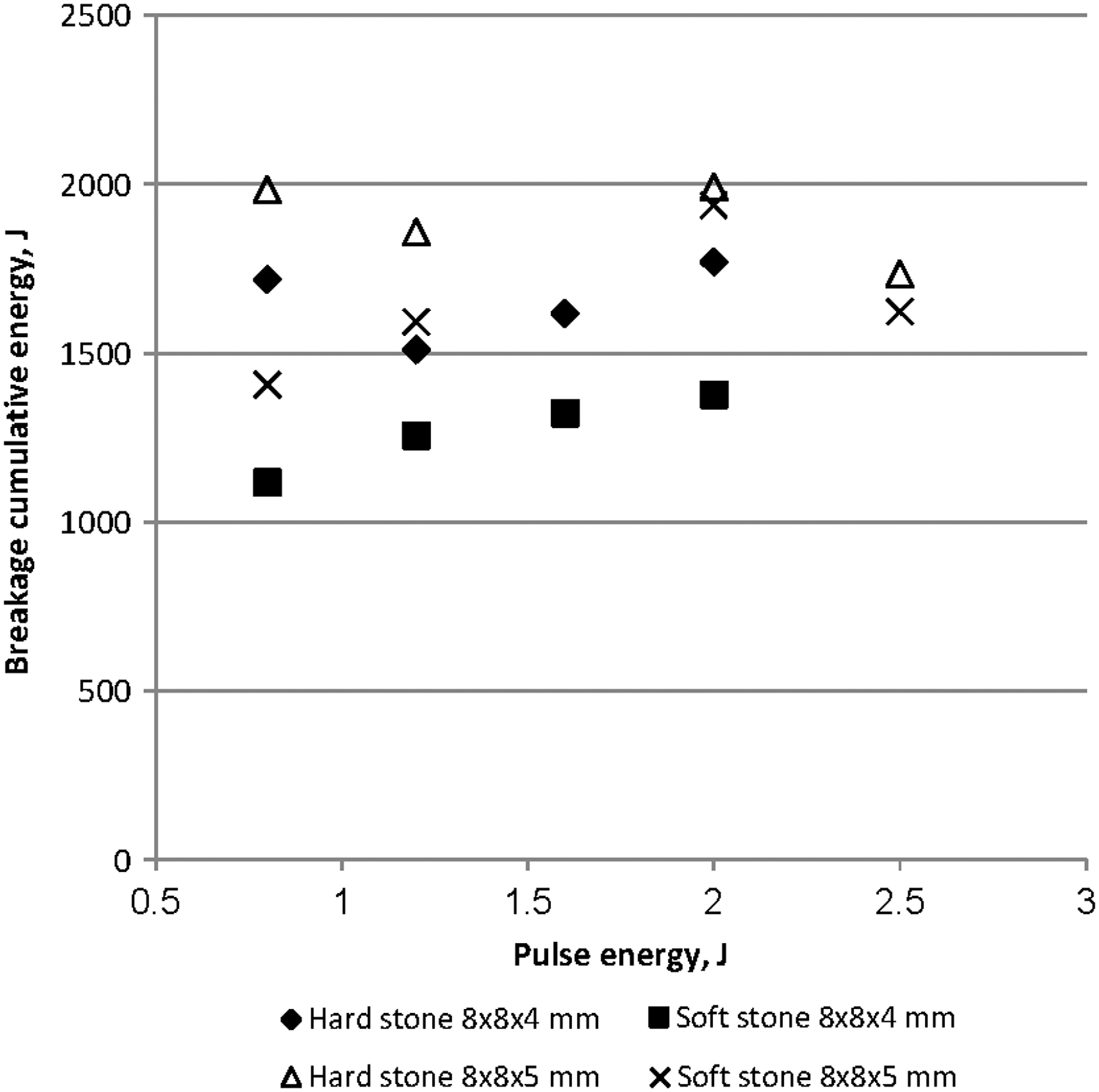
Dependency of cumulative energy required for the destruction of the stone as a function of the pulse energy of the laser lithotripter.
The results of further tests conducted with the Holmium laser at higher-power settings (see Appendix Table A3) are shown in Figure 7. The standard deviation values of data are not shown in these graphs, as they impair a clear reading of the results. The standard deviation data of the experiments results can be seen in Appendix Tables A1–A3.
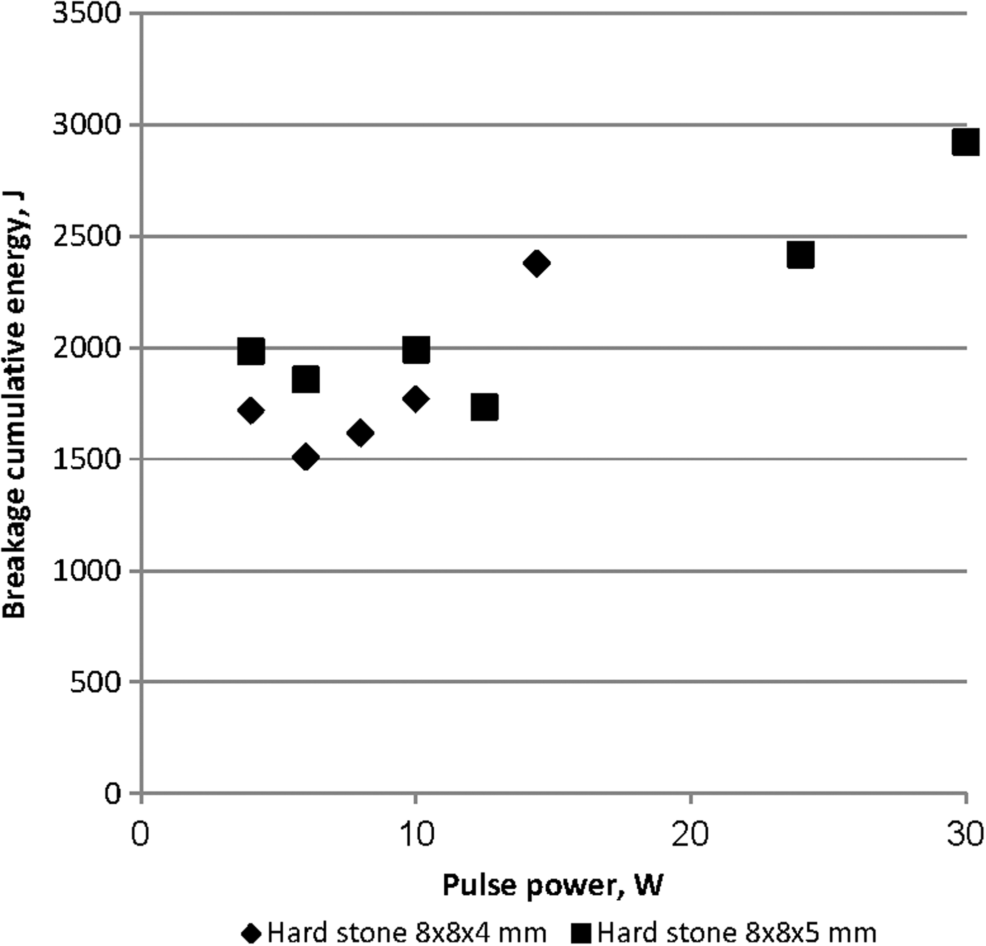
Dependence of the cumulative energy required for the destruction of stone phantoms on laser lithotripter output power.
Comparing the results received for cumulative energy in a pulse energy range of 0.8–2.5 J for LL (Fig. 6), we can determine whether it is possible to obtain results similar to those for NEPL (Fig. 5).
Thus, from Figure 7, it follows that an increase in Holmium laser output power leads to an increase in the cumulative energy required for fragmentation of the samples in the range above 14 W.
Discussion
The results demonstrate that NEPL in all cases investigated in the given work requires significantly less cumulative energy and correspondingly less time in order to destroy artificial stone phantoms than a Holmium laser lithotripter at all its investigated settings. Differences between the devices in cumulative energy until disintegration, number of pulses, and fragmentation time reach nearly one order of magnitude. However, we should take into account some limitations of this in vitro study, because it does not precisely reproduce real clinical conditions. In any case, both devices successfully completed the test objective, that is, all stone samples were fragmented to less than 2 mm size.
Statistical analysis confirmed significant differences in the sample data of the obtained results. In addition to significant differences in the measured values themselves, the deviation of their values is also considerably lower for NEPL as compared with that of LL.
With that, NEPL consistently expended significantly less cumulative energy to destroy the soft stones as compared with the energy required to break hard stones. On other hand, LL often required approximately the same or sometimes even higher cumulative energy in order to disintegrate soft stones than hard stones (see Figs. 5 and 6).
Another derived parameter of interest is specific volumetric energy needed for stone fragmentation, (E sum/V, where V is the sample volume) and its dependence on lithotripter probe size and stone density. Data of specific volumetric fragmentation energy for each lithotripter, probe size, and stone density/hardness are shown in Figure 8.
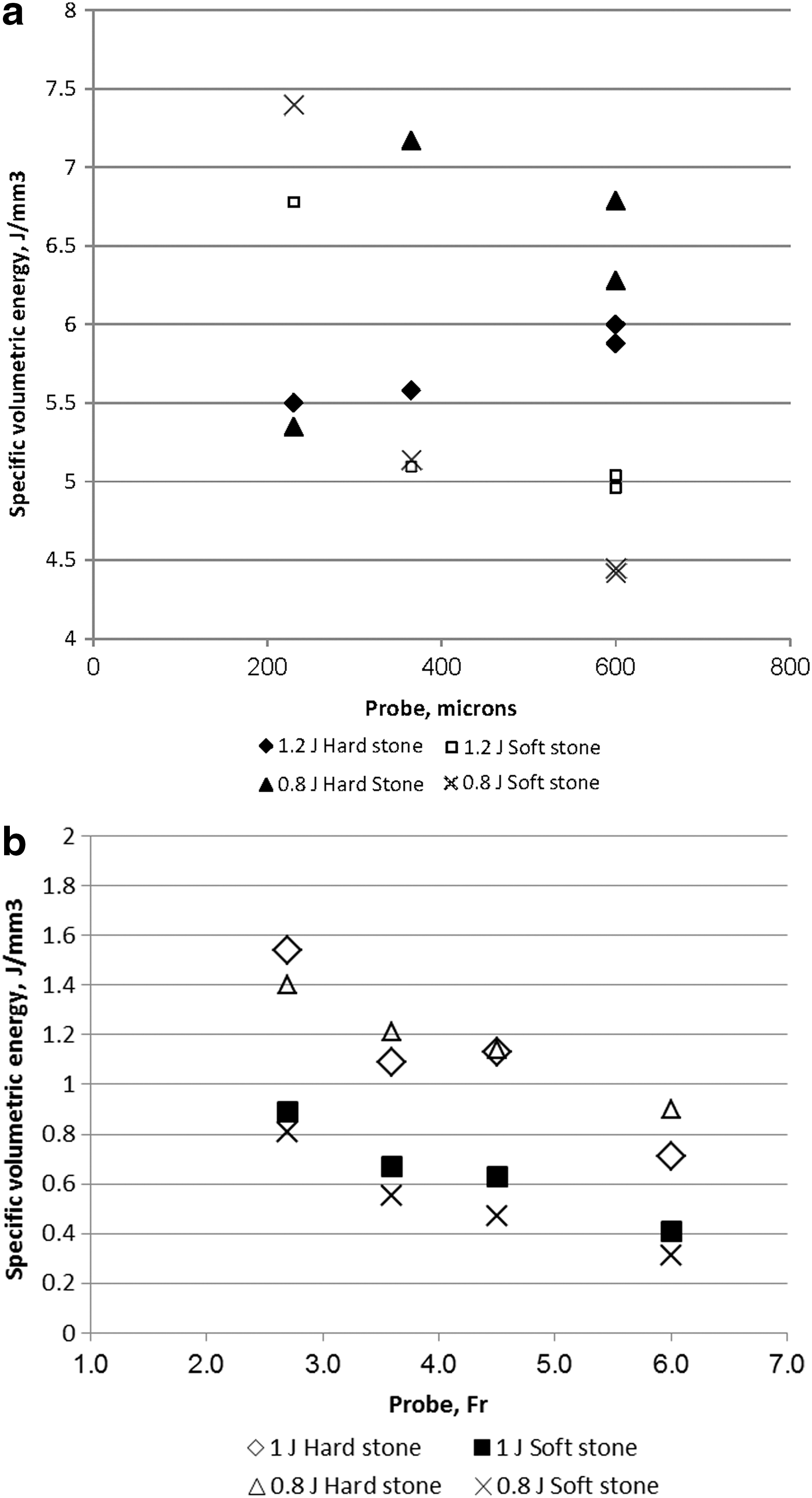
Dependence of the specific volumetric energy required for stone fragmentation on stone density and lithotripter probe's size.
The results obtained clearly indicate very different correlations between the specific volumetric energy required to destroy the stone phantoms based on probe diameter and stone density for both lithotripters. As for LL, there is no visible correlation between the specific volumetric energy, the density of the stone phantom, and the diameter of the probe, which seems to confirm the prevailing opinion as to the relative “indifference” of the laser to the properties of the fragmented material. In the case of NEPL, there is a clear dependency of the specific volumetric energy on the density of the stone phantoms and an inverse correlation between probe size and specific volumetric energy needed to break the stone.
Such observed differences in the results can be understood by taking into account the mechanisms of stone destruction for the investigated devices. For LL, photothermal and photomechanical effects are associated only with the laser beam's ability to create very high temperature at a local point as described earlier and do not have an apparent dependence on the properties of the stone. In contrast, the breakdown on the distal end of NEPL probe creates a plasma channel in the solid state, which, in turn, produces tensile thermomechanical stresses. Breakdown also depends on the length of the arc discharge, which is a function of distance between the probe electrodes and corresponding probe diameter. In addition, the ability of cumulative residual stresses to turn micro-cracks into a macro-crack leading to cracking of the sample depends on the density (or porosity) of the sample. Samples with lower density need less energy in order to be destroyed.
Generally and on average, the specific volumetric energy required for stone fragmentation with the use of the NEPL is more than 6 times less than with Holmium LL (Fig. 9).
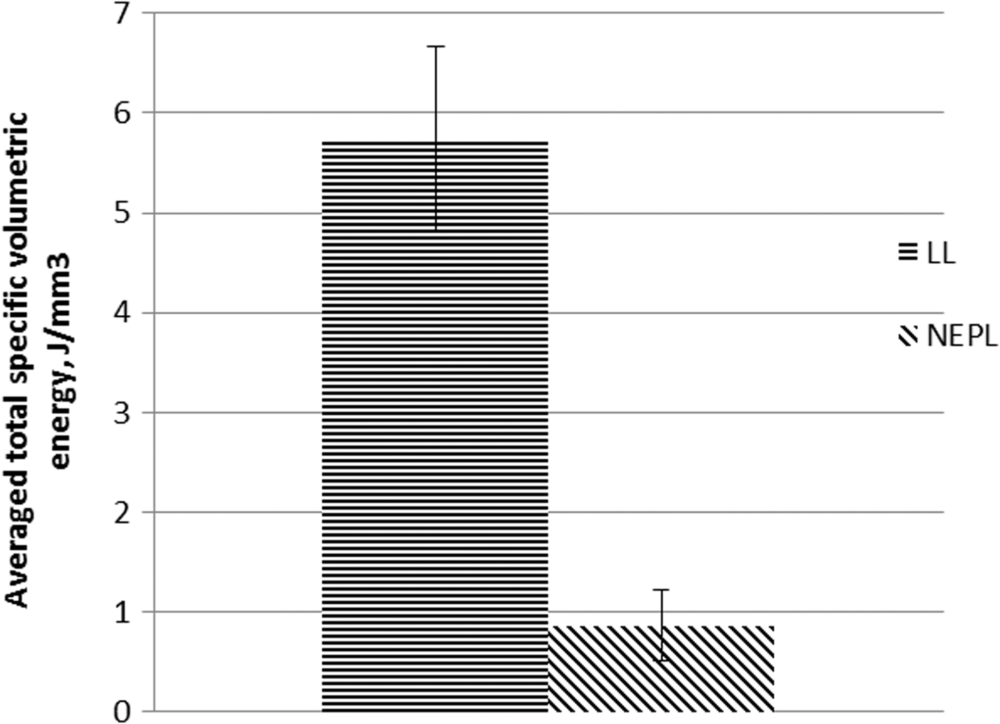
Total specific volumetric energy required to break the experimental samples.
Thus, the NEPL requires significantly lower total energy and, consequently, less time to break stones than the Holmium laser at comparable pulse parameters.
Furthermore, as shown in Figure 6, an increase in the pulse energy of the Holmium laser by more than thrice led only to minor changes in the cumulative energy required for the destruction of the stone phantoms. Moreover, in some cases, even a slight increase in cumulative energy with an increase of pulse energy (Fig. 6) can be observed for stone fragmentation. Note also that an increase in laser output power by applying pulses with higher frequency leads to an increase in the energy required for fragmentation of the stone samples (Fig. 7).
This observation leads to the clinical conclusion that starting fragmentation at higher energies in order to produce smaller fragments with Holmium LL is not recommended (especially for retrograde endoscopic lithotripsy in the renal pelvis and calyces).
On the contrary, if the condition of the procedure allows maximum pulse energy to be applied to a stone using NEPL, it can increase the efficacy of the lithotripsy and shorten stone destruction time. This can also be explained based on the electropulse mechanism, through which increasing pulse energy, E, achieves fragmentation by increasing high voltage applied to the NEPL probe (E=CV 2 /2, where C is capacity and V is voltage). Increasing voltage, in turn, facilitates conditions of discharge penetration into the solid and, thus, raises the efficacy of the electropulse mechanism.
Therefore, the different stone fragmentation mechanisms explain the essential differences in the data obtained for NEPL and LL as well as the effect of density (hardness) of the stone phantoms, energy in the pulse, and diameter of the investigated probes on the resulting efficacy of the device.
Conclusion
The results of the present study confirm that for all types of stone samples and pulse settings used in this experiment, the NEPL requires significantly less total energy and time for stone disintegration than the Holmium laser lithotripter and is, therefore, more effective for stone fragmentation than the Holmium laser.
The two compared lithotripters differ according to the mechanisms at work in the destruction of stone, which explains the differences between the received results based on the density and hardness of the stone phantoms.
NEPL has always required significantly less energy for the destruction of soft stones, as compared with the energy required to break hard stones. At the same time, the Holmium laser often required approximately the same or even higher energy on soft stones than on hard stones.
Correlations between pulse energy and the properties of the stone phantoms used for fragmentation testing have been confirmed experimentally for NEPL.
As for LL, there is no observed correlation between specific volumetric energy, density of the stone phantom, and diameter of the probe. It, therefore, confirms hypotheses regarding the relative “indifference” of the laser to the properties of fragmented material. In contrast, in the case of NEPL, the specific volumetric energy is clearly dependent on the density of the stone phantoms, with an inverse correlation observed between probe size and specific volumetric energy needed to break the stone.
Footnotes
Disclosure Statement
No competing financial interests exist.
| Hard “stone” | ||||||||
|---|---|---|---|---|---|---|---|---|
| Pulse energy 0.8 J, sample 8×8×4 mm, frequency 18 Hz, power 14.4 W | Pulse energy 3 J, sample 8×8×5 mm, frequency 8 Hz, power of 24 W | Pulse energy 2.5 J, sample 8×8×5 mm, frequency 12 Hz, power of 30 W | ||||||
| Esum, J | Qty of pulses | Time, sec | Esum, J | Qty of pulses | Time, sec | Esum, J | Qty of pulses | Time, sec |
| 2379±138 | 2974±173 | 165±10 | 2418±294 | 806±98 | 101±12 | 2922±318 | 1169±127 | 97±11 |