
Abstract
Background and Purpose:
Laparoscopy is a common approach to manage varicoceles in both the adult and pediatric population. The purpose of this study is to report our experience and compare outcomes between conventional laparoscopy and laparoendoscopic single-site (LESS) surgery for varicocelectomy in the pediatric population.
Patients and Methods:
A retrospective cohort study was performed of all patients who underwent conventional laparoscopic varicocelectomy (LV) and laparoendoscopic single-site varicocelectomy (LESSV) at a single pediatric institution from December 2007 to March 2012. Patient demographics, intraoperative details, narcotic use, and complications were reviewed.
Results:
LV was performed in 32 patients and LESSV in 11 patients. None had conversion to open surgery. Median age was 16 years for LV (range 12–23) and 15 years for LESSV (range 12–20), P=0.061. Median operative time was 55 minutes for LV (range 28–90) and 46 minutes for LESSV (range 33–59), P=0.037. Nine (81.8%) patients in the LESSV group and 10 (31.2%) patients in the LV group were administered narcotics in the recovery room, P=0.005. One (3.1%) patient in the LV group was administered ketorolac in the recovery room, P=1. Five patients in each group, LESSV (45.5%) and LV (15.6%), received acetaminophen in the recovery room, P=0.092. All procedures were performed on an outpatient basis except for one because of a concomitant procedure. Median follow-up was 22 months in LV and 15 months in LESSV, P=0.015. One (3.1%) postoperative hydrocele was noted after LV and 1 (9.1%) after LESSV, P=0.451. All varicoceles were clinically resolved in both groups.
Conclusions:
LESSV is comparable to LV in the pediatric population. Our initial experience indicates that the LESS approach may be more painful in the immediate postoperative period than conventional laparoscopy. The LESS technique warrants further evaluation to determine if one approach is clearly more advantageous.
Introduction
V
As minimally invasive technologies improve, alternative modalities are implemented to expand on conventional laparoscopy. 5 The laparoendoscopic single-site (LESS) technique was introduced several years ago and has been performed successfully for a variety of procedures, including nephrectomies, cholecystectomies, and appendectomies. 6 –9 Initially, LESS surgery was not widely accepted because of limited maneuverability and clashing of instruments, 10,11 but introduction of flexible laparoscopes and instruments has facilitated interest in the LESS technique. The advent of LESS surgery has the potential to improve cosmesis, decrease postoperative pain, and shorten convalescence in appropriately selected patients. 12 In the pediatric population, shorter recovery time may lead to decreased loss of work productivity for parents. The method has been proven to be feasible and effective, with high patient satisfaction for urologic diseases. 13
Kaouk and Palmer 11 first reported pediatric LESS varicocelectomy (LESSV) in 2008. 11 The authors described the surgical technique and potential complications, but a comparative analysis in a pediatric cohort has yet to be published. We report our experience and compare outcomes between a conventional laparoscopic approach and LESS surgery for varicocelectomy.
Patients and Methods
Medical records of all patients who underwent laparoscopic varicocelectomy (LV) or LESSV at a single pediatric hospital from December 2007 to March 2012 were retrospectively reviewed. Operative indications included scrotal pain, testicular hypotrophy, grade increase, and patient's decision. Data included patient demographics, intraoperative details, recovery room narcotic use, and complications, including hydrocele formation and recurrence. Narcotic use was converted to morphine sulfate equivalents. Follow-up visits included physical examinations to evaluate for resolution of varicocele and development of hydroceles.
Statistical significance was set at P<0.05 using the Wilcoxon-Mann Whitney for continuous and Fisher exact and chi-square tests for categorical variables. Statistical calculations were performed with PASW statistics gradpack (version 20.0, SPSS Inc., Chicago, IL).
Surgical procedure
Each patient, regardless of surgical technique, was placed supine on the operating room table under general anesthesia with his lower abdomen and genitalia prepped and draped in the usual sterile fashion. The LV procedure was performed with a 5-mm umbilical trocar, 3 or 5-mm instrumentation through two additional skin punctures or 5-mm trocars, and a standard 5-mm 30-degree laparoscope. The LESSV procedure was performed with an Olympus Endoeye flexible tip laparoscope or standard 5-mm laparoscope, standard 3- or 5-mm instrumentation, and an Olympus TriPort (Fig. 1), which is a multiport device that is placed via an open technique through a skin and fascial incision. The Olympus Endoeye (Fig. 2) is a 5-mm flexible tip slaparoscope that delivers a 100-degree angulation and an 85-degree field of view. Ligation and division of spermatic vessels were achieved with suture ligations or energy sources.

Olympus Triport.
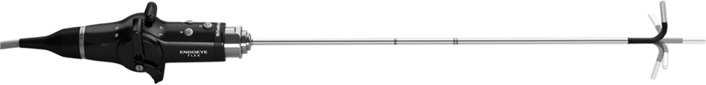
Olymus Endoeye.
Access was obtained via a midline umbilical incision during LESSV. The size of the umbilical fascial incision was made by clinical judgment. It was not marked or measured. The Olympus Triport and scope were introduced. Standard 3- or 5-mm instruments were used. Sharp and blunt dissection was used to open the posterior peritoneum and mobilize gonadal vessels. An energy source, including LigaSure, Enseal Trio, or Olympus Thunderbeat, was used to seal and divide the gonadal vessels. Artery and/or lymphatic sparing was performed based on surgeon preference. The umbilical fascia was closed with absorbable suture.
Access was obtained via a midline umbilical incision or infraumbilical curvilinear incision during LV. A 5-mm camera port was placed, and a laparoscope was introduced. Subsequently, two other stab incisions or 5-mm trocars were placed, typically on each side of the abdomen to triangulate the instruments. The LV procedure was then performed with the same dissection used for LESSV. The fascia of all three ports was closed with absorbable suture.
Results
During the study period, a total for 43 patients (32 LV and 11 LESSV) were identified. The first LESSV was performed in February 2011. A single surgeon performed all LESSV procedures. Multiple surgeons performed laparoscopic procedures, including the surgeon who performed the LESSV procedures. Demographic factors are presented in Table 1. There was no significant difference in age, weight, or body mass index between the two groups, P=0.061, 0.54, 0.266, respectively. A difference was noted, however, in terms of height, P=0.046. The two cohorts were comparable in terms of varicocele side, type of varicocelectomy, and concomitant procedures, P=0.451, 0.092, and 0.637, respectively. No accessory incisions were used. All procedures were performed on an outpatient basis except for one patient who was admitted for 2 days because of a concomitant procedure (laparoscopic pyeloplasty).
LESSV=laparoendoscopic single-site varicocelectomy; LV=laparoscopic varicocelectomy; BMI=body mass index.
Perioperative parameters are presented in Table 2. The operative time was shorter in the LESSV group compared with the LV group, P=0.037. There were no intraoperative complications and no difference in median blood loss, P=0.401. Recovery room narcotic administration was higher in LESSV patients (81.8%) than in LV patients (31.2%), P=0.005. There was no difference, however, in immediate postoperative ketorolac and acetaminophen administration, P=1 and 0.092, respectively. Median follow-up was shorter in the LESSV group compared with the LV group, P=0.015. One (3.1%) patient after LV and one (9.1%) patient after LESSV experienced a postoperative hydrocele, P=0.451. Both hydroceles were managed with an open scrotal approach. There was no difference in complication rate between the groups. None of the patients in either group experienced testicular atrophy. All varicoceles were clinically resolved in both groups.
LESSV=laparoendoscopic single-site varicocelectomy; LV=laparoscopic varicocelectomy; EBL=estimated blood loss.
Discussion
The laparoscopic technique was first described in 1988 14 and has gained increased popularity because of the relative ease of the operation. It is now commonly performed for pediatric and adolescent patients. At one point, a major shortcoming of laparoscopic artery-sparing varicocelectomy was persistent or recurrent varicoceles (3.6%–37.5%). 1 Increasing experience and development of new instruments has led to improvement of surgical outcomes. LV allows for clear visualization of the surgical field and relatively easy access to the surgical site with minimal dissection. In addition, LV is safe and effective in patients with previous inguinal procedures or bilateral varicoceles. 1
Improvements in technology and evolution of minimally invasive surgical techniques have led to the advent of LESS surgery. The LESS approach provides intracorporeal access while reducing the number of skin incisions to one that can be hidden in the umbilicus. 15 Fewer incisions may lead to lower incidences of port site infection and pain, which may potentially lead to faster recovery when compared with traditional laparoscopic surgery. 1 As seen with our experience, LESS surgery allows the surgeon to use a single incision to perform bilateral procedures. Neither additional ports, nor position changes, are needed when performing a bilateral varicocelectomy.
There still remain obstacles to single-port surgery, however, especially in the pediatric population, including limitations because of instrumentation, hand collisions, triangulation difficulties, and the potential for a steep learning curve. External hand collisions between the two working instruments or between the camera holder and surgeon pose the greatest challenge. 15 To eliminate hand collisions, the use of two instruments of different lengths may be considered. It may be difficult to maintain an optimal view because of crowding and an in-line view with instruments. The introduction of 5-mm flexible laparoscopes has provided enhanced views of the operative field. Prebent and flexible instruments may also be considered, in an effort to mitigate the challenges of a single entry point for the laparoscope and instruments. Significant coordination between the surgeon and the camera holder is essential because of the small working space in a pediatric patient. 16
It is important to confirm that any potential advantage of LESS does not compromise surgical outcome. Our experience supports the surgical effectiveness and suitability of LESSV. None of the LESS procedures needed additional incisions, and they were performed without compromise in operative time compared with LV. We found LESS to be comparable to LV in terms of perioperative outcomes and success rates in a pediatric population. Evaluation revealed a discrepancy in analgesic utilization, however, although pain management was not addressed with a standard protocol. During the immediate postoperative period, the LESSV group received more narcotics, and no difference was found for ketorolac and acetaminophen administration. Our analysis indicates that LESSV may be more painful than LV in the immediate postoperative period.
To the best of our knowledge, our study is the first report to compare LESSV and LV in the pediatric population. Limitations of our study include a small sample size with respect to the LESSV group, a single surgeon experience with LESSV, lack of randomization, and lack of standardization for pain management.
Conclusion
LESSV is comparable to conventional LV in the pediatric population. Our initial experience indicates that the LESS approach may be more painful in the immediate postoperative period than traditional laparoscopy. The LESS technique warrants further evaluation to determine if one approach is clearly more advantageous.
Footnotes
Disclosure Statement
No competing financial interests exist.