
Abstract
Purpose:
To evaluate whether CT-identified Randall plaques can be used to foresee the recurrence of stone disease (SD); to define a cut point that could identify a high-risk population.
Materials and Methods:
A retrospective study of patients attended for SD from January 2004 to December 2009 was conducted. Study population was patients with a first episode of calcium SD that was diagnosed by abdominal CT. Papillae tip attenuation was measured in Hounsfield units (HU) on unenhanced abdominal CT images. Patients with recurrent SD were identified; t test, Pearson correlation, and receiver operating characteristic (ROC) curve analysis were used.
Results:
A total of 543 patients were evaluated; 187 fulfilled the criteria and were included, and 49 (26.2%) had recurrent SD. Mean follow-up: 5 years (3–7 years). Papillae tip attenuation was significantly higher in the recurrent group (46.2 HU vs 40.1 HU, P=0.01) and correlated well with the possibility of developing SD (R=0.83). Attenuation >43 HU showed a ROC curve area under the curve=0.87 with sensitivity=77% and specificity=84% separating patients with a RR=8.7 of development of recurrent SD. The number of papillae >43 HU correlated with recurrent SD (RR=11.2 for ≥3 papillae vs <3 papillae with density >43 HU).
Conclusions:
The presence of the Randall plaques can be used as a marker for predicting SD recurrence. A cut point of 43 HU could be used to identify a high-risk population.
Introduction
The European Guidelines on Urolithiasis 1 recommend that after stone passage, every patient should be assigned to a low- or high-risk group of stone formers. The evaluation is based on stone composition analyzed by infrared spectroscopy or X-ray diffraction, medical history, associated genetically determined metabolic diseases, acquired diseases, and anatomic abnormalities. Only high-risk stone formers need specific metabolic evaluation.
The presence of small calcifications over the tip of the renal papillae is considered of utmost importance for the formation of calcium stones 2 –5 that, in turn, represent the vast majority of stones. 1,6 Idiopathic calcium oxalate stone formers, brushite stone formers, and patients with enteric hyperoxaluria represent almost 100% of calcium stone formers. In all of these groups, there have been described microcalcifications over the tip of the renal papillae like the Randall plaques or Bellini ducts plugs, 7 which are calcium phosphate or oxalate deposits in the papillary tip and within the collecting ducts that further evolve by perforating the urothelium covering the renal papillae.
Traditionally, the presence of microcalcifications at the level of the tip of the papillae should be considered a risk factor both for the initial episodes and for the recurrence of SD. 3,8,9 Endourologic evaluation of the presence of these microcalcifications demonstrated that stone activity is proportional to the calcification surface, and it was proposed as a possible screening tool. 3,4,8,10 According to previous studies, the presence of microcalcifications in the tip of the renal papillae increases the CT attenuation and can be identified in unenhanced abdominal CT images. 11 –15
Our objective was to evaluate whether the presence of high attenuation regions over the tip of the renal papillae identified in abdominal CT images, corresponding to microcalcifications present at this level, can be used to foresee the recurrence of SD and thus to be used as a risk criterion that could trigger a metabolic evaluation of the patients and prophylactic treatment.
Materials and Methods
We performed an evaluation of patients with recurrent SD comparing the density of the papillae of recurrent stone-forming patients vs patients with nonrecurrent SD. A retrospective review of our database of patients with urinary stones was performed. We reviewed 543 patients treated between January 2004 and December 2009. We aimed at having at least 3 years of follow-up with a mean follow-up of 5 years.
The inclusion criteria were: • Patients with a first episode of SD. • Stone with calcium composition. • An abdominal CT performed in our center during the first episode of SD. • Patients who achieved stone-free status after the first SD episode, confirmed with an abdominal CT.
The study adheres to local regulations and standards and was approved by the Institutional Review Board. Informed consent was deemed unnecessary according to the local rules of our center for retrospective studies. All CT studies had been performed for a clinical or diagnostic purpose as part of the standard protocol of our center.
Papillae attenuation was measured in Hounsfield units (HU) on the abdominal CT performed during the first stone episode.
Unenhanced abdominal CTs were performed using Siemens (Erlangen, Germany) multidetector CT equipment (Emotion Duo or Sensation 64). Detailed CT acquisition parameters for a standard nonenhanced abdominal scan are outlined in Table 1. In those patients who also had nephrographic and excretory phases, these were used together with the unenhanced series for better localization of renal papillae. The examinations were performed using 5-mm slice acquisition and reconstruction. In patients for whom additional 2-mm and 1-mm slice images were available, we only analyzed the 5-mm images to preserve compatibility and homogeneity with the 5-mm-only CT studies.
The CT images were reviewed using the Raim Alma 2010 (© ALMA IT SYSTEMS, Barcelona, Spain) software.
Renal papillary Hounsfield density was measured by placing regions of interest (ROIs) with a mean size of 0.1 cm2 over the very tip of the papilla (Fig. 1).
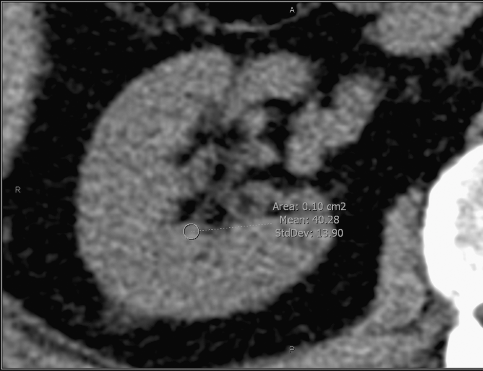
Renal papilla Hounsfield density measuring by placing a region of interest over the tip of the papilla.
The images were magnified to 5× to prevent contamination of the ROI with fat within the renal sinus. Each kidney was divided into three parts corresponding to the three caliceal groups; in each one, the attenuation of the renal papillae was recorded. A total of six measurements for each patient were recorded, three in each kidney using the unenhanced CT images. In every kidney, one papilla from each caliceal group was measured, choosing the easiest to identify. The mean attenuation of the papillae was calculated for each kidney.
The patients who fulfilled the inclusion criteria were evaluated, and we identified those patients who had at least one more episode of active SD. All stones were obtained either by spontaneous expulsion or after a surgical intervention (ureterorenoscopy, percutaneous nephrolithotomy, laparoscopic ureterolithotomy). For the evaluation of stone composition, we used the laboratory result of stone analysis (spectrometry). In all patients, a follow-up abdominal CT confirmed the “stone-free” status after the initial SD episode.
Two statistical groups were established: Recurrent stone formers (RSF) and nonrecurrent stone formers (NRSF). We defined recurrent disease in patients who had at least two active SD episodes and nonrecurrent in patients who had only one initial active SD episode.
The two-tailed independent sample t test and the Wilcoxon signed-rank test were used to test the significance of differences between groups. A P value less than 0.05 was considered significant from a statistical point of view. Pearson correlation coefficient was used to evaluate the correlation between the renal papillae attenuation and the possibility of development of SD. The recever operating characteristic (ROC) curve analysis was used to define a cut point between RSF and NRSF that could help characterize a high-risk population. The relative risk to develop a new episode of calcium SD was calculated for this population.
Results
We reviewed 543 patients who were treated for SD between January 2004 and December 2009. A flowchart of the inclusion algorithm can be seen in Figure 2.

A flowchart of the inclusion algorithm.
A total of 187 patients fulfilled the criteria and were included in the study. All patients had available stone analysis from the first SD episode showing calcium composition. All patients underwent regular follow-up in our center. Of the 187 patients included, 49 (26.2%) had at least another active stone episode. All patients with recurrence underwent either medical or surgical treatment. In all patients, stones were obtained either by spontaneous expulsion after medical treatment or external shockwave therapy or after surgical interventions (ureterorenoscopy, percutaneous nephrolithotomy, laparoscopic ureterolithotomy). All patients with recurrence had calcium stones. The mean follow-up was 5.1 years (3–7 years). Patients' demographics and attenuation measurements results are found in Table 2.
RSF=recurrent stone former; NRSF=nonrecurrent stone former; HU=Hounsfield unit.
Analysis of papillae tip attenuation
The mean attenuation of renal papillary tips of the RSF patients was significantly higher when compared with the NRSF group (46.2 HU vs 40.1 HU, P=0.01).
There was a good correlation between the attenuation of the renal papillae and the possibility of developing SD (R=0.83).
In the ROC curve analysis, a cut point of 43 HU separated with a sensitivity of 77% and specificity of 84% the patients with high risk of recurrence showing an area under the curve (AUC) of 0.87 (Fig. 3).

The receiver operating characteristic (ROC) curve distribution.
High papillae attenuation values (>43 HU) were associated with a higher risk of development of another SD episode (RR=8.7).
Analysis of the number of papillae with attenuation >43 HU
RSF patients had a significantly higher number of papillae with tip density >43 HU than NRSF (4.5 vs 2.1, P=0.003).
There was a good correlation between the number of papillae with density >43 HU and the possibility of development of a second episode of SD (R=0.82).
In the ROC curve analysis, a cut point of three papillae with attenuation >43 HU separated with a sensitivity of 81% and specificity of 86% the patients with high risk of recurrence showing an AUC of 0.91.
The relative risk of development of recurrent SD was 11.2 for those that had ≥3 papillae vs <3 papillae with density >43 HU.
Discussion
The abdominal CT is the gold standard for the diagnosis of SD. 1 Previous studies demonstrated that the abdominal CT could be used for the evaluation of the attenuation of the tip of the renal papillae, 11 a region traditionally linked with the development of Randall plaques and Bellini duct plugs and implicitly with the development of calcium stones. 3,8,9 The evaluation of the attenuation of renal papillae tip in stone-free patients, previous to the development of SD, seems to be able to identify a high-risk group for the development of SD. 16
In a similar fashion, the present study evaluates the attenuation of the tip of the renal papillae in patients who already had a SD episode. We noted that patients who are going to develop another episode of SD over a mean of 5 years have a higher attenuation of the renal papillae than nonrecurrent patients. Furthermore, the number of affected papillae correlates with the risk of development of further episodes of SD. A cut point of 43 HU can be used with good sensitivity and specificity to separate recurrent and non-recurrent calcium stone formers. It seems that this cut point might represent a critical accumulation of calcium in the renal papillae, which could be “the point of no return” for the formation of calcium stones. Also, patients who present an involvement of more papillae show higher risk for development of recurrent SD.
We verified that all patients achieved stone-free status after the initial episode of SD using abdominal CT images. Nonetheless, no method, even CT, can detect submillimeter stones that may be already growing on papillary tips. The presence of such small stones would raise the HU value of the papilla tip, without being able to distinguish that case from the presence of Randall plaques or Bellini ducts plugs. On the other hand, from a practical point of view, the CT attenuation of a papillae with microcalcifications because of the Randall plaques, ductal plugging, or submillimeter stones would be higher than that of a normal papillae, signalling the presence of any of these conditions, all three representing a higher risk for recurrent SD.
Our study provides radiologists and urologists with a new noninvasive marker that can be used for the evaluation of the possibility of recurrence of calcium stones. In more and more patients, the diagnosis of SD is performed with abdominal CTs, so the population targeted by our study is increasing. Furthermore, the abdominal CT is an exploration used more and more frequently for the evaluation of almost any abdominal problem, and so the number of stones incidentally diagnosed in abdominal CT images is growing. This information can be easily extracted from the abdominal CT images because the evaluation of the papillae tip is simple to perform and reproducible in most radiologic settings. It is noninvasive, as opposed to earlier articles suggesting minimally invasive endoscopic examination. 3,4,8,10
One of the limitations of our study could be the fact that we included patients who were scanned using two different CT scanners. Nonetheless, an inclusion criterion was the requirement that all CTs were performed in our center. Our scanners are fine-tuned by the manufacturer (the same for both machines) to produce similar/identical results, which suggests that attenuation measured on a CT scan acquired by any of our scanners should generate the same result. Furthermore, the use of two different CT scanners allowed us to include a higher number of patients. Nonetheless, external validation studies are necessary for a standardization of the protocol of CT software to assess the presence of microcalcifications over the tip of the papillae applicable to different CT machines.
In addition, the use of 5-mm thick slides could be criticized. Analyses from previous studies of patients who had 1-mm and 5-mm slides available, however, demonstrated that there were no significant slice thickness-dependent differences. 11
Recent studies have converted the abdominal CT in a new Swiss knife of stone diagnosis. Abdominal CT images can be used to differentiate between stone composition, either by dual energy analysis or using an attenuation cut point of 600 HU to separate uric acid and calcium stones. 17 Also, a cut point of 41 HU used for the attenuation of the tip of the renal papillae separates patients with high risk of development of an initial episode of SD. 16 Our study comes to complete the diagnosis of SD performed by CT by adding another cut point of 43 HU that identifies patients with high risk of recurrence. In this fashion, urologists can dispose of preliminary information about the recurrence probability and the need for metabolic evaluation of selected patients even before the treatment of the initial stone.
Conclusion
The presence of a higher papillae tip attenuation identified in unenhanced abdominal CT images can be used as a marker for predicting SD recurrence. A papillae tip attenuation cut point of 43 HU could be used to identify a high-risk population.
Footnotes
Disclosure Statement
No competing financial interests exist.