
Abstract
Introduction:
Recent technological advancements have led to the introduction of new three-dimensional (3D) cameras in laparoscopic surgery. The 3D view has been touted as useful during robotic surgery, however, there has been limited investigation into the utility of 3D in laparoscopy.
Materials and Methods:
We performed a prospective, randomized crossover trial comparing a 0° 3D camera with a conventional 0° two-dimensional (2D) camera using a high definition monitor (Karl Storz, Tuttlingen, Germany). All participants completed six standardized basic skills tasks. Quality testing scores were measured by the number of drops, grasping attempts, and precision of needle entry and exiting. Additionally, resolution, color distribution, depth of field and distortion were measured using optical test targets.
Results:
In this pilot study, we evaluated 10 medical students, 7 residents, and 7 expert surgeons. There was a significant difference in the performance in all the six skill tasks, for the three levels of surgical expertise and training levels in 2D vs 3D except for the cut the line quality score and the peg transfer quality score. Adjusting for the training level, 3D camera image results were superior for the number of rings left (p=0.041), ring transfer quality score (p=0.046), thread the rings (no. of rings) (p=0.0004), and thread the rings quality score (p=0.0002). The 3D camera image was also superior for knot tying (quality score) (p=0.004), peg transfer (time in seconds) (p=0.047), peg transfer pegs left (p=0.012), and for peg transfer quality score (p=0.001). The 3D camera system showed significantly less distortion (p=0.0008), a higher depth of field (p=0.0004) compared with the 2D camera system.
Conclusion:
3D laparoscopic camera equipment results in a significant improvement in depth perception, spatial location, and precision of surgical performance compared with the conventional 2D camera equipment. With this improved quality of vision, even expert laparoscopic surgeons may benefit from 3D imaging.
Introduction
L
Technological advancements have led to the introduction of three-dimensional (3D) cameras to laparoscopic surgery. One of the first prototypes was developed in 1992 at the Nuclear Research Center Karlsruhe, Germany and was tested in various ex vivo experiments and clinically during laparoscopy. 4 Early 3D endoscopic systems created apparent binocular disparity by using a 2D monitor and alternating the image at high frequency (50–60 hz). Unfortunately, this resulted in poor image quality. Additionally, many subjects suffered dizziness, ocular fatigue, and nausea performing 3D surgery. 5,6
Technical advancements have improved 3D vision and image quality since these initial designs. Recently, newer 3D systems have been demonstrated as successful during laparoscopic skill task testing. 7,8 Other reports are less enthusiastic about the 3D laparoscopic camera system citing it not to be superior to the 2D camera system with regard to efficiency and performance. 6
We evaluated a novel 3D laparoscopic optical system that incorporates two objective lenses and video imagers at the distal tip of the laparoscope. Signals of the two imagers are processed and merged into one 1080p 60 hz video signal with alternating lines, one for each eye. These alternating lines are displayed by a 3D monitor as a left and right hand polarized interlace image. The viewer wears glasses feeding the images to each eye generating stereoscopic viewing.
This novel optical system may yield improved depth perception, spatial location, and precision, when compared to conventional 2D laparoscopic equipment. Hence, we sought to compare the 3D and 2D imaging on the surgeon's performance in a laboratory setting using six standardized laparoscopic surgical tasks and the study subjects stratified by the level of laparoscopic experience.
Materials and Methods
Population, randomization, and skills testing
We recruited participants from the University of California, Irvine under an Institutional Review Board (IRB) approved educational protocol. We targeted enrollment to novice medical students, surgical trainees, and expert laparoscopic surgeons. We directed participants to complete six basic skills tasks utilizing laparoscopic tools: transferring rings on a peg, passing a suture thread through rings, cutting a paper circle, performing a surgeon's knot, suturing along a hexagon, and transferring blocks on a peg board. The participants were randomly assigned to start either with the 3D or the 2D system. After finishing with the first testing session, the participants entered a cool-down period of ∼10 minutes to reduce the warm-up effect. Subsequently, they would crossover and perform the same skill tasks with the alternate imaging system.
The 3D system (Karl Storz, Tuttlingen, Germany) consisted of a 0° 10-mm laparoscope with a novel TipCam design and a 24” HD 3D monitor (Sony United States, San Diego, CA). Participants wore passive, polarized glasses for viewing (Fig. 1). The conventional 2D system used a 0° 10-mm Karl Storz laparoscope (Karl Storz) with a conventional HD Storz camera (Karl Storz) on the same 24” HD 3D monitor, but in a 2D mode. After adjustment of light intensity and white balancing for both optical systems, all tasks were performed using a pelvic trainer box with two working ports and one camera port placed at the same position for all participants.
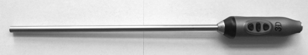
Karl Storz 0° 10-mm three-dimensional (3D) laparoscope with novel TipCam design.
Skill tasks
Task 1: Transferring rings on a peg was performed using six rings on six pegs. Subjects were given 2 minutes to pick up all six rings from the pegs, place the rings on the table, and then put the rings back onto the pegs. The task was stopped at 2 minutes regardless of the amount of completion. The number of rings on the pegs was used to determine the quantity score. The number of dropped rings was used to calculate the quality score from 1 to 4.
Task 2: Passing a suture thread through the rings was performed using a 2.0 prolene suture. Subjects were given 2 minutes to pass the suture through as many rings as possible using laparoscopic grasping forceps. The number of rings was recorded for the quantity score and the number of missed attempts and past pointing errors were used to calculate the quality scores from 1 to 4.
Task 3: Cutting a paper circle, a fundamental of laparoscopic surgery (FLS) approved task, was performed using laparoscopic scissors. The subjects were given 2 minutes to complete the task. Quantity scores were determined by the total distance cut after 2 minutes. Quality scores of 1–4 were determined by the precision of cutting on line.
Task 4: Tying a surgeon's knot was performed using a 2.0 silk suture on a 0 vicryl SH needle on a silicone suture slab marked with dots for needle entry and exit. Subjects were given 1 minute to complete the full knot. The number of ties within the knot was used to measure the quantity score. The quality score from 1 to 4 was measured by how close the suture was to the designated needle entry and exit dots.
Task 5: Suturing along the hexagon was performed using a 4.0 silk suture on a RB1 needle on a suture slab marked with dots surrounding a hexagon figure (sides=1 cm). Subjects were given 3 minutes to complete as many suture throws as possible using the entering and exiting dots around the hexagon. The quantity score was measured by the number of suture throws completed. The quality score from 1 to 4 was measured by how close the suture was to the entering and exiting dots for each suture placement.
Task 6: Transferring blocks on a peg board, an FLS-approved task, was performed using six blocks placed on a board with 12 pegs. Subjects were given 2 minutes to transfer the blocks. The subject was instructed to pick up the block with the left grasping forceps, transfer the block from the left hand to the right hand grasping the forceps, and place the block down with the right hand instrument on the contralateral side of the peg board, and when all blocks were transferred, to reverse the process to replace the blocks on the side of the board, where they were positioned at the initiation of the task. All six blocks had to be picked up and placed on both the left and right side of the board for completion. The number of blocks transferred to both sides in 2 minutes determined the quantity score. The number of dropped blocks determined the quality score from 1 to 4 during the transfer process.
The mean quality and quantity scores with standard deviations were calculated for each participant group. The mean quality and quantity scores were averaged for all three groups for each task. These average mean scores with standard deviations were then compared for the 2D vs the 3D imaging systems and for differences between the three groups.
Optical tests
Optical resolution, color, and contrast evaluation
A resolution was measured using a United States Air Force (USAF) resolving test pattern (Edmund Optics, Barrington, NJ) in accordance with the manufacturer's instructions. The resolution was defined as an imaging system ability to distinguish object details. The test target measures a resolution in terms of line pairs (LP) per millimeter. The USAF 1951 resolution target uses a repeating series of parallel bars decreasing in size. These bars are separated into group and element numbers. A system resolution is defined as the highest group and element in which the three bars can still be distinguished. 9
Color representation was measured using a Gregtag Macbeth Color Checker Target (Edmund Optics). The laparoscopes were evaluated at a distance of 10 and 20 mm away from the test target. Each test was performed in triplicate.
Contrast was evaluated by using a grayscale gradient having 15 density steps from a density of 0.07 (low) to 1.5 (high) corresponding to optical density increments of 0.10. The difference between density steps is linear, which leads to a logarithmic change in diffuse reflectivity. The laparoscopes were positioned 20 mm above the target, directly over the darkest square and were then moved slowly along the scale toward the lightest square, until the observers noted an inability to distinguish a difference between two steps of contrast. 9
Depth of field
Depth of field, the distance between the nearest and farthest objects in a scene that appear acceptably sharp in an image was assessed using an Edmund optics depth of field target (Edmund Optics). The laparoscopes were evaluated 10 mm away from the test target in triplicate.
Distortion
We measured distortion with a multifrequency grid distortion target and calculated the difference between the actual and theoretical dot location. Measurement was performed in triplicate. 10
Statistical analysis
The 2D and 3D imaging data were analyzed separately by ANOVA using pairwise t-tests between training levels. Significance levels for pairwise t-tests were adjusted for multiple comparisons using the Tukey method. The 2D and 3D imaging data were analyzed together using two-way ANOVA to test for differences in the training level and 2D vs 3D effect simultaneously. All analyses were performed with SPSS version 19 (IBM Corporation, Armonk, NY) and a p-value <0.05 was considered as statistically significant.
Results
Skill tasks
We recruited 24 participants, 10 medical students, 7 urology residents, and 7 expert urology surgeons for evaluation. The results of the skill task testing using the 2D and 3D imaging systems are summarized in Tables 1 and 2. We documented a significant difference in performance in all six skill tasks between the three levels of surgical experience in both the 2D and 3D imaging. No differences were detected in the cut the line quality score and the peg transfer quality score. Skills in 2D vs 3D were adjusted for the training level (expert 2D vs expert 3D, resident 2D vs resident 3D, and medical student 2D vs medical student 3D) and showed significant differences in the number of rings left (0 vs 0, 0.29 vs 0, 2.4 vs 0.5, p=0.041), ring transfer quality score (3.86 vs 4, 3.43 vs 3.71, 2.3 vs 3.3, p=0.046), thread the rings (no. of rings) (3.29 vs 4.86, 1.57 vs 4.14, 1.2 vs 2.6, p=0.0004), and thread the rings quality score (2.57 vs 3.57, 1.71 vs 3.29, 1.5 vs 2.6, p=0.0002). 3D imaging was also superior for knot tying (quality score) (5.86 vs 6.57, 4.43 vs 6.57, 1.7 vs 3.5, p=0.004), peg transfer (time in seconds) (101.29 vs 86.71, 115.29 vs 104.57, 117.4 vs 111.9, p=0.047), peg transfer pegs left (1.29 vs 0, 1.57 vs 1, 3.4 vs 1.5, p=0.012), and for peg transfer quality score (2.71 vs 3.71, 2.57 vs 3.14, 1.9 vs 3.0, p=0.001). We noted no difference for the ring transfer (time in seconds), cut the line (distance in cm), cut the line quality score, suturing hexagon number of throws and quality score, and for knot tying number of knots (Table 1).
2D=two dimensional; 3D=three dimensional; SE=standard error.
The significant differences between the different training levels for the 2D and 3D imaging are shown in Table 2.
Optical resolution, color, and contrast evaluation
The 2D imaging system provided a higher resolution at 20 mm distance—5.66 LP/mm compared with 5.04 LP/mm for the 3D imaging system. At a distance of 10 mm, it was not possible to conduct the optical resolution test with the 3D laparoscope because of blurry vision and loss of the 3D vision. The 2D laparoscope showed a resolution of 4.00 LP/mm at the distance of 10 mm. Contrast evaluation and color representation were similar for both laparoscopes (Table 3).
LP=line pairs.
Depth of field
The 3D imaging system provided a higher depth of field for 5 LP parallel—40 mm vs 32 mm and an almost identical result for 5 LP vertical—32 mm vs 33 mm at a distance of 20 mm (p=0.004). At a distance of 10 mm, the 3D imaging system showed a depth of field of 50 mm (5 LP parallel), 40 mm (5 LP vertical), and 2 mm for both 15 LP parallel and vertical at a distance of 10 mm to the target. Measurements for the 2D imaging system could not be conducted because of blurry vision.
Distortion
The 3D imaging system showed a maximum distortion of 15% compared with 18% for the 2D imaging system (p=0.0008).
Discussion
Over the last two decades several clinical and laboratory studies compared 3D vision with 2D laparoscopic vision with contradictory results. 5,6,11 –13 Some study groups reported superior performance, while others showed identical results for task or surgical performance and vision.
McDougall and colleagues performed one of the first 3D studies comparing the two different dimension modalities for advanced technical maneuvers, such as suturing, knot tying, kidney dissection, and renal vessel securing, during a laparoscopic skills training pig lab. The participants' subjective evaluation showed that the 3D imaging system did not provide improved vision or better surgical performance. 6 This negative result might be associated with limitations of the first generation 3D imaging systems that alternate the image at a frequency of 50–60 hz on a 2D monitor, and require the surgeon to wear special glasses that provide the alternating images to only one eye at a time.
To address these limitations, modern 3D imaging systems, such as the Storz 3D TipCam system, have two distal chip sensors on the tip of the laparoscope present an optimal stereotactic image in high definition on a 24” or 32” HD LCD monitor. Observers are required to wear lightweight passive polarized glasses for polarization of light for one eye different to the other. These refinements in optical technique improve the image quality and prevent side effects of the early 3D imaging systems such as dizziness, nausea, and eye fatigue. 5,6
All our participants reported favorable subjective evaluations during the study, and did not show any of the historical side effects for 3D imaging systems. We demonstrated significant laparoscopic skill performance improvements with the 3D imaging system for ring transfer (rings left), ring transfer quality score, thread the rings (no. of rings) and quality score, for knot tying quality score, peg transfer (time, pegs left, and quality score) compared with the 2D imaging system. However, tasks, such as cutting the line, suturing along a hexagon, and knot tying (three of knots) did not show any significant difference with either the 3D or 2D imaging system. This might be related to the small number of subjects in each group of expertise: 10 medical students, 7 residents, and 7 experts. For skill task times, we saw a trend for ring transfer, and a significant improvement for peg transfer, a standardized FLS task. In support of our findings, several studies have similarly indicated improvement in task times during advanced applications, such as suturing or knot tying with the 3D viewing. 8
The quality score (number of misses, number of dropped pegs, number of missed rings, accuracy of grasping, number of correctional moves) for four of six tasks showed a significant improvement when subjects used the 3D imaging system. The improvement in quality represents the expected advantages of the 3D imaging system in depth perception and stereopsis for all three levels of laparoscopic experience. The quality improvement scores show a significant reduction in correctional moves and decrease in past pointing errors for all levels of laparoscopic experience. All participants rated the 3D imaging system superior to the standard 2D imaging system largely due to the improved depth perception and spatial orientation. Additionally, the Storz 3D TipCam system offers a substantial lighter weight compared with the traditional endoscope with a camera head and this might be an advantage during surgery.
With regard to image quality, the 3D optical system provided a significantly better depth of field, less distortion, and a slightly better color representation compared with the standard 2D optical system. One of the limitations of the 3D system is related to the distance of the two distal chip sensors at the tip of the laparoscope. As a function of the separation of these chips, at a distance of 1.5 cm to the test target, the vision gets blurry and at a distance of 1 cm, we noticed a complete loss of 3D vision.
The current trial has its limitations in the small sample size, and the in vitro nature of the study, but it demonstrated substantial advantage for surgeons of all levels on validated in vitro surgical tests. Whereas clinical data is pending, we believe that the 3D vision will likely decrease the operative time by improved depth perception, spatial location, and precision especially during advanced technical maneuvers like renal vessel dissection, suturing, and kidney reconstruction during partial nephrectomies. The advantages of 3D visualization could therefore potentially increase the efficacy and reduce complication rates for patients. Certainly, clinical evaluation is needed to prove these statements, and we are currently in the process of initiating a clinical evaluation of 2D vs 3D vision for laparoscopic renal procedures, which will evaluate operative times and complications.
Conclusion
The introduction of 3D laparoscopic imaging results in a significant improvement in depth perception, spatial location, and precision compared with the conventional 2D laparoscopic imaging. In the laboratory environment, the 3D imaging system resulted in a reduction of correctional moves, and a higher degree of accuracy during grasping, and suturing skill task. With this improved quality of vision even expert laparoscopic surgeons may benefit from 3D imaging. Clinical correlation is in progress.
Footnotes
Disclosure Statement
No competing financial interests exist.