




Abstract
Purpose:
To assess the feasibility and report early outcomes of robot-assisted anatrophic nephrolithotomy (RAN) as a treatment modality for patients with complex staghorn calculi.
Patients and Methods:
In this single-center prospective study, seven consecutive patients underwent RAN for complex staghorn calculi. After dissection to the renal hilum and clamping of the renal vessels, a nephrotomy was made along the Brodel line and dissection carried through the collecting system to the calculus. The stone was extracted, and the collecting system and parenchyma were closed in layers; no cooling of the kidney was performed.
Results:
The mean patient age was 47±16 years, mean body mass index was 31.9±10.0 kg/m2, and five of seven patients had complete staghorn calculi. Mean warm ischemia time was 35±7 minutes, mean robotic time was 158±51 minutes, and mean estimated blood loss was 121±39 mL. Mean length of stay was 3.0±1.7 days, and there was one perioperative complication. Five of seven patients had >90% reduction in stone burden, and two (29%) patients were completely stone free. Mean follow-up time was 5.1±4.3 months, and there was no decrease in postoperative estimated glomerular filtration rate compared with preoperative values.
Conclusions:
Our preliminary experience with RAN demonstrates a safe procedure with encouraging outcomes as a minimally invasive treatment modality for patients with extensive stone burden. Longer follow-up to determine the effect of RAN on renal function is needed.
Introduction
S
Laparoscopy has recently offered an alternative minimally invasive treatment modality for complex kidney surgery. Laparoscopic anatrophic nephrolithotomy (LAN) has been reported as an alternative to PCNL for managing staghorn calculi. 4 –7 At our center, as in many centers in the United States and Europe, robotic surgery is replacing laparoscopy as the preferred minimally invasive approach to kidney surgery. Robot-assisted anatrophic nephrolithotomy (RAN) has previously been reported in two studies, totaling four patients. 8,9 The objective of this study was to report our experience of RAN for treating complex staghorn calculi in seven patients at a single center. Our definition of complex staghorn calculi is complete staghorn where the stones fill the entire renal collecting system or partial staghorn with unfavorable anatomy for PCNL.
Patients and Methods
Patients
In this single-center prospective study, between October 2011 and June 2013, seven consecutive patients with staghorn calculi underwent RAN by a single surgeon (RM). Six of seven patients had no previous surgical intervention for nephrolithiasis on the side of the current treatment. Before surgery, all patients had a noncontrasted CT of the abdomen and pelvis to evaluate stone burden and delineate renal anatomy (Fig. 1). Preoperative variables of interest included age, sex, body mass index (BMI), symptoms, laterality, complete vs partial staghorn calculus (complete, all renal calices and renal pelvis containing calculus; partial, <all renal calices containing calculus or renal pelvis without calculus), mercaptoacetyltriglycine-3 (MAG-3) scan, indication for surgery, preoperative creatinine and preoperative estimated glomerular filtration rate (eGFR) calculated by the Modification of Diet in Renal Disease study equation. Operative and postoperative variables included warm ischemia time (WIT), robotic time, operative time, estimated blood loss (EBL), length of stay (LOS), percentage of residual stone burden, postoperative day 1 creatinine level, most recent postoperative creatinine level, most recent postoperative eGFR value, and follow-up time (months).

Representative preoperative three-dimensional computed tomography demonstrating staghorn calculi.
Surgical technique
All procedures were performed transperitoneally using the standard robotic approach for kidney surgery. In summary, patients were positioned in the lateral decubitus position with the affected side up. A 12-mm camera port was placed lateral and superior to the umbilicus and three 8-mm robotic working ports were placed under direct vision in the ipsilateral upper quadrant, lower quadrant, and lateral abdomen (Figs. 2A,B). A 12-mm assistant port is usually placed close to the midline, midway between the camera port and the robotic ports.

Representative patient positioning and port placement for RAN.
The kidney was mobilized and the renal hilum exposed in a similar fashion as for a robot-assisted partial nephrectomy. Subsequently, 12.5 g of mannitol was administered intravenously before clamping the renal hilum. Both renal arteries and veins were clamped using the robotic bulldog vascular clamps. Alternatively, the laparoscopic Satinsky clamp was used in one case secondary to complex renal hilum anatomy. After controlling the renal hilum, as in open surgery, a vertical incision was made along the Brodel line of the kidney using cold monopolar scissors through the renal parenchyma and the collecting system until the stones were exposed (Fig. 3A). Robotic forceps were then used to carefully dislodge and remove the stones (Fig. 3B, Fig. 4). After removing the stones, the collecting system was closed using a running 3-0 absorbable suture (Fig. 3C), and the kidney parenchyma was closed using a running horizontal mattress 2-0 V-Loc™ suture (Covidien, Mansfield, MA) (Fig. 3D).

Robotic view of the nephrotomy and collecting system dissection
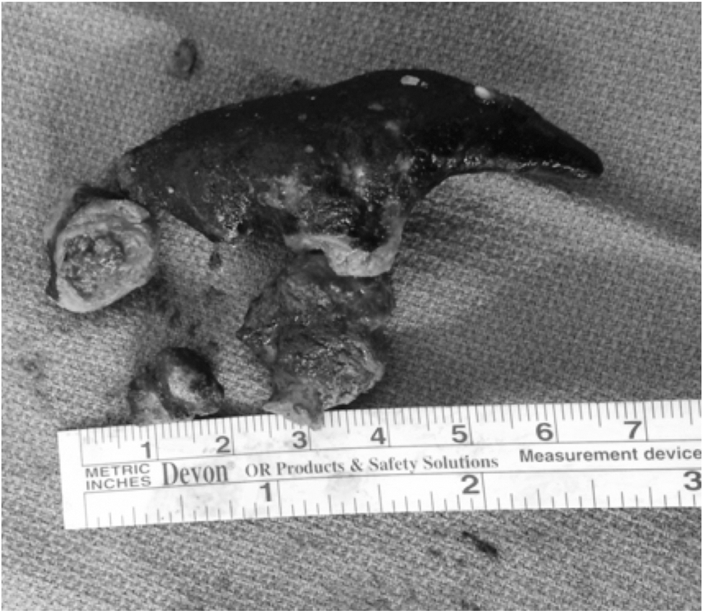
Representative image of the extracted stone.
One patient had significantly thinned parenchyma, and only one layer of suture was used to close the parenchymal defect. No new drains or stents were placed for any of the patients; however, two patients had a previously placed ureteral stent and nephrostomy tube that were kept during surgery. Hemostatic agents, such as Floseal™ (Baxter Healthcare Corporation, Deerfield, IL) and Surgicel™ (Ethicon, Somerville, NJ) were used to achieve hemostasis at the nephrolithotomy site. The stone fragments and bulldog clamps were then placed in an Endo Catch™ bag (Covidien, Mansfield, MA), the robot was undocked, and the Endo Catch bag was delivered through the camera port.
Results
Complete patient characteristics, preoperative variables and indications for surgery are listed in Table 1. The mean age was 47±16 years, five patients were female, and mean BMI was 31.9±10.0 kg/m2. The most common symptom was flank pain, and four out of seven patients had a documented history of repeated urinary tract infection. Five patients had complete staghorn calculi; the other two had partial staghorn calculi. The mean preoperative creatinine level was 1.10±0.68 mg/dL, and all but one patient had a preoperative eGFR of >60 mL/min/1.73 m2.
Calculated using the Modification of Diet in Renal Disease study equation.
SD=standard deviation; BMI=body mass index; UTI=urinary tract infection; MAG-3=mercaptoacetyltriglycine-3; NR=not recorded; MR=mental retardation; PCNT=percuntaneous nephrostomy tube; Cr=creatinine; eGFR=estimated glomerular filtration rate.
All seven procedures were performed as planned. There were no intraoperative complications and no conversions to open surgery. The mean warm ischemia time was 35±7 minutes, mean robotic time was 158±51 minutes, mean operative time was 222±48 minutes, and mean EBL was 121±39 mL (Table 2). The mean LOS was 3.0±1.7 days; patient 1 had a prolonged stay secondary to social factors—severe mental retardation and difficult clinical assessment, and patient 7 had postoperative gross hematuria necessitating continuous bladder irrigation and two units of packed red blood cell transfusion. There were no hospital readmissions.
Mean eGFR follow-up 4.4±4.9 months.
Calculated using the Modification of Diet in Renal Disease study equation.
SD=standard deviation; WIT=warm ischemia time; EBL=estimated blood loss; LOS=length of stay; Cr=creatinine; eGFR=estimated glomerular filtration rate.
On postoperative imaging (abdominal radiography vs CT), two patients were stone free, two patients had small fragments of residual stone (less than 1 cm representing 5% total stone burden), and three patients had between 20% and 40% residual stone burden. Among the three patients with 20% to 40% residual stone burden, one patient (patient 2) elected to have no further treatment, one patient (patient 3) elected to have serial ureteral stent exchanges, and one patient (patient 4) elected to have PCNL and ureteroscopy. The patients with 5% residual stone, to our knowledge, did not seek further treatment. The mean postoperative day 1 creatinine level was 1.19±0.57 mg/dL, and the mean most recent postoperative creatinine level was 1.06±0.47 mg/dL; eGFR was unchanged in five of six patients and improved in one patient (19 mL/min/1.73 m2 preoperative vs 25 mL/min/1.73 m2 postoperative). Mean follow-up time was 5.1±4.3 months.
Discussion
We investigated the feasibility of RAN as an alternative minimally invasive treatment modality for complex staghorn calculi in an attempt to decrease the number of procedures necessary for complete stone removal. To our knowledge, this is the third and largest study reporting outcomes of RAN 8,9 with the longest follow-up to date. In our preliminary experience, we demonstrated that this procedure may achieve complete stone-free rates with a single procedure with minimal operative blood loss and morbidity.
The Clinical Research Office of the Endourological Society (CROES) PCNL Global Study 2 and the British Association of Urological Surgeons Section of Endourology 3 have reported the largest studies to date regarding the efficacy of PCNL for treatment of patients with staghorn calculi. The CROES study group analyzed outcomes of 1466 patients with staghorn compared with 3869 patients with nonstaghorn stones undergoing PCNL. They found that patients with staghorn stones more frequently underwent multiple punctures (16.9% vs 5.0%) and had lower complete stone-free rates (56.9% vs 82.5%). 2 The United Kingdom study group reported on 299 patients who underwent PCNL for staghorn calculi demonstrating an intraoperative complete stone-free rate of 59% and 47% on formal postoperative imaging. 3
Laparoscopic techniques have previously been applied to anatrophic nephrolithotomy in an attempt to recreate the stone-free rates of open surgery with less morbidity. Initial reports of LAN are promising. Giedelman and associates 4 were able to completely render 4 of 8 patients stone free, and Simforoosh and colleagues 5 achieved similar results in 3 of 5 patients treated with LAN. Zhou and coworkers 6 performed LAN on 11 patients using warm ischemia rendering 91% of patients stone free; a postoperative urine leak developed in three patients, however.
The drawbacks of laparoscopy include the two-dimensional flat image, rigid instruments with fulcrum effect, decreased dexterity, and surgeon fatigue. The majority of these drawbacks have been overcome with robotic instruments, allowing surgeons to perform more complex cases including robot-assisted partial nephrectomies, 10 pyeloplasties, 11 and RAN. 8,9
In the current study, we performed RAN using warm ischemia; by comparison, Ghani and colleagues 8 performed RAN in three patients using cold ischemia. Although there were five of seven patients who had a relatively prolonged WIT of >30 minutes, postoperative creatinine and eGFR levels were comparable to their preoperative renal function in all patients. Admittedly, the long-term renal function of these patients remains to be determined. In the future, we plan to use preoperative and postoperative renal scan with differential renal function to better quantify the effect of long WITs. One short-term advantage of performing RAN with warm ischemia vs cold ischemia is that the procedure can be performed with small minimally invasive incisions, avoiding a larger incision needed for inserting a hand port for placing ice-slush around the kidney.
While RAN for the management of staghorn calculi is able to accomplish the tenets of open surgery in a less morbid fashion, there are pitfalls associated with the procedure. The reported stone-free rate associated with OAN is 71% 1 and, in our experience, 29 % of patients were stone free. These stone-free rates, however, are similar to those of Ghani and associates 8 (33%) and comparable to large PCNL studies 2,3 (47%–57%). This may be explained by an early learning curve for RAN and by the lack of proprioception with the robot and inability to assess for residual stone burden during the procedure. Fluoroscopy is used during PCNL to evaluate for residual stones, which is not feasible using the robotic system. Laparoscopic ultrasonography may be feasible for locating fragments of stone left during extraction; this is an imaging modality that has been reported to be successful 8 and one we will explore in the future if there is concern for residual stone burden.
We performed RAN using WIT; historically, the OAN technique and that of Ghani and coworkers 8 have used cold ischemia, which is accepted as being renoprotective. Finally, whether a robotic procedure for managing staghorn calculi proves to be cost-effective remains to be determined. If mean LOS is used as a surrogate for cost-effectiveness, however,the mean LOS of 3.0 days in our RAN series suggests a potential benefit compared with current LAN studies (mean LOS 3.54 and 5.4 days 5 ), although the sample size is small. With improvement and refinement of the technique, the goal of robotic surgery for treatment of patients with staghorn calculi is a safe, efficacious modality that decreases the number of procedures needed to achieve complete stone-free rates in a cost-effective manner.
Conclusions
Based on our initial experience of seven patients, RAN is a safe procedure with encouraging outcomes as a minimally invasive treatment modality for patients with extensive stone burden. We recommend that robotic surgeons who are comfortable with robot-assisted partial nephrectomy consider RAN in the appropriate patient, because there are duplicated steps common to both procedures including renal hilum exposure, renal vessel clamping, nephrotomy, collecting system closure, and parenchymal closure. Using intraoperative imaging techniques, appropriate patient selection, and continued experience with RAN will improve WIT, decrease robotic console time, and ultimately improve stone-free rates. Longer follow-up to determine the true effect of the procedure on renal function and further studies reporting technical refinements will be beneficial for legitimizing RAN as a mainstream treatment modality for staghorn calculi.
Footnotes
Disclosure Statement
No competing financial interests exist.