
Abstract
Background and Purpose:
Proper cleaving of reusable laser fibers is needed to maintain optimal functionality. This study quantifies the effect of different cleaving tools on power output of the holmium laser fiber and demonstrates morphologic changes using microscopy.
Materials and Methods:
The uncleaved tips of new 272 μm reusable laser fibers were used to obtain baseline power transmission values at 3 W (0.6 J, 5 Hz). Power output for each of four cleaving techniques—11-blade scalpel, scribe pen cleaving tool, diamond cleaving wheel, and suture scissors—was measured in a single-blinded fashion. Dispersion of light from the fibers was compared with manufacturer specifications and rated as “ideal,” “acceptable,” or “unacceptable” by blinded reviewers. The fiber tips were also imaged using confocal and scanning electron microscopy. Independent samples Kruskal-Wallis test and chi square were used for statistical analysis (α<0.05).
Results:
New uncleaved fiber tips transmitted 3.04 W of power and were used as a reference (100%). The scribe pen cleaving tool produced the next highest output (97.1%), followed by the scalpel (83.4%), diamond cleaving wheel (77.1%), and suture scissors (61.7%), a trend that was highly significant (P<0.001). On pairwise comparison, no difference in power output was seen between the uncleaved fiber tips and those cleaved with the scribe pen (P=1.0). The rating of the light dispersion patterns from the different cleaving methods followed the same trend as the power output results (P<0.001). Microscopy showed that the scribe pen produced small defects along the fiber cladding but maintained a smooth, flat core surface. The other cleaving techniques produced defects on both the core and cladding.
Conclusion:
Cleaving techniques produce a significant effect on the initial power transmitted by reusable laser fibers. The scribe pen cleaving tool produced the most consistent and highest average power output.
Introduction
I
These fibers, however, need additional care that is not required with single-use fibers. Specifically, reusable fibers must be properly handled by operating room and support staff to prevent mechanical trauma, because excess bending may lead to inefficient energy transfer and fiber fracture. 4,5 The fibers must also be maintained with routine stripping of the outer jacket and cleaving of the silica core between cases. 3 A number of laser fiber cleaving tools are commercially available; however, the optimal method of cleaving laser fibers has not been clearly defined. In addition, common surgical tools such as suture scissors and scalpels may be used for both reusable and single-use fibers, especially if intraoperative cleaving is performed because of tip damage.
The purpose of this article is to quantify the effect of different cleaving tools on energy output of reusable laser fibers and to demonstrate changes in tip morphology with microscopy.
Materials and Methods
A prospective, randomized, single-blinded trial was conducted using SlimLine-200 (272 μm diameter) reusable laser fibers (Lumenis Ltd., Yokneam, Israel). To obtain power measurements, the fibers were connected to a Lumenis VP100 holmium laser (Lumenis Ltd.) that was set at a power output of 3 W (0.6 J at 5 Hz), which is the standard initial setting for laser lithotripsy at our and other institutions. 6 –8 The fiber tip was secured in a standard position (Fig. 1) with the tip flush to the entrance of a PM150X Air-Cooled Thermopile power sensor (Coherent Inc., Santa Clara, CA). A FieldMaxII-TOP Energy Meter (Coherent Inc.) was used to measure and record the initial power output transmitted by the silica fibers, which was obtained by calculating the average of 50 power measurements recorded during a 5 second interval. Power measurements were performed in air because of equipment constraints that did not allow for measurement in a liquid environment.
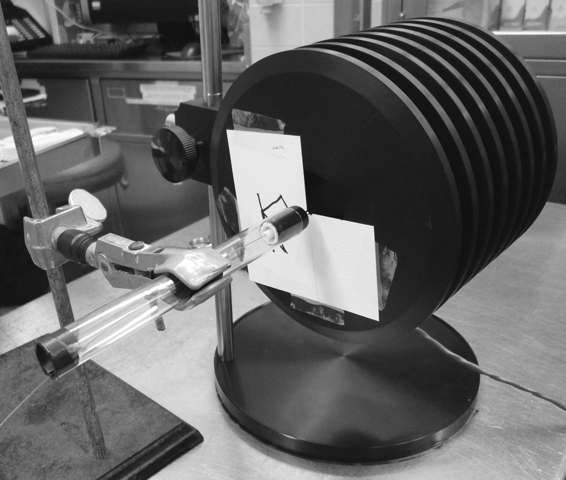
Setup for measuring laser power transmission.
The fiber was cleaved 2 cm proximal to visible defects using each of four commonly available tools: An 11-blade scalpel, scribe pen cleaving tool (Sancliff Inc., Worcester, MA), diamond cleaving wheel (Cook Medical, Bloomington, IN), and straight Mayo suture scissors (Fig. 2). For the scribe pen and scalpel, the fiber was placed on an operating table and scored perpendicularly through the jacket. The diamond cleaving wheel was placed onto the table, and the fiber was pressed down perpendicularly onto the edge of the wheel and then scored along the wheel's axis. The fiber was then grasped on either side of the score line and pulled straight apart without bending the fiber. The middle of the suture scissor blades were used to transect the fiber perpendicularly to the long axis of the fiber. Following each cleave, a standard 200 μm fiber stripper (Micro Electronics Inc., Hilliard, OH) was used to remove 5 mm of the colored jacket. All cleaving and stripping was performed by the same person. A second investigator, blinded to the method of fiber cleaving, measured power output.
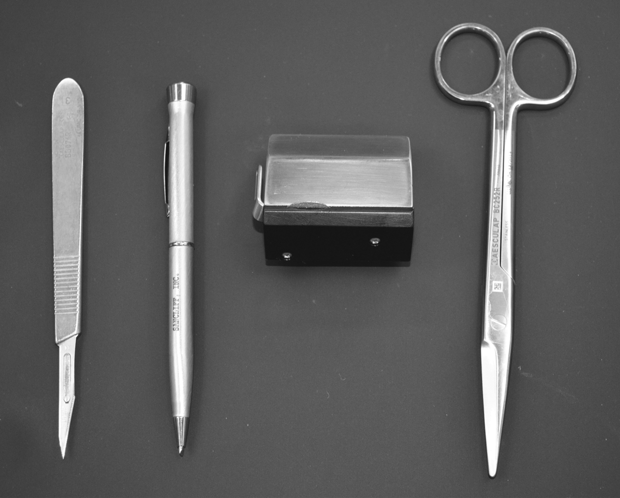
Left to right: 11-blade scalpel, scribe pen cleaving tool, diamond cleaving wheel, straight Mayo suture scissors.
To characterize the adequacy of cleaving, the fibers were cleaved 10 times with each of the four techniques, and the laser aiming beam was projected onto a dark surface at a distance of 1 cm from the fiber tip. The pattern of light dispersion was photographed using a Nikon D5100 16.2 megapixel camera (Nikon Corporation, Tokyo, Japan), and each image was then classified by a group of 10 blinded urologists as “ideal,” “acceptable,” or “unacceptable” according to manufacturer recommendations.
To measure power output, each of the four cleaving techniques—scalpel, scribe pen, diamond cleaving wheel, and suture scissors—was performed seven additional times. Three power measurements were taken for each trial, for a total of 21 power measurements per cleaving technique. These were then compared with baseline power output from two new, uncleaved SlimLine 200 reusable laser fibers (Lumenis Ltd.), which acted as controls.
After power measurements were taken, the silica fiber tips from each trial were imaged for visual characterization. A 5 megapixel confocal microscope was used to inspect the profiles of the cleaved tips at a 90-degree angle from the long axis of the laser fibers. Scanning electron microscope (SEM) images of the fiber ends were also prepared, which allowed high-resolution viewing of the cleaved silica fiber core and cladding surfaces. To obtain the SEM images, the fiber tips were placed vertically onto aluminum Pin Stub Mounts covered with PELCO Tabs™ Carbon Conductive Tabs (Ted Pella, Inc., Redding, CA). The tips were plated using a Cressington 108Auto Sputter Coater (Cressington Scientific Instruments Ltd., Watford, United Kingdom) for 15 seconds three times with gold/palladium, rotating each sample 30 degrees between coats. Fiber tips were imaged at 400× magnification by a VEGA LSH SEM (Tescan USA, Cranberry Township, PA) operated at an accelerating voltage of 10 kV. Composition analysis was also run on a new fiber tip by a NSS Spectral Imaging System (ThermoFisher Scientific Inc., Waltham, MA) using energy dispersive X-ray spectrometry (EDX).
Data were analyzed with SPSS Statistics (IBM, version 20). Independent samples Kruskal-Wallis test and chi square were used for statistical analysis, with significance at α<0.05. Bonferroni corrections were applied to pairwise comparisons to account for multiple tests.
Results
The SlimLine-200 laser fiber has three visible components: The outer colored jacket, thin cladding, and fiber core. EDX analysis distinguished the major component of the cladding as niobium and the core as silicon dioxide (Fig. 3).
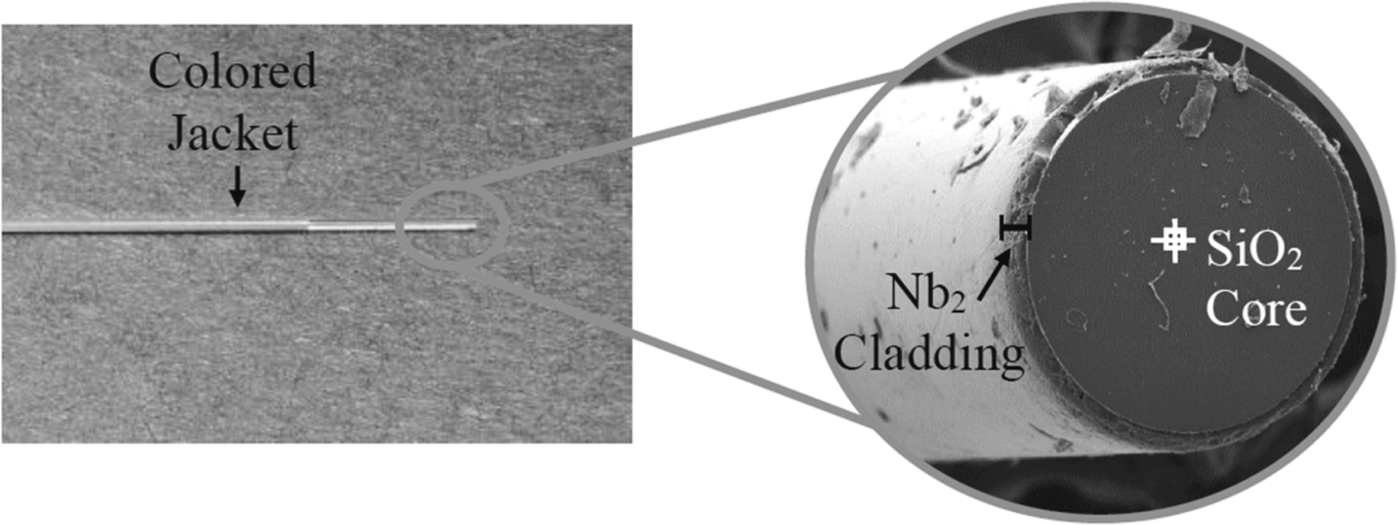
The SlimLine-200 reusable laser fiber is composed of an outer colored jacket, thin cladding, and fiber core. Analysis by NSS Spectral Imaging System clearly distinguishes the silica-based core from the niobium-based cladding.
Cleaving of the core and cladding by the tested techniques was associated with a significant difference in power output (P<0.001), as seen in Figure 4. The new, uncleaved fiber tips transmitted 3.04±0.04 W of power and were used as a reference (100%). The scribe pen cleaving tool had the next highest average output at 2.95±0.11 W (97.1% of reference), which was similar to the output of the uncleaved tips (P=1.0). The fiber tips cleaved with the scalpel had the second-highest power output at 2.53±0.22 W (83.4% of reference; P=0.007 vs control). Fibers cleaved with the diamond cleaving wheel (2.34±0.52 W, 77.1% of reference; P=0.002 vs control) and the suture scissors (1.87±0.64 W, 61.7% of reference; P<0.001 vs control) showed the lowest power output as well as the highest standard deviation.

With the laser set at a power of 3 W, the measured power output of the uncleaved (control) fibers was 3.04 W, while the fibers cleaved with suture scissors transmitted only 1.87 W.
On pairwise comparison, the fiber tips cleaved with the scribe pen tool had similar power transmission to the new, uncleaved fiber tips (P=1.0). The remaining cleaving methods using the scalpel, diamond cleaving wheel, and suture scissors yielded power output results that were statistically different from the uncleaved fibers (all P values <0.008) and scribe pen fiber tips (all P values <0.002), and were not statistically different from each other (P values 0.16–1.0).
The adequacy of fiber cleaving based on light dispersion patterns followed the same pattern as the power output results (Fig. 5), with a significant difference seen in the proportion of adequate cleaves based on cleaving method (P<0.001). All uncleaved fiber images were rated as adequate for use, with 95% being rated as “ideal” and 5% being “acceptable.” Fibers cleaved with the scribe pen, scalpel, and diamond cleaving wheel were rated as adequate 82%, 69%, and 73% of the time, respectively. The suture scissors produced the worst cleaves based on light dispersion, with 59% being rated as “unacceptable” and only 2% rated “ideal.” On pairwise comparison, the suture scissors were statistically inferior to each of the other cleaving methods tested (P<0.001).

Characterization of cleaving adequacy based on light dispersion from the cleaved fiber tips.
Representative images showing the light dispersion, confocal microscopy, and SEM images for each of the cleaving techniques are shown in Figure 6. Both confocal microscopy and SEM images showed that the uncleaved fibers had smooth, flat tips that were oriented perpendicular to the length of the fiber. High-resolution photographs with the SEM showed a smooth core and uniform cladding.
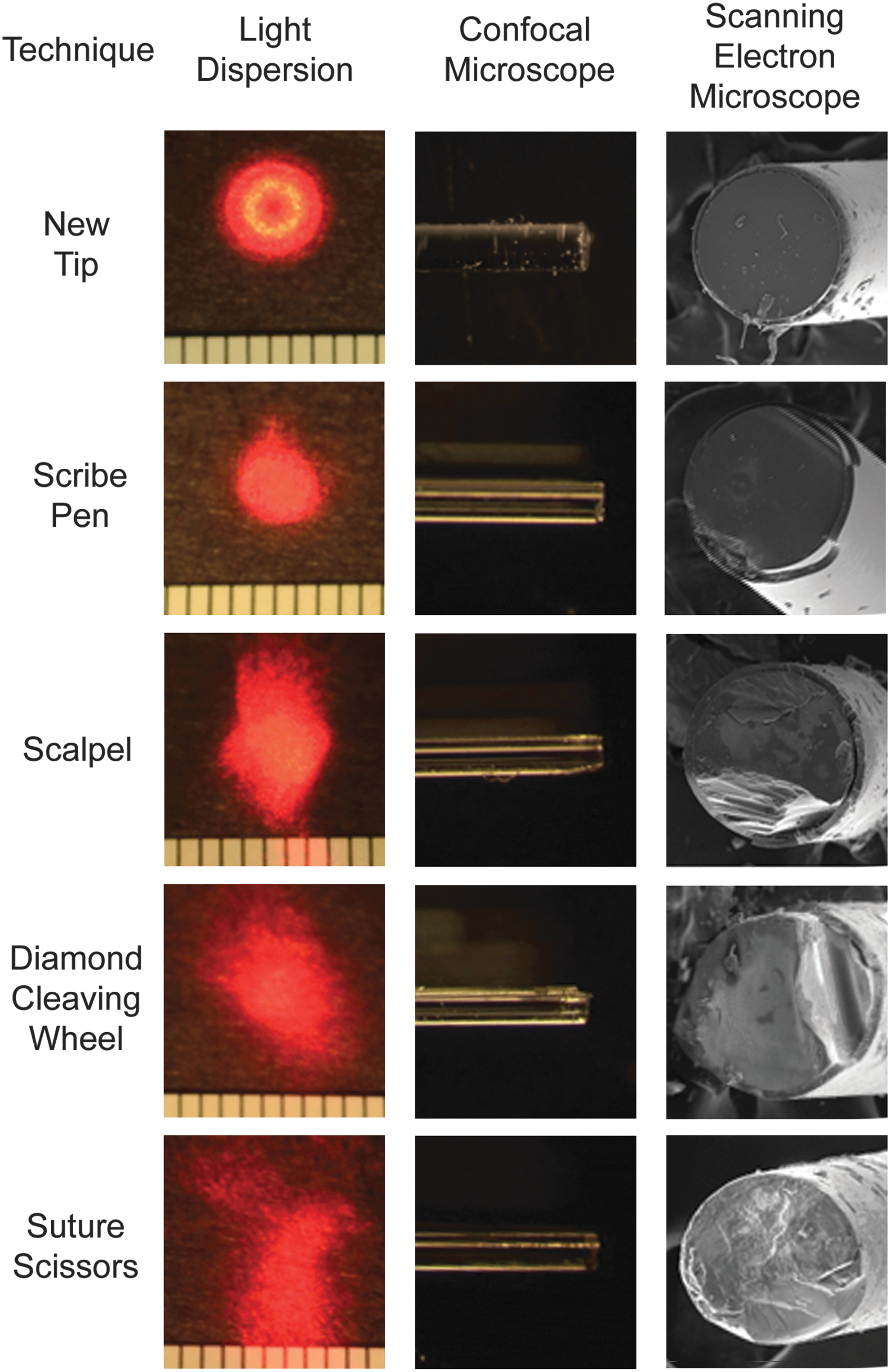
Light dispersion from fiber tips and images with confocal microscopy and scanning electron microscopy.
As seen in Figure 7, cleaving with the scribe pen produced a similarly oriented perpendicular tip with a few samples showing a small raised edge. The SEM revealed a smooth central core surface with one to two defects along the periphery and cladding. The scalpel-cleaved fibers were slanted with a rounded raised edge as seen by confocal microscopy. The SEM images showed fibers with somewhat smooth cores; however, approximately one fourth to one third of the cross-sectional area of the core material had been removed with cleaving, presumably at the location where the scalpel contacted the fiber. This produced defects along the core and cladding as seen with microscopy. Similar to the scalpel-cleaved fibers, the fibers cleaved with the diamond cleaving wheel showed defects at one edge of the fiber, presumably at the site of contact with the instrument. The core surface of the diamond-cleaved fibers was more variegated, however, and when viewed in profile with confocal microscopy, the fiber tips were seen to have more uneven and jagged tip edges. Finally, the tips cleaved with suture scissors did not show focal defects along the rim of the core but instead exhibited a diffusely uneven surface. Some of the fibers showed a protrusion of circumferential cladding while others showed a loss of cladding with only a bare core. This group exhibited the most variation in the appearance of the cleaved tips with microscopy and also showed the greatest variation in power output.
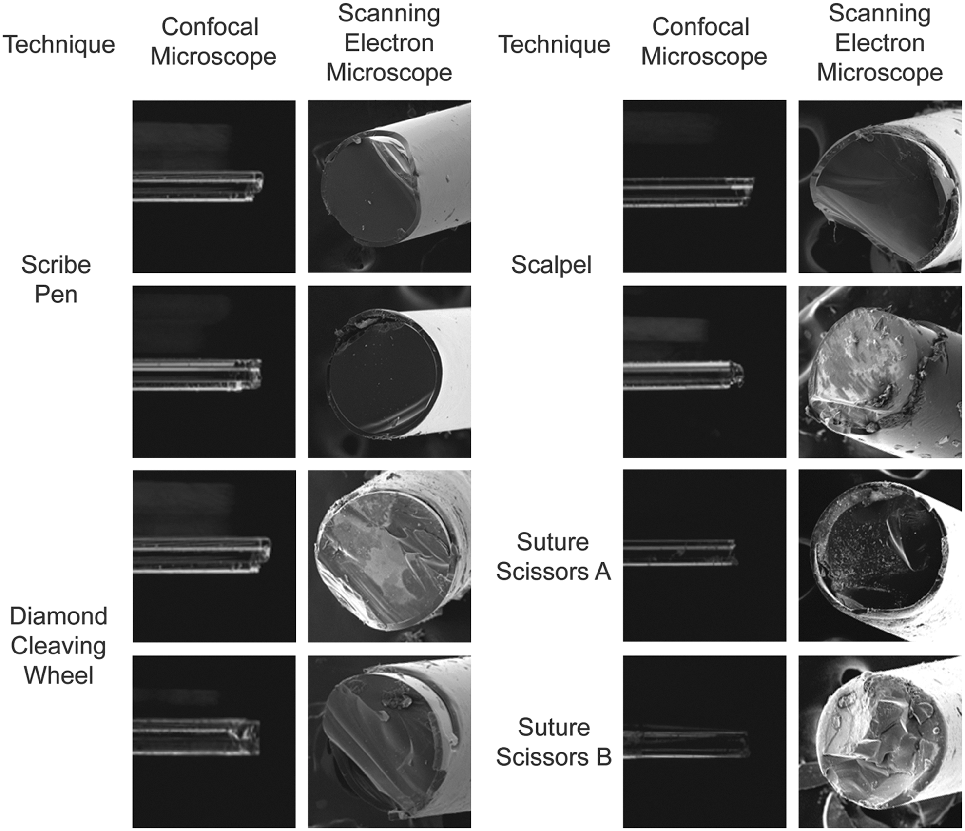
Additional fiber tips cleaved with the scribe pen, scalpel, diamond cleaving wheel, and suture scissors. Fibers cleaved with suture scissors demonstrated circumferential protrusion of cladding (
Discussion
Ureteroscopy with laser lithotripsy is a cost-effective and efficacious treatment for patients with urolithiasis. 9,10 There remains significant cost associated with these procedures, however, and in particular with the holmium laser fibers and ureteroscopes. Reusable laser fibers have been shown to reduce the cost of materials for ureteroscopy by an average of $118 per procedure after taking into account the reprocessing cost, which has been reported at $25 per use for material costs alone. 3 Unlike disposable fibers, however, which are opened new and are ready to use without any additional preparation, reusable laser fibers must be reprocessed between each use. This involves cleaving of the silica core, stripping of the outer protective colored jacket, and sterilization and packaging. The manufacturer-provided instructions for proper care of the reusable laser fiber also include visualization of light dispersion through the fiber to ensure proper cleaving. According to the product manual for the SlimLine fiber, the ideal pattern of light dispersion should be circular and well-defined, similar to that seen with the new uncleaved fiber. Acceptable cleaves show an ovoid dispersion pattern, and unacceptably cleaved fibers show poorly defined patterns or irregular circumference.
Light dispersion patterns parallel the laser energy transmission from the fiber to the target. With smooth planar surfaces, the light dispersion patterns are uniform and easily predicted by the Snell law of refraction; this takes into account the index of refraction between the silica glass and subsequent material. Rough surface dispersion patterns, however, are estimated by more complex formulations such as the microfacet theory, which analyzes a surface from the perspective of repeating microunits. Studies exploring this theory have shown that light traveling through samples with increasingly rough surfaces exhibit greater proportions of light transmission that is scattered and consequently shifted away from the ideal refraction angle given by the Snell law. 11 Therefore, cleaving methods that produce rough surfaces will result in more diffuse light dispersion patterns and less energy reaching the desired target (Fig. 8).

Incident laser beams are transmitted through smooth surfaces while maintaining good collimation and minimal scatter. Transmission through rough surfaces, however, leads to dispersion of optical output and uneven energy distribution.
In an experimental benchtop setting, optical scatter from rough fiber surfaces is reflected in lower energy reaching the meter. In a clinical setting, the laser fiber is fired in close proximity or direct contact to the target substance, thus attenuating the degree of optical scatter. Other articles investigating the effects of fiber tip damage during lithotripsy, however, have raised concerns of inefficient stone fragmentation, 12,13 decreased fiber longevity, 14 and accidental ureteral mucosal damage 8 based on the principle of optical scatter. We hypothesize that the same underlying mechanism of noncollimation of photons from a rough surface will produce similar effects whether the rough surface is created from tip damage sustained during lithotripsy or from suboptimal cleaving technique.
Little is known about the effects of fiber cleaving on power output from the Ho:YAG laser. Our results show that fiber cleaving technique has a substantial effect on the amount of power that is transmitted from the laser generator to the target. As expected, the new fiber tips demonstrated a power output that was nearly identical to the generated power, with low variability. Tips cleaved with the scribe pen produced the most similar results to the new fiber, transmitting the highest power (97% of the output of the new fibers) with the lowest variability. Pairwise comparison showed that the power output from the fibers cleaved with the scribe pen was similar to that seen with the uncleaved fibers (P=1.0). This was the only cleaving tool that did not significantly reduce power output. The other cleaving methods using the scalpel, diamond cleaving wheel, and suture scissors all yielded statistically similar power output results to each other (P values 0.16–1.0) yet were statistically different from both the scribe pen and the new fibers (all P values <0.008). When compared with the new tips, the fibers cleaved with the scalpel, diamond cleaving wheel, and suture scissors all transmitted significantly less power (61.7%–83.4%). The light dispersion, confocal microscopy, and SEM imaging corresponded with the power output results. The fibers cleaved with the scribe pen had the most focused light dispersion pattern and smoothest core while the remaining cleaving methods resulted in diffuse light dispersion patterns and defects along the core and cladding. Thus, our results suggest that the scribe pen cleaving tool is the optimal fiber cleaving method among those tested.
Although no previous study to our knowledge has studied power output associated with laser cleaving techniques, several studies have investigated the changes in laser fiber energy or power output over time. Vassar and associates 12 performed in vitro Ho:YAG lithotripsy on human calcium oxalate monohydrate stones and found that energy output after 0.15 kJ had been applied to stones was significantly lower than energy output from new laser fibers. Greater energy loss was seen when 1.5 J pulses were used than when 1.0 J pulses were applied. In addition, the energy loss at 1.0 J/pulse with 272 μm fibers (59.6%) was significantly greater than the corresponding energy loss seen with larger diameter (365–940 μm) fibers (1.0%–5.1%). These authors cite noncollimation of optical output because of damaged fiber tips as the cause of inefficient energy transmission. 12 This is in agreement with the microfacet theory and the results of the current study, because fiber tips with the smoothest silica cores on the SEM had the highest power transmission while those with significant surface defects exhibited decreased power output. Similarly, studies of optical fiber tip damage have shown that small-diameter (<300 μm) fibers tend to exhibit a greater degree of burn-back and reduction of energy transmission during lithotripsy compared with larger-diameter fibers. This may contribute to less efficient stone fragmentation and a shorter working life of the fiber. 14 Grant and colleagues 13 also performed an in vitro study in which a 600 μm neodymium:yttrium-aluminum-garnet fiber was used for tissue ablation at varying power settings. After 300 seconds of laser ablation, loss of power transmission ranged from less than 5% at the 10 W setting to greater than 45% at 20 W. Significantly, no study has demonstrated a gain in power or energy output with time.
Loss of energy and power transmission through surgical laser fibers has been demonstrated with both lithotripsy and tissue ablation. This appears to be most pronounced with small diameter fibers and higher power settings. We have shown that higher initial power output can be preserved when reusable laser fibers are cleaved with the scribe pen cleaving tool, whereas the greatest power loss was observed when suture scissors were used for cleaving. Adequacy of fiber cleaving as assessed by light dispersion patterns mirrored the relative power output for the techniques tested, indicating that light dispersion may be used as an important quality control measure. This should be assessed after each fiber cleave, because fibers with unacceptable light dispersion patterns may be recleaved to preserve maximal initial power output. Because power output can be expected to either remain stable or decrease with use, proper fiber cleaving may have implications for surgical efficiency.
Our study does have several limitations. The power measurements and imaging were obtained from a single brand of reusable laser fiber using one laser generator. Although many fibers have similar composition, it is possible that differences may exist between fiber types and their responses to cleaving. For example, it may be more difficult to produce a uniformly smooth cleave for larger-diameter fibers. The SlimLine-200, however, was selected for testing because small caliber laser fibers are commonly used in flexible ureteroscopy and are subjected to higher mechanical stresses. In addition, more rapid burn-back may prompt the need for more frequent cleaving and stripping of the laser fiber. 12 Finally, although power output was found to be greatly affected by the method of fiber cleaving in this benchtop test, it is unknown whether this difference in power transmission affects clinical efficacy during procedures using the holmium laser. Additional in vitro and in vivo testing may help to further characterize the effects of laser fiber cleaving on surgical outcomes.
Conclusions
Cleaving techniques produce a significant effect on the power transmitted by 272 μm reusable laser fibers. The scribe pen cleaving tool produced the most consistent and highest average power output, whereas suture scissors produced the worst outcomes. Utilization of the scribe pen for fiber cleaves may preserve higher initial power output during use of the holmium laser. In addition, the light dispersion pattern of the aiming beam closely correlates with power output. This can be used as a proxy to judge adequacy of fiber cleaving and subsequent power output.
Footnotes
Acknowledgments
Wayne Kelln provided assistance with acquiring the scanning electron microscope images. Dr. Kerby Oberg assisted with acquiring the confocal microscopy images.
Disclosure Statement
Dr. D. Duane Baldwin serves as a consultant for Terumo, Boston Scientific, and a lecturer for Cook Medical. For the remaining authors, no competing financial interests exist.
Abbreviations Used
References
Supplementary Material
Please find the following supplemental material available below.
For Open Access articles published under a Creative Commons License, all supplemental material carries the same license as the article it is associated with.
For non-Open Access articles published, all supplemental material carries a non-exclusive license, and permission requests for re-use of supplemental material or any part of supplemental material shall be sent directly to the copyright owner as specified in the copyright notice associated with the article.