
Abstract
Purpose:
To determine whether shock wave lithotripsy (SWL) may be a risk factor for renal functional impairment in a swine model of metabolic syndrome (MetS).
Materials and Methods:
Nine-month-old female Ossabaw pigs were fed an excess calorie atherogenic diet to induce MetS. At 15 months of age, the MetS pigs were treated with 2000 SWs or an overtreatment dose of 4000 SWs targeted at the upper pole calyx of the left kidney (24 kV at 120 SWs/min using the unmodified Dornier HM3 lithotripter; n=5–6 per treatment group). Serum creatinine (Cr) and blood urea nitrogen (BUN) levels were measured in conscious pigs before and ∼60 days after SWL to provide a qualitative assessment of how well both kidneys were filtering (glomerular filtration rate [GFR]). Bilateral renal function was assessed at ∼65 days post-SWL in anesthetized pigs with GFR and effective renal plasma flow (ERPF) quantified by the renal clearance of inulin and para-amino hippurate, respectively.
Results:
Cr and BUN values were within normal limits before SWL and remained unchanged after lithotripsy in both the 2000 SW- and 4000 SW-treated pigs. GFR and ERPF of kidneys treated with SWL at either SW dose were similar to the contralateral nontreated kidney. Chronic histological changes in the SW-treated pole of the kidney included interstitial fibrosis, sclerotic glomeruli, and dilated and atrophic tubules.
Conclusions:
Our results are consistent with the view that a single SWL session does not result in renal impairment, even in the presence of MetS.
Introduction
M
Shock wave lithotripsy (SWL) is an effective noninvasive treatment modality for renal and ureteral stones. 12 However, SWL can have undesirable side effects such as tissue injury, which can lead to loss of renal mass and functional impairment. 13 Despite reports that SWL may be associated with long-term complications, the consensus in the urologic community is that SWL is safe and that benefits outweigh the risks when used appropriately in treating the general urolithiasis population. 14,15 The few reports addressing the long-term outcome of SWL in stone patients with chronic renal insufficiency would also suggest that SWL is safe in such a vulnerable cohort. 15 –17 The etiology of CKD in these patients is thought to be obstructive uropathy with SWL treatment providing relief from obstruction and generally improving renal function. However, there is limited information on whether SWL treatment may exacerbate the risk for renal functional impairment in patients with MetS—obesity, hypertension, insulin resistance, and other features of MetS being risk factors for CKD. 4,18 We examined this issue in a swine model of diet-induced obesity and MetS that develops similar features of human MetS. 19,20
Materials and Methods
Animal studies were conducted in accordance with the National Institutes of Health Guide for the Care and Use of Laboratory Animals and were approved by the Institutional Animal Care and Use Committees of Indiana University School of Medicine and Methodist Hospital. Seven to 9-month-old female Ossabaw pigs (n=5–6 per treatment group) were fed an excess calorie, atherogenic diet of 6,000 kcal/day to induce obesity and other features of MetS. 19,21 After 6 months on the high-fat diet, the MetS pigs underwent SWL treatment using the unmodified HM3 lithotripter.
Pigs were anesthetized (induction with ketamine [20 mg/kg] and xylazine [2 mg/mL]; maintenance with 1–3% isoflurane) and the urinary collecting system of the left kidney visualized using contrast medium (injected into a ureteral catheter) and X-ray fluoroscopy. SWs were targeted to the upper pole calyx and delivered at 2000 SWs or 4000 SWs (24 kV, 120 SWs/min) with X-ray verification of SW targeting done every 500 SWs on-the-fly, and with SW treatment paused every 1000 SWs to replace the electrode and check targeting (∼1 minute).
Intravenous blood samples were collected from conscious, fasted pigs a few days before SWL and at ∼60 days post-SWL. Samples were assayed for serum chemistries including serum creatinine (Cr) and blood urea nitrogen (BUN).
Approximately 65±1 days after SWL, pigs were reanesthetized for bilateral renal function measurements. Glomerular filtration rate (GFR) and effective renal plasma flow (ERPF) were estimated by the renal clearance of inulin and para-amino hippurate, respectively, using previously described colorimetric methods. 22 Cardiovascular and renal function measurements were begun ∼30 minutes after the end of all surgery, and consisted of two to four 30-minute clearances. Upon completion of all functional measurements, pig kidneys were perfusion fixed in situ, harvested, sectioned, and prepared for histologic analysis. 22
The pigs in the present series of experiments had robust MetS and are a subset of the animals assessed for glucose tolerance and insulin resistance in a recently published study. 21
Statistical analysis
Variables were summarized by means and standard errors of means. Paired Student's t-tests were used to compare renal function (GFR, ERPF, urine flow rate [UV], absolute sodium excretion rate [UNaV], and fractional excretion of sodium [FENa]) between the SWL-treated kidney and contralateral nontreated kidney (control) for both 2000 SW and 4000 SW groups. Paired Student's t-tests were also used to compare pre- and post-SWL serum Cr and BUN levels for the 2000 SW and 4000 SW groups separately. Independent two-sample t-tests were used to compare renal function between 2000 SW- and 4000 SW-treated groups. Two-sided p-values <0.05 were considered significant.
Results
Cardiovascular and renal function
Conscious animals
Serum Cr and BUN values were within normal limits and similar before and ∼60 days after SWL in both groups of pigs (Table 1).
Anesthetized animals
Mean arterial blood pressure and heart rate in the 2000 SW-treated pigs (71±3 mm Hg and 132±12 beats/min, respectively) were nearly identical to those measured in the 4000 SW-treated pigs (73±2 mm Hg and 130±11 beats/min, respectively). Renal hemodynamics (GFR and ERPF) and excretion (UV, UNaV, and FENa) were similar in the SWL-treated and contralateral nontreated kidney regardless of SW dose delivered (Table 2). The slightly lower renal hemodynamic values and much higher urinary fluid and sodium excretion rates in the kidneys treated with 4000 SWs did not reach a level of statistical significance compared to their 2000 SW counterparts (p≥0.1185). Overall, cardiovascular and renal functions were similar within and across groups.
T=SWL-treated kidney; CNT=contralateral, nontreated kidney; GFR=glomerular filtration rate; ERPF=effective renal plasma flow; UV=urine flow rate; UNaV=absolute sodium excretion rate [urine sodium concentration×urine flow rate]; FENa=fractional excretion of sodium [UNaV/(plasma Na concentration×GFR)].
Renal histology
Figure 1 shows the histology of kidneys treated with 2000 SWs (Fig. 1A, C, E) or 4000 SWs (Fig. 1B, D, F). The most obvious and consistent chronic change noted in the upper pole of the left kidney of SWL-treated animals was distinct linear streaks/bands of interstitial fibrosis that in most cases extended from the papilla to the cortex. These bands were detected on both the anterior and posterior surfaces of the kidney (Fig. 1A, B). Occasionally, the cortical fibrotic bands occurred as multiple, wide regions that spanned the entire width of a renal lobule and involved the accompanying renal papilla as is noted in Figure 1A (within circle). These larger fibrotic bands were commonly associated with dimpling of the capsular surface. Within the cortical fibrotic bands most tubular segments are atrophic, many glomeruli show sclerosis, and the interstitial space is extensively fibrotic (Fig. 1C–F). Dilated tubular segments were occasionally seen within and adjacent to the fibrotic bands (Fig. 1D–F). The 2000 and 4000 SW-treated kidneys showed similar changes. Large regions of cortical tissue adjacent to the fibrotic bands appear normal (Fig. 1C, D).
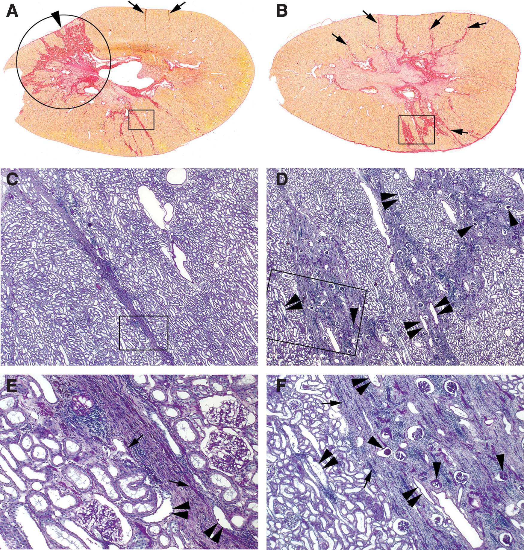
Chronic cortical changes in SW-treated kidneys.
Histological scoring of the renal cortex from a subset of SW-treated pigs quantified the degree of obliterated and partially sclerotic glomeruli, tubule atrophy, and interstitial fibrosis. No arteriosclerosis or hyaline arteriosclerosis was observed. More tubules in the 4000 SW-treated group appeared dilated compared to the control MetS pigs not treated with SWs (Table 3).
Kidney sections stained with Jones' silver and PAS. Histologic scoring is of the renal cortex. Linear fibrotic steaks of varying lengths with most extending from medulla to cortex.
TA=tubular atrophy; IF=interstitial fibrosis; AS=arteriosclerosis; HA=hyaline arteriosclerosis; PAS=periodic acid-schiff; 1+= mild (<10%); 2+= moderate (11–25%); 3+= moderately severe (25–75%); 4+= severe (>75%).
SWL-induced chronic changes in renal papillae were always associated with regions of cortical damage (circle, Fig. 1A). Figure 2 shows papillary histology from a 2000 SW-treated (Fig. 2A, B) and 4000 SW-treated (Fig. 2C, D) animal. The degree of papillary damage varied from sparse regions of interstitial fibrosis with minimal tubular damage and moderate tubular dilation to widespread interstitial fibrosis and extensive tubular enlargement (Fig. 2A–D). The two primary changes included interstitial fibrosis and tubular dilation. Tubular changes included extensively dilated outer and inner medullary collecting ducts and an occasional thick ascending limb (Fig. 2B, D). A greater number of these tubules were dilated in regions with extensive interstitial fibrosis and the degree of dilation was also more pronounced. Another tubular change was atrophy, which was localized to sites of interstitial fibrosis. A few medullary-collecting ducts possessed a unique configuration of its lining cells that resembled a polyp-like structure projecting into the tubular lumen suggesting cell proliferation (Fig. 2B).

Chronic medullary changes in SW-treated kidneys.
Histologic evidence of tissue injury to the untreated lower renal pole was rare or absent in SWL-treated pigs.
Discussion
Patients who have kidney stones, MetS, or both are at increased risk for CKD. 4,6,10,11,18 The modality of stone removal could also add to this risk if it results in deterioration in renal function due to loss of renal parenchyma.
SWL is a noninvasive treatment often used to remove uncomplicated kidney stones. 12 Treatment of the kidney with SWs causes trauma to the renal parenchyma and can reduce microcirculatory function. 13 The general consensus is that SWL is safe and that such acute complications are transient with no long-term sequelae in the normal urolithiasis population. However, the functional outcome of SWL in patients with CKD or comorbidities for CKD is less clear. Early work suggested that the long-term effect of SWL on renal function is influenced by the degree of renal insufficiency at the time of treatment—patients with mild renal insufficiency showed an improvement in glomerular function, whereas patients with moderate to severe renal insufficiency showed initial improvement and then further deterioration in renal function that may have been related to SW treatment or the natural progression of the disease. 14 A retrospective analysis of 131 nephrolithiasis patients with CKD followed up for about 3 years after SWL found that stone removal and relief of obstruction was associated with delayed deterioration of renal glomerular function compared with those patients not treated with SWL or failed SWL. 17 A 19-year follow-up retrospective study of 288 patients found that the rate of renal insufficiency (∼5%) was unchanged after SWL despite higher rates of obesity and diabetes. 23 In contrast, a 15-month follow-up prospective study on 134 patients reported that SWL was associated with a higher prevalence of CKD after treatment, and that BMI was a risk factor for CKD after SWL. 24
The Ossabaw pig provides a unique large-animal model of human MetS. 19,20 This animal model has features of MetS (e.g., obesity, hypertension, insulin resistance, hyperglycemia, and dyslipidemia), which show a strong association with human CKD. 4,18 A recent study characterized juvenile MetS Ossbaw pig kidneys as having several risk factors for the development and progression of CKD: glomerular hyperfiltration; hyperperfusion; microvascular proliferation; adiposity; increased oxidative stress; inflammation; marked proximal tubule vacuolization; and mildly elevated interstitial fibrosis. 25 However, the nontreated pole of the SW-treated kidney or control (no SWs) kidneys of our adult MetS pigs did not show any renal pathology. That is, non-SW-treated regions of the kidney did not show proximal tubule vacuolization or renal fibrosis consistent with a pathological process. Further investigation is needed to explain these inconsistencies in juvenile and adult MetS pig renal pathology.
Animal studies examining the renal effects of SWL in the setting of MetS are lacking. We are aware of only two reports showing that kidneys of alloxan-induced diabetic rats were more susceptible to the injurious effects of SWL than nondiabetic rats—greater degree of parenchymal hemorrhage and apoptosis, which was particularly evident after repeated SWL sessions. 26,27 If the damage to the renal parenchyma is permanent, and of sufficient magnitude, then undoubtedly there will be functional consequences. And, the potential for functional deterioration following SWL may be evident in the setting of CKD or compromised renal function. Our results show that MetS pigs treated with a clinical dose or over-treatment dose of SWs results in injury to the kidney with permanent loss of functional renal mass. Despite such localized pathology, we found no significant intermediate-term effects on glomerular filtration, renal perfusion, or urinary fluid and electrolyte excretion in the SWL-treated kidney compared to the contralateral nontreated kidney. That is, global function was preserved in the SWL-treated kidney. Also, serum Cr and BUN levels measured in conscious MetS pigs were within normal limits 28 and unchanged after SWL treatment. This implies that the integrated GFR of both kidneys was normal, even in the presence of MetS and following SWL. Therefore, our animal data would support the view that a single session of SWL does not increase the intermediate-term risk of renal functional impairment in stone formers with MetS who have normal renal function.
Footnotes
Acknowledgment
This work was supported by PHS grant P01-DK43881.
Disclosure Statement
No competing financial interests exist.