
Abstract
Purpose:
We compared the flow characteristics of novel three-dimensional (3D) printed ureteral stents with four conventional double-pigtail stents in an ex vivo porcine model.
Materials and Methods:
In six ex vivo porcine urinary systems with kidneys and ureters intact, we deployed a 5F occlusion catheter in an interpolar calix. We tested each system with antegrade irrigation with a 0.9% saline bag placed 35 cm above the renal pelvis. We evaluated four standard stents (6F Universa® Soft, 7F Percuflex,™ 7/10F Applied Endopyelotomy, 8.5F Filiform Double Pigtail) and compared them with a 9F 3D printed prototype stent. For each stent, we measured the total, extraluminal, and intraluminal flow rates.
Results:
The mean total flow rates for 3D printed stents were significantly higher than the 6F, 7F, and 7/10F stents (P<0.05). No significant difference was seen in the total flow rate for the 3D printed stent and the 8.5F stent. The mean extraluminal flow rates for the 3D stents were similar to those of 7F stents, but significantly lower than 6F stents (P<0.001) and 8.5F stents (P<0.05) and higher than 7/10F stents (P<0.001). The mean intraluminal flow rates for the 3D printed stents were significantly higher than the 6F, 7F, 7/10F, and 8.5F stents (P<0.05).
Conclusions:
In this pilot study, 3D printed stents manifested a mean total flow rate comparable to the flow rates of contemporary stents. Continued advances in technology and material may permit functionally feasible 3D printed ureteral stents.
Introduction
T
Stent size selection is well known to be important for optimizing stent comfort, stent migration, and other complications. A longer stent can result in urinary irritation and voiding symptoms, while a shorter stent can result in stent migration. 4 –11 Because standard contemporary manufacturing processes do not allow for variation or patient customization, three-dimensional (3D) printing of ureteral stents may prove useful in the creation of personalized medical technology. 3D stent printing also has the potential to dramatically decrease the cost of stents and could reduce the carbon footprint of stent manufacture because stents created in the operating theater would need no transportation or packaging.
Recently, we reported the development of a novel ureteral stent using three-dimensional printing technology. 12 In a pilot study conducted by our research team, we determined that the 3D printing of ureteral stents was feasible, and these stents could be successfully deployed in porcine and cadaver models. Laboratory evaluations of the flow and short-term drainage characteristics of this novel stent, however, have yet to be performed. As such, the aim of this study was to evaluate the flow characteristics of the novel 3D printed ureteral stents and to compare these findings with those of three conventionally used double-pigtail stents in an ex vivo porcine model.
Materials and Methods
Stents evaluated
We evaluated five double-pigtail ureteral stent models: The 6F Universa® Soft (Cook® Medical, Bloomington, IN), the 7F Percuflex™ (Boston Scientific, Boston, MA), the 7/10F Applied Endopyelotomy (Applied Medical, Rancho Santa Margarita, CA), the 8.5F Filiform Double Pigtail Stent Set Black Silicone (Cook® Medical), and the 9F 3D printed 12 (Fig. 1). Stents were evaluated in a randomized sequence. After randomization, we conducted flow measurements.
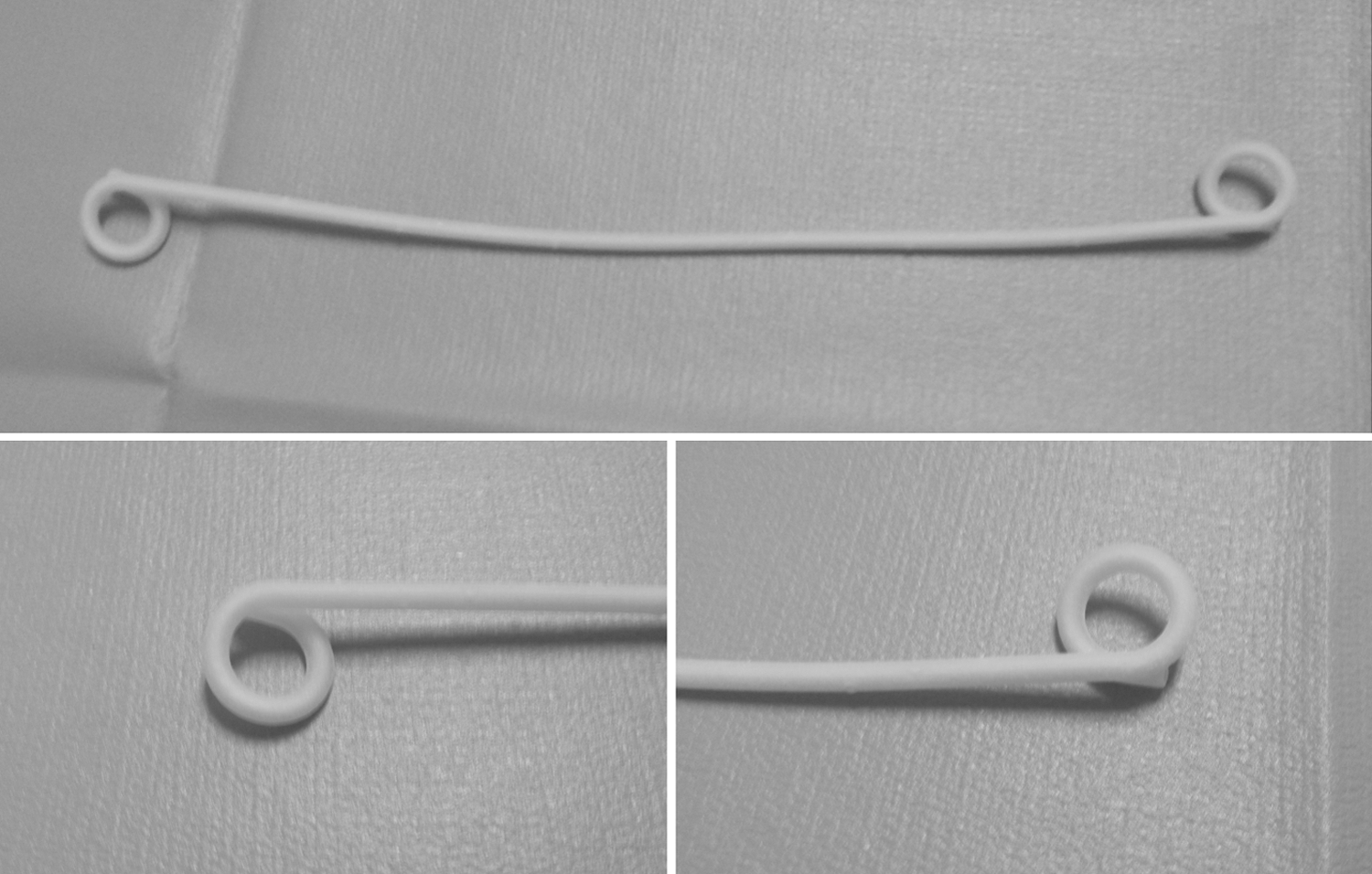
9F 3D printed ureteral stent.
3D stent design characteristics
Details on the 3D printed stent were published previously. 12 Briefly, the 3D printed ureteral stents were printed with an EOSINT P 395 (EOS e-Manufacturing Solutions, Krailing, Germany) printer and composed of a thermoplastic, nylon polyether block amide (Elasto Plastic, Shapeways, Eindhoven, Netherlands). Additional holes along the body of the stent were included in the design. The current prototype design lacks the typical tapering of the ends featured on commonly used stents. As a result of their manufacturing and materials, the printed stents show higher friction between the guidewire and stent lumen as well as the stent and the ureteral mucosa. Moreover, the stent “pigtails” have a limited shape memory and often return back to a looser, yet functional coil shape than their original fully coiled position after deformation of the material such as the insertion and removal of a guidewire.
Study design
Six ex vivo porcine urinary systems with the kidneys and ureters intact were used during the duration of this study. In an antegrade fashion, we inserted a 0.035 Amplatz superstiff guidewire (Boston Scientific) through the renal parenchyma into the renal pelvis. Over the wire, we deployed a 5F ureteral occlusion balloon catheter (Boston Scientific). The occlusion balloon was inflated with 0.5 mL of saline as described by Hafron and colleagues 13 to secure the catheter within an interpolar calix. We then removed the guidewire and connected the occlusion catheter to a 1L 0.9% saline bag positioned 35 cm above the renal pelvis to establish antegrade flow. The distal end of the ureter drained into a 100 mL graduated cylinder to measure flow 14
To establish a baseline flow rate before stent placement, we allowed 0.9% saline to flow antegrade to the graduated cylinder via the occlusion catheter for a 5-minute calibration period. Next, we performed 15 consecutive 3-minute antegrade flow tests on the unstented ureter and recorded the outflow after each trial.
Flow measurement technique
We introduced a 0.035 guidewire into the renal pelvis in a retrograde manner, and an independent observer selected a stent at random that we advanced over the guidewire. With the guidewire positioned within the internal lumen of the stent, we calibrated the collecting system for 5 minutes, as described previously. We performed 15 consecutive 3-minute flow tests with the guidewire in place and recorded the results as extraluminal flow. For total flow (combined extraluminal and intraluminal), we removed the guidewire from the stent and repeated the 5-minute calibration period. We then obtained 15 consecutive 3-minute flow measurements. For intraluminal flow, we reinserted the guidewire into the stent (to prevent stent occlusion) and tied two silk ligatures around the midureter.
After removing the guidewire, we then proceeded with system calibration and obtained 15 consecutive three-minute flow measurements. All measurements were collected in the same fashion for all stents. An 11F access sheath (Cook Medical), however, was used for initial deployment of the 3D printed stent (cannot be deployed with standard Seldinger technique). The 3D printed stent was placed through the 11F access sheath given that its rougher surface was a limiting factor in an attempt to use the Seldinger technique for insertion. Once the stent was placed, the access sheath was removed before commencement of flow testing. Deployment with the access sheath was tested on the contemporary stents in the ex vivo kidney model and did not result in any changes in flow rates.
Statistical analysis
Head to head comparisons between all five ureteral stents were performed using the Mann Whitney U test. A P value of less than 0.05 was considered statistically significant. All statistical analysis was performed using IBM SPSS statistical package, version 21.0 (IBM Corporation, Armonk, NY).
Results
We used a total of 6 ex vivo renal collecting systems, with one renal unit for a test without stent and one renal unit per stent, ranging from 156 to 178 g each with a mean weight of 162.83 g. The mean ex vivo flow measurements are demonstrated in Table 1. The mean flow rate for the ureter without a stent was 4.97±0.062 mL/min. All five stent groups had superior total flow when compared with this control group (Table 1). The total flow rate for the four stent groups ranged from 5.13 mL/min to 5.68 mL/min. Extraluminal flow rate ranged from 2.52 mL/min to 4.40 mL/min, and intraluminal flow rate ranged from 4.79 mL/min to 5.51 mL/min (Table 1).
Combined extraluminal and intraluminal.
Direct comparisons of ureteral stent groups showed that the mean total flow rate was greatest for the 3D printed stent followed by the 8.5F, 7F, 6F, and the 7/10F stents. The stents tested showed a significant difference in total flow rates (P<0.05) compared with the 3D printed 9F stent with the exception that there was no significant difference between the 8.5F and 9F stents with regard to total flow rate (P>0.05).
The mean extraluminal flow rate was highest for the 6F stent followed by the 8.5F, 7F, 3D printed, and 7/10F stents, respectively. There was no significant difference between the 7F and 3D printed stents with regard to extraluminal flow rate (P>0.05). All other stents showed a significant difference in extraluminal flow rate (P<0.05) compared with the 9F stent. The mean intraluminal flow rate was highest for the 3D printed stent followed by the 8.5F, 7F, 6F, and 7/10F stents, respectively. All stents showed a significant difference in intraluminal flow rates compared with the 9F stent (P<0.05).
Discussion
We recently demonstrated the feasibility of 3D printing of medical equipment including ureteral stents and laparoscopic trocars. 12 3D printing of operating room equipment offers the potential advantages of personalizing equipment to the patient, lower cost, and diminished environmental impact. 12 In an effort to incrementally take a prototype to clinical fruition, we are working to assess functional metrics including flow (as a surrogate for ability to decompress the ureter), sterility issues, biocompatibility, and administrative challenges.
In the current study, we evaluated the flow characteristics of the 3D printed stents. We demonstrated that the novel 3D printed ureteral stents provided adequate upper urinary tract drainage in an ex vivo porcine model. Specifically, direct comparison of ureteral stent groups showed that the mean total flow rate for the 3D printed stents was favorable when compared with selected contemporary stents.
Three stent characteristics are known to affect drainage of the collecting system: (1) An increased inner diameter of the stent, 13,15 –17 (2) an increased stiffness of the stent, 18 and (3) drainage holes along the body of the stent. 17,19 –21 After discussion with our collaborating engineers, we aimed to 3D print a ureteral stent that met these parameters.
Modern 3D printing technology provides a wide variety of options from which the stents could be printed. We selected a material composition that increased the stiffness of the stent compared with contemporary stent material. In addition to increased stiffness, the material chosen also allowed for the greatest inner diameter with the lowest outer diameter compared with other flexible 3D printing build materials that were available at the time this study was performed. This allowed for an increased inner diameter of the stent but with a greater outer diameter compared with the contemporary stents used in this study.
Unlike traditional ureteral stents, we designed additional holes along the body of the stent to facilitate improved drainage of the collecting system. The 3D printed stent was compared with different sizes of standard stents as in previous studies 15 to assess for possible effects of the advantages of 3D printing and the limitations of this process on the flow characteristics of ureteral stents.
Brewer and colleagues 15 described an in vivo model to assess the flow characteristics of indwelling ureteral stents and compared the flow characteristics of five ureteral stents in porcine collecting systems. The authors demonstrated that the intraluminal and total flow increased as the inner diameter of the stent increased. In addition, the authors found that the inner diameter of the stent, rather than the external diameter, was the critical factor in determining luminal and total flow characteristics. Further, Ramsay and associates 16 and Hafron and coworkers 13 concluded in their studies that increased inner stent diameter was directly related to increased flow in a ureter with a stent. Similarly, our results showed that total ureteral flow in a ureter with a stent increased as the inner diameter of the stent increased, with the 7F and 9F 3D printed stents (1.50 mm and 1.40 mm, respectively) showing greater total flow rather than the 6F and 7/10F stents (1.33 and 1.27/1.52, respectively (Table 2).
ID=inner diameter; OD=outer diameter.
Moreover, in a study comparing the flow characteristics of three unique ureteral stents, Olweny and colleagues 17 concluded that intraluminal flow may correlate with the inner diameter of a standard stent with uniform extraluminal and intraluminal size throughout their length. For specialty stents, however, with variable internal or external diameters, flow characteristics were not predicated on lumen or stent size. In our study, the 7/10F stents deviated from the expected trend, which parallels the findings of the aforementioned group. Thus, the discrepancy in flow rates may be attributed to the varying internal diameter along the length of the specialty stents.
While intraluminal flow may correlate with the inner diameter of a stent, Hubner and colleagues 18 demonstrated that the stiffness of the stent, rather than its inner diameter is the most important factor to influence flow rates. After subjecting stents to compression and kinking forces, Hubner and coworkers 18 reported that flow resistance in softer stents was notably greater than in harder stents. Their data demonstrated that the greater flow rates of the hard stents could likely be attributed to the increased stiffness of the stent, which reduced kinking and luminal compression. Thus, it appears ureteral flow may potentially increase as inner stent diameter and stent stiffness increase 18
Furthermore, Mardis and colleagues 19 showed that greater flow rate occurred in stents with larger inner diameters and with holes along the side of the stent. In a study comparing 9F prototype mesh stents to 7F polyurethane stents, Olweny and coworkers 20 describe that the mesh stents experienced equal or greater flow dynamics than the 7F polyurethane stents. They suggested that the overall area of the side holes in the mesh stent far exceed the openings in the 7F stent. In a separate study, Olweny and associates 17 stated that additional fluid drainage through side holes of the stents may have affected the overall channel properties, resulting in different flow dynamics. Other studies, however, report that holes alongside the body of the stent may be inactive during indwelling periods. 21
As such, the relationship between flow rates and stent wall openings remains mixed. Our 3D printed stents had additional side holes along the body of the stents, which increased the overall surface area when compared with the conventional stents we evaluated. Future assessment of the 3D printed stent side holes is necessary, however, for drawing casual relationships with respect to its superior flow rates.
Several limitations in the study exist. Although the 3D printed stents had comparable flow rates, they needed the use of an 11F access sheath (Cook® Medical) for deployment while other stents tested did not need the same dilation process. Previous studies have used similar techniques. Papatsoris and colleagues 22 evaluated a novel thermoexpandable ureteral metal stent with a shaft diameter of 10.5F where a dilation-insertion sheath was used to aid in insertion of the stent. The outer surface of the 3D printed stents was rough, which may result in higher friction against the ureteral mucosa during deployment. We speculate that incorporating the application of a lubricous hydrophilic coating to the stent material during the printing process in future studies could reduce friction and ease deployment.
In addition, in this study, 15 consecutive measurements were made for each ex vivo model that resulted in a greater number of continuous runs, which may affect change in flow rates, compared with previous studies that evaluated ureteral stent flow rate in an ex vivo model. 13,14 Also, our 3D printed stents did not possess a tapered end. This may have a significant effect clinically given that stents are placed more easily over guidewires when they are tapered. To our best knowledge, however, the relationship between flow rates and tapering of a stent has not been studied. In addition, a small sample size of one renal unit per stent was used for this study and a larger sample size in future studies is needed. Also, the antegrade irrigation with a 0.9% saline bag placed 35 cm above the renal pelvis used in this study may not be representative of normal intrapelvic pressures. Futures studies must include tests at varying levels of intrapelvic pressure that may be a more ideal model.
There remain several questions that need to be evaluated in future studies. First, in our study, after ureteral stent placement, stents did not remain indwelling for extended periods. This may affect the performance and flow characteristics of all tested stents. Second, potential irritation, migration, encrustation, and biocompatibility of the ureteral stents were not examined, and these should be addressed in future studies. Third, physiologic and histologic responses after the insertion of the 3D printed ureteral stent were not evaluated. Future studies are needed to evaluate the 3D printed ureteral stents in an in vivo model to evaluate the long-term effects of the stent on ureteral function. Any modification to the 3D printed stent design in the future made to address these issues may affect the flow rates and must be further tested.
Finally, this study was aimed at gaining an improved understanding of the ex vivo flow characteristics, without considering their clinical efficacy. As such, further development and testing is needed before introduction into clinical urology practice.
Conclusions
In this pilot study, 3D printed stents manifested mean total flow rates comparable to the flow rates of contemporary stents. Continued advances in technology and material may permit a functionally feasible 3D printed ureteral stent.
Footnotes
Author Disclosure Statement
No competing financial interests exist.