
Abstract
Background and Purpose:
Stereoscopic imaging systems have improved the surgical accuracy and patient safety but have induced unwanted visual disturbance, nausea, and ocular symptoms simultaneously. We measured and compared visual discomfort and visual fatigue induced by three-dimensional (3D) surgical imaging system and two-dimentional (2D) surgical imaging system, respectively.
Methods:
This study compared ocular symptoms and visual functions immediately after four laparoscopic tasks including pick beans, paper cut, pass the curved needle, and knot tying. Ten participants started with 3D laparoscopy, 9 participants with 2D laparoscopy on the first day, and reversed the laparoscopy for the participants on the second day. Before performing the tasks and immediately after performing the tasks for 1 hour, the participants underwent an interview with questions on ocular symptoms, and then received the systematic measurements of the visual functions objectively. The ocular symptoms were compared between the two groups, and the visual functions were compared in each group and between the two groups.
Results:
When comparing the 3D laparoscopy group with the 2D laparoscopy group, symptom scores showed statistically significant differences in blurred vision during the task (z=−3.64, P=0.00), irritated or burning eyes (z=−2.17, P=0.03), dry eyes (z=−2.72, P=0.01), eyestrain (z=−3.11, P=0.00), headache (z=−3.20, P=0.00), discomfort in eyes (z=−3.74, P=0.00). The objective visual functional parameters such as distance exophoria (P=0.83), near exophoria (P=0.88), distance esophoria (P=0.93), near esophoria (P=0.80), the fusion range (P=0.09), the accommodative convergence/accommodation (P=0.56), and the tear film breakup time (P=0.48) had no significant difference between the two groups.
Conclusions:
When the passively polarized 3D surgical imaging system was compared with the 2D surgical imaging system, although subjective feelings were uncomfortable, there was no objective evidence to indicate that the 3D surgical imaging system resulted in an increment of visual fatigue. The visual fatigue and discomforts were moderate and could be tolerated by the surgeons.
Introduction
S
There has not been any report, however, on the impacts of the passive polarized 3D display on ocular symptoms of a surgeon who conducts laparoscopic operations. This study aims to address the effect of passive polarized stereoscopic display on a surgeon's ocular symptoms after performing laparoscopic surgical tasks.
Methods
The observational case-control study was performed on 19 subjects with a median age of 25 years (range 24–30 years). All subjects did not have any laparoscopic experience and had habitual visual acuity or corrected visual acuity of at least 6/6 in each eye. None of them had strabismus or any manifest ocular disease; all of them had normal stereopsis and no eye irritation before the tests. The Medical Ethics Committee of the First Affiliated Hospital of Anhui Medical University approved the study protocol, and the study followed the tenets of the Declaration of Helsinki.
The participants were randomly assigned into two study groups and asked to perform standardized tasks in the same operating room. Ten participants started with 3D laparoscopy, 9 participants with 2D laparoscopy on the first day, and reversed the laparoscopy for the participants at the second day. The 3D system was the passive polarized stereoscopic laparoscopic operating systems (Viking 3D high definition [HD], Viking Systems), with a screen resolution of 1920×1200, a screen size of 24 inches; the 2D system was the Karl Storz 2D HD laparoscopic system, with a screen resolution of 1920×1080, a screen size of 26 inches. The degree for tasks for both lenses was 30 degrees. The working distance for both is 1.5 m (Fig. 1).
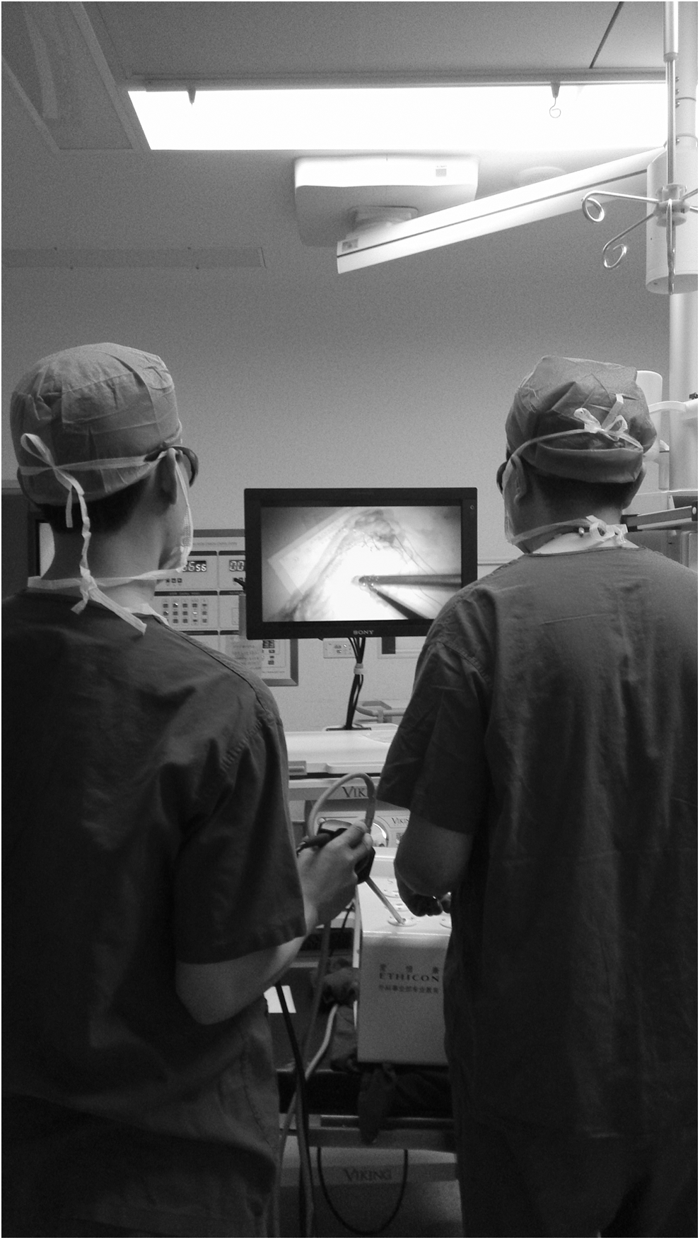
Using three-dimensional laparoscopy systems to perform surgical tasks.
Before performing the tasks and shortly after performing the tasks for 1 hour, the participants underwent an interview with questions on ocular symptoms, based on previous studies, 10,11 including questions on blurred vision, difficulty in refocusing from one distance to another, irritated or burning eyes, dry eyes, eyestrain, headache, tired eyes, sensitivity to bright lights, discomfort in eyes. General scores (ranging from 0 to 10) for the symptoms were assessed by the study participants themselves. The scores were calculated by subtracting the scores of the examination before the tasks from the scores of the examination after the tasks.
The horizontal phoria is a tendency of the eyes to deviate from the parallel, which is characterized by a deviation that can take various forms according to its relative direction, such as esophoria, exophoria, etc. In general, it does not give rise to symptoms, but if persons do a sustained near-viewing task, it will arouse symptoms such as blurred vision, diplopia, burning eyes, headache or dizziness, insomnia, and so on. The fusion range actually is the vision range of the monocular sensory area; it reflects the function fusing two slightly different images (parallax) created by each of the eyes.
The accommodative convergence/accommodation (AC/A) ratio means the accommodative convergence changes (in prism diopters) to the stimulus to accommodation (in diopters). The change of fusion range and AC/A ratio may cause visual fatigue, blurred vision, and other symptoms. The tear film breakup time (BUT) is an indicator of dry eyes, and patients with dry eye syndrome also reported visual fatigue. Therefore, visual function was examined by measuring the horizontal phoria, the fusion range, the AC/A ratio, and the tear film BUT before performing the tasks and immediately after performing the tasks for 1 hour.
The horizontal phoria was measured by phorometer (Flucrum Pharmaceutial Co., Zhengzhou, China); the fusion range and the AC/A ratio were measured by synoptophore (TSJ-IV-A, Photoelectric Instrument Co., Ltd. Changchun, China); and the BUT was measured by slit lamp corneal microscope (YZ5FI,66 vision Technologies Inc, Suzhou, China).
The ocular symptoms were compared between the two groups, and the visual functions were compared in each group and between the two groups. Statistical analysis was performed using SPSS Statistics 10.0 software (IBM-SPSS Inc. Chicago, IL). The Kolmogorov–Smirnov test was applied to assess whether the parameters followed a normal distribution, the raw questionnaire data of symptom scores were not normally distributed, nonparametric statistical tests (Wilcoxon two-sample tests) were used, and the data were presented as median (quartile range).
Because the tests of homogeneity of variances were equal in visual functions, we chose one-way analysis of variance (least significant difference method) to perform the multiple comparisons for the parametric mean values obtained before performing the tasks and the values measured after performing the tasks. The data were presented as mean±standard deviation. All P values were two-sided and were considered statistically significant when the P values were <0.05.
Results
The Wilcoxon two-sample tests were used to compare the symptom scores. Median symptom scores and interquartile ranges for the 3D and 2D groups are shown in Table 1. Wilcoxon two-sample tests indicated that there were significant differences between the two groups in the symptom scores reported for blurred vision during the task (z=−3.64, P=0.00), irritated or burning eyes (z=−2.17, P=0.03), dry eyes (z=−2.72, P=0.01), eyestrain (z=−3.11, P=0.00), headache (z=−3.20, P=0.00), discomfort in eyes (z=−3.74, P=0.00), as shown in Table 1.
3D=three dimensional; 2D=two dimensional.
P value for comparison between three-dimensional and two-dimensional groups.
Symptoms were reported on a scale from 0 (none) to 10 (very severe), with a score of 5 representing a moderate response. The data are presented as median and percentiles (25th–75th).
When the values before the tasks were compared with values obtained after performing the tasks in both 3D and 2D groups, statistically significant differences were found. The fusion function (P=0.00) and tear film BUT (P=0.00) were higher during performing the tasks in the 3D group, and the tear film BUT (P=0.00) was higher in the 2D group. Nine subjects showed varying degrees of exophoria, four subjects showed varying degrees of esophoria, and six subjects were normal before the tests. The subjects had no significant changes in the magnitude of the phoria when compared before and after the tests (P>0.05) whether it was in distance or in near horizontal position, as listed in Table 2.
3D=three dimensional; 2D=two dimensional; AC/A=accommodative convergence/accommodation; BUT=breakup time.
P value (1) for comparison between before and after 3D operation.
P value (2) for comparison between before and after 2D operation.
P value (3) for comparison between after 3D and 2D operations.
The values after performing the tasks between the 3D and 2D groups revealed that the parameters had no significant difference between the two groups: the horizontal exophoria (distance: P=0.83, near: P=0.88), the horizontal esophoria (distance: P=0.93, near: P=0.80), the fusion range (P=0.09), the AC/A (P=0.56) and the tear film BUT (P=0.48), as shown in Table 2.
Discussion
As shown in Table 1, imaging system use itself likely causes general ocular and physical symptoms associated with performing sustained visual tasks no matter whether it is a 3D or 2D imaging system. The symptom scores in the 3D group were worse than those in the 2D group at the similar work conditions, mainly in blurred vision, dry eyes, eyestrain, headache, discomfort in the eyes. Accordingly, the ocular symptoms may be associated with the use of the 3D vision system, a result that was consistent with previous reports. 12,13 All the mean scores both in the 3D group and the 2D group were generally no greater than 5 on a 10-point scale, which indicated that the ocular discomforts were existent and very moderate, and also could be tolerated by the surgeons. It should be noted, however, that the contents of our test were laparoscopy surgical skills (beans picking, paper cut, pass the curved needle, and knot tying). Comparing to laparoscopic surgical tasks, surgeons are more nervous while performing laparoscopic operations. In addition, strong-arm psychological pressure and highly concentrated attention will make the subjective visual symptoms more serious. 14
To comprehensively understand whether the visual function was changed, we examined the horizontal phoria, the fusion range, the AC/A, and the tear film BUT according to the ocular symptoms before and when the tasks were performed for 1 hour.
In most persons, a small phoria (latent strabismus) is common (70%–80% of the population), and the need to compensate for phoria by sensory-motor fusion can cause asthenopic complaints, such as blurred vision, diplopia, burning eyes, and headache with prolonged reading. We should discriminate whether the 3D vision aggravates the phoria. In our study, 9 subjects showed a exophoria, four subjects a esophoria, and six subjects were normal before the tests. We compared the data gained after the tests with data before the tests in both groups and between the two groups; there was no significant difference. The 3D vision did not cause an increased phoria, and the symptoms differences between the two groups may not be caused by phoria.
In stereoscopic 3D display viewing, the images for both eyes are projected as two separated images on a screen, in contrast to natural conditions, in which the distance at which the eyes converge is the same as the distance at which the eyes should focus. It indicates that in 3D images, the object is in focus on the screen, which is behind the convergence point. This discrepancy between the convergence point and the focus point leads to the vergence-accommodation conflict. 15 –17 In our study, the fusion range was reduced in the 3D group—namely the vergence function declined, which caused the vergence-accommodation conflict, the latter leading to the visual fatigue. In the 2D display viewing, there was no vergence-accommodation conflict, so there was no significant change when comparing before and after the tasks. When we compared the 3D group with the 2D group (P=0.09), the changes had no difference; both subjective and objective parameters of visual fatigue are mild.
The tear film BUT is an indicator of dry eyes. Toda and associates 18 reported that 51.4% of patients reporting visual fatigue also had dry eye syndrome, and that 71.3% of patients with dry eye syndrome also reported visual fatigue. Although the dry eyes symptoms could not exclude interference factors, how the laparoscopic operation affected the dry eyes was our concern. In our study, no matter whether in the 3D group or in the 2D group, the tasks could cause decreased tear film BUTs, and the results were consistent with dry eye symptoms. When the tear film BUTs in the 3D groups were compared with those in the 2D groups, no significant difference was found.
The reasons for dry eyes in laparoscopic operations studies 19,20 suggest that reduction of the spontaneous eye-blink rate (SEBR) during video display terminal use is primarily determined by marked visual attention, resulting in an exacerbation of dry eye symptoms. The reduction of SEBR leads to the eyedrop not being uniformly distributed in the surface of the cornea; at the same time, it causes the increment of tear evaporation, and then again, the low humidity in operation room increases tear evaporation.
Potential limitations of our study should be mentioned. In this study, the performing time was 1 hour; a longer working time may have caused more definitive changes, but taking into account that most clinical surgery time is about 1 hour, we performed the study at 1 hour. In addition, Yum and colleagues 21 reported that viewer age may have an effect on watching a 3D display; the accommodation and convergence abilities of older persons are generally lower than those of younger persons. In our study, all the subjects were relatively young, with a mean age of 25 years, so that it has remained elusive whether the results of our study are also valid for all the surgeons.
In addition, the impacts of the 3D surgical imaging system on the visual discomfort and visual fatigue were in a short time. Currently, there is a lack of reports on the effects of long-term 3D surgical imaging system usage on visual functions. This will be the focus of our next studies. This trial is a single-center study with a small sample of 19 subjects. Currently, we are short of a multicenter study with a large sample. We will be committed to cooperate with hospitals nationwide for further exploration.
Conclusion
The laparoscopic operations induced unwanted visual discomfort and dry eyes. The passive polarized 3D surgical imaging system compared with 2D surgical imaging systems resulted in increasing visual fatigue as assessed subjectively but did not deteriorate the visual functions in short-tem laparoscopy tasks as measured objectively. It suggests that the passive polarized 3D surgical imaging system leads to a mild visual fatigue and discomfort subjectively, but it could provide an excellent stereoscopic vision that improved task performance during laparoscopy. The 3D surgical imaging systems will facilitate the expansion of laparoscopic surgery to more complex interventions and help to expand the field of endoscopy-assisted surgery.
Footnotes
Acknowledgments
This work was supported by the Clinical Key Subjects Program of the Ministry of Public Health (Urology), the National Natural Science Foundation of China (81170698, 81370856, 81401518), the Key Science and Technology Program of Anhui Province (12010402128), the Anhui Provincial Natural Science Foundation (1408085QH180) and the cultivation project for NSFC at Anhui Medical University (2013KJ14). The funders had no roles in study design, data collection and analysis, decision to publish, or preparation of the manuscript.
Author Disclosure Statement
No competing financial interests exist.