
Abstract
Purpose:
Small renal masses (SRM) can be managed via a variety of nephron-sparing procedures (NSPs), but the association between choice of NSP and renal parenchymal volume (RPV) preservation is not well understood. We sought to examine RPV preservation after partial nephrectomy (PN) performed via open, robotic, or laparoscopic approaches and thermal ablation (TA) performed via cryoablation (CA) or radiofrequency ablation (RFA).
Patients and Methods:
The study was a retrospective review of three institutional databases of patients with a SRM <4 cm treated via one of the five NSPs (open PN, laparoscopic PN, robotic PN, percutaneous CA, or percutaneous RFA). The 30 most recent consecutive cases treated via each NSP were selected to obtain a total of 150 cases for analysis. Patient characteristics were obtained via manual chart review, and tumor characteristics were assessed via the R.E.N.A.L. nephrometry score. Using three-dimensional rendering software, preoperative and postoperative RPV was calculated for the tumor-bearing kidney, excluding the tumor itself (for preoperative images) or the postsurgical/ablative defect (for postoperative images). The percent change in RPV was compared between the procedure types.
Results:
One hundred fifty cases were included in the final analysis, with 30 cases from each NSP category. While preoperative tumors were larger in the PN group, there was no difference in the mean nephrometry score between groups. The TA group was found to have a lower mean RPV loss (−8.1% vs −16.5%, p<0.005). There was no difference in the RPV loss between modalities of TA (CA vs RFA) or between approaches to PN (open, laparoscopic, robotic). Matched-pair analysis based on the tumor size and multivariate analysis indicated TA vs PN was independently associated with less RPV loss.
Conclusions:
TA is associated with less RPV loss than PN in the management of SRM, but there is no difference between modalities of TA (CA vs RFA) or between approaches to PN.
Introduction
P
Patients and Methods
Following Institutional Review Board approval, we retrospectively reviewed records from 2007 to 2013 from three participating institutions of patients with an SRM less than 4 cm in maximum diameter treated by one of the five NSPs: open partial nephrectomy (OPN), laparoscopic partial nephrectomy (LPN), robot-assisted partial nephrectomy (RPN), percutaneous CA, and percutaneous RFA. Each NSP was performed by a single high-volume urologist specializing in that particular procedure.
Power analysis performed on preliminary data indicated a minimum sample size of 19 cases per group to obtain a β=0.80 at an α=0.05. To further increase power, we chose to review the 30 most recent consecutive cases for each NSP, to make a final cohort of 150 total cases available for analysis. Cases were excluded if preoperative or postoperative contrast-enhanced imaging was not available, the patient had previous surgery to the ipsilateral kidney, multiple tumors were treated simultaneously, or there was discordance between preoperative and postoperative imaging modality (computed tomography [CT] vs magnetic resonance imaging [MRI]).
OPN was performed via a flank incision with retroperitoneal access to the kidney with clamping the renal hilum under cold ischemia conditions. Both LPN and RPN were performed via a transperitoneal approach with clamping of the renal hilum under warm ischemia conditions. RFA and CA were performed via percutaneous access and under CT guidance, as previously described in detail. 4,5 Briefly, CA was performed using a standard double freeze/thaw cycle and the ice ball geometry and extension were actively monitored with CT guidance to assure extension 1 cm beyond the tumor margin. RFA was performed using double ablative cycles and complete ablation was confirmed on immediate postprocedural CT scan. Patient demographics and comorbidities were collected via manual chart review. Tumor characteristics were classified according to the R.E.N.A.L. nephrometry scoring system and stratified into complexity groups as follows: low (<7), moderate (7–9), and high (>9). 6
The primary endpoint was the percent change in RPV after the procedure. Secondary endpoints were changes in renal function and an analysis of factors that correlated with the RPV loss following treatment.
All patients underwent preoperative and postoperative CT or MRI scans with intravenous contrast to facilitate clear delineation of renal parenchyma and tumor tissue. Preoperative imaging was performed a maximum of 3 months before the procedure, while postoperative imaging was performed between 1 and 6 months after the procedure, and when multiple imaging assessments were available, the time point closest to 6 months was chosen for volume assessment. The imaging modality (CT vs MRI) was consistent between the preoperative and postoperative scans for any particular patient. The imaging was loaded onto 3D reconstruction software (VitreaView, Vital Images, Inc., Minnetonka, MN), which utilizes a disc summation technique of volume estimation where the borders of the kidney and tumor are traced on two-dimensional axial images and then compiled to form a 3D image. 7 For the preoperative volume analysis, measurements were obtained for normal appearing, nonmalignant RPV, and tumor volume, with care taken to exclude the collecting system, hilum, or nonfunctional appearing regions of the kidney (i.e., cysts). For postoperative volume analysis, similar measurements were performed to obtain measurement of the normal appearing nonmalignant RPV, with care taken to exclude the collecting system, hilum, and areas of necrosis, postsurgical scare, or postablative defect (Fig. 1). Radiologists blinded to the nature of the procedure performed all volume calculations, which were validated by a single genitourinary radiologist.
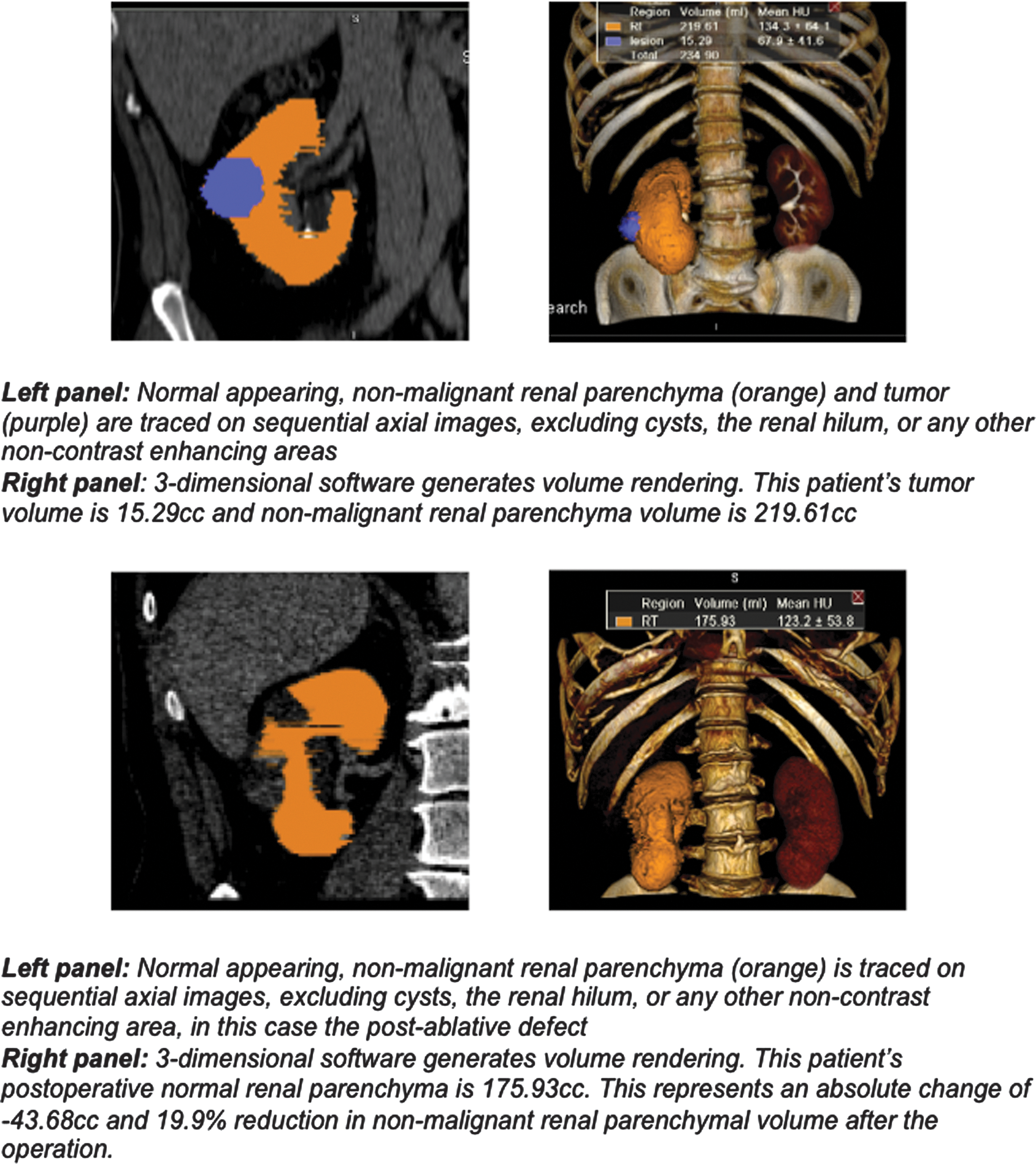
Example of pre- and postoperative renal parenchymal volume and tumor volume measurement.
Renal function data were collected for each patient by estimating the glomerular filtration rate (GFR) from serum creatinine levels via the Modification of Diet in Renal Disease equation. 8 The GFR was calculated on the preoperative outpatient consultation visit and again at the time on the first postoperative outpatient visit, typically 1–3 months after the procedure.
Continuous variables are described as mean±standard deviation or median and interquartile range. For comparisons between treatment cohorts, a Student's t-test and Mann–Whitney U test were used for means, and Chi-square analysis was used for proportions. When there were more than two variables, one-way ANOVA and the Kruskal–Wallis test were used. A matched-pair analysis based on tumor size to compare RPV loss following TA vs PN was performed. Matching was specified in a 1:1 manner using the parameter of tumor size based on intervals of 5 mm from 0.5 to 4.0 cm. Additionally, a multivariate analysis was performed using linear regression modeling to determine the independent association between treatment modality and relative loss of kidney parenchyma, while accounting for measurable tumor characteristics. Two separate models were created to account for the two classification schemes for treatment modality: (1) TA vs PN and (2) RFA, CA, OPN, LPN, or RPN. The other variables tested in the regression were all the other measured factors that could be related to the difficulty of the procedure, namely, tumor diameter, E-score, N-score, A-score, and L-score. Although ischemia time was measured for PN cases, it was not included in the multivariate analysis as it is a variable only applicable to PN and would be inappropriate to include this variable in an analysis of the effects of TA vs PN due to perfect multicollinearity. Analysis was performed with SPSS version 21.0 software (IBM, Armonk, NY), with significance assumed at p<0.05.
Results
A total of 307 NSP cases were reviewed for inclusion in our study. One hundred fifty-seven cases were excluded due to the following: tumor size >4 cm (n=23), lack of available preoperative or postoperative contrast-enhanced imaging (n=107), multiple tumors treated simultaneously (n=7), and discordance between preoperative and postoperative imaging modality (n=20). One hundred fifty cases were available for final analysis, 30 cases in each of the five NSP groups.
Table 1 compares the patient characteristics between the different treatment groups. Comparing TA vs PN groups, patients who underwent TA were older (p=0.001) and more likely to suffer from diabetes mellitus (p=0.001) and hypertension (p=0.038). Otherwise, the groups were similar with regard to gender, race, preoperative GFR, and other measured comorbidities (p>0.05). When analyzing patient characteristics between the specific NSPs (CA, RFA, OPN, LPN, and RPN), differences were noted in the same categories (age, incidence of hypertension, and incidence of diabetes mellitus), but again there were no statistically significant differences in the preoperative GFR (p=0.346) or other measured variables (p>0.05).
TA=thermal ablation; PN=partial nephrectomy; CA=cryoablation; RFA=radiofrequency ablation; OPN=open partial nephrectomy; RPN=robot-assisted partial nephrectomy; SD=standard deviation; GFR=glomerular filtration rate; LPN=laparoscopic partial nephrectomy; BMI=body mass index.
Table 2 compares kidney and tumor characteristics between the treatment groups. While preoperative nonmalignant RPV was similar, the mean preoperative tumor volume and tumor diameter were lower in the TA group compared to the PN group; 8.2 cc vs 18.2 cc (p<0.005) and 2.2 cm vs 2.8 cm (p<0.005), respectively. There was no difference in the R.E.N.A.L. nephrometry score sum (p=0.797) or complexity groupings between the TA and PN groups (p=0.659). The groups only differed with regard to the A-score parameter with a higher proportion of patients with anterior tumors treated by PN (p=0.012). When analyzing tumor characteristics between the specific NSPs, the mean preoperative tumor diameter and volume were largest in the OPN group (diameter=3.0 cm and volume=18.6 cc), while the CA group had the smallest preoperative tumor diameter (2.1 cm) and volume (8.0 cc). The mean ischemia time for OPN, LPN, and RPN was 22.0±11.2, 23.4±10.6, and 16.3±3.7 minutes, respectively.
RPV=renal parenchymal volume.
Figure 2 demonstrates the mean change in renal function and nonmalignant RPV of the tumor-bearing kidney following a NSP. The mean time interval between a NSP and assessment of postoperative RPV was 4.0±2.3 months following TA and 5.4±1.6 months following PN (p<0.005). RPV loss was significantly lower after TA (−8.1%±8.0%) than after PN (−16.5%±11.9%; p<0.005). However, mean RPV loss was similar between modalities of TA: CA (−8.6%±8.3%), RFA (−7.6%±7.7%; p=0.615) and approaches to PN: OPN (−16.4%±14.4%), LPN (−16.1%±12.1%), and RPN (−16.9%±9.1%; p=0.558).

Postoperative change in renal parenchymal volume and function. TA=thermal ablation; PN=partial nephrectomy; CA=cryoablation; RFA=radiofrequency ablation; OPN=open partial nephrectomy; LPN=laparoscopic partial nephrectomy; RPN=robot-assisted partial nephrectomy; GFR=glomerular filtration rate.
There was no difference in the mean time interval between the NSP and assessment of postoperative renal function between the TA and PN groups (p=0.435). The mean change in the GFR was significantly lower after TA (−8.2 %±13.9%) compared to PN (−13.7%±14.4%; p=0.004). However, the change in postoperative GFR was similar between modalities of TA: CA (−8.9%±13.4%) and RFA (−7.2±15.0, p=0.581) and approaches to PN: OPN (−14.0%±15.7%), LPN (−12.6%±13.5%), and RPN (−14.9%±15.3%, p=0.508).
In the matched-pair analysis comparing using the parameter of tumor size, 98 cases were included, 49 after TA and 49 after PN. TA was associated with a significantly lower degree of RPV loss (−8.7%±7.8%) compared to PN (−14.9%±11.8%, p=0.003).
Multivariate linear regression analysis was performed to identify preoperative tumor characteristics and treatment modality variables independently associated with the relative change of RPV. In multivariate model #1, which categorized the NSP broadly as TA or PN, larger tumors (p=0.043) and more endophytic tumors (p=0.019) were associated with a greater RPV loss, while treatment by TA was independently associated with less RPV loss (p<0.005). In multivariate model #2, which categorized the NSP as CA, TA, OPN, LPN, or RPN, larger tumors (p=0.029), more endophytic tumors (p=0.014), and extirpative surgery, by any approach, were associated with a greater RPV loss compared to CA or RFA (Table 3). There was no independent association between approach to PN or modality of TA and RPV loss (p>0.05).
β=regression coefficient in a multivariate regression analysis; CI=confidence interval.
Discussion
The treatments available in the management of an SRM have expanded greatly and resulted in considerable debate within the field of urology. Urologists must now counsel their patients about a variety of treatment options, including extirpative surgery and TA. Furthermore, within the broad category of extirpation, the options further expand to account for the various approaches (open, laparoscopic, or robot assisted) and the extent of surgery (radical vs PN). A sizeable body of evidence suggesting comparable oncologic control and improved renal functional preservation with PN compared to radical nephrectomy supports the basis for the American Urological Association (AUA) guidelines recommendation that PN be considered the standard for the management of T1a tumors (<4 cm). 1,2,9
The more recent emergence of minimally invasive TA provides a less invasive alternative to the technically challenging PN. In general, TA by either modality (RFA or CA) results in comparatively low perioperative complications rates, quick recovery, and short or no inpatient hospitalization. 10 While TA may provide certain advantages, this approach still lags behind extirpative surgery in terms of important criteria of oncologic control, in which studies suggest a higher local recurrence rate for the ablative strategies. 2
Secondary to oncologic control, treatment strategies should attempt to preserve renal function whenever feasible and safe to do so, as chronic kidney disease is associated with an increased risk of cardiovascular morbidity, hospitalizations, and overall mortality. 11 Numerous studies have attempted to compare the relative effects of each of the NSPs on renal function, based on serum creatinine and GFR estimations; however, the review of the literature yields conflicting reports. Several studies have reported no difference in renal functional outcomes between PN and RFA or CA. 12 –15 However, Turna et al. reviewed their institutional experience of patients with solitary kidneys treated by the variety of NSPs and found that PN was associated with a larger decrease in renal function compared to either CA or RFA. 16
The difficulty with using serum creatinine measurements or GFR calculations to estimate RPV change following a NSP is that these methods are too crude to accurately reflect the degree of nephron preservation. Studies from transplant literature illustrate this point, whereby the removal of 50% of functional nephrons through donor nephrectomy does not result in a 50% loss of renal function. 17 This blunted impact of unilateral nephrectomy is thought to be due to hyperfiltration initially, leading to compensatory hypertrophy of the remaining kidney. 18 This situation also occurs after a NSP for the management of SRM, whereby the remaining renal parenchyma (both from the contralateral kidney and remaining ipsilateral kidney) is able to compensate for the loss of nephrons and the affect on serum creatinine and GFR is blunted. We postulate that this is why studies focusing on the endpoint of serum creatinine or GFR may be missing clinically relevant differences between surgical approaches to localized renal masses. Several well-designed studies suggest that RPV is a key determinant of function. 19,20 Therefore, we view RPV as a more accurate indicator of the degree of renal preservation following a NSP.
The results of our study demonstrate a significantly higher level of renal parenchymal preservation with TA compared to PN. Although tumor size was greater in the PN groups and represents a confounder on univariate analysis, we performed a matched-pair analysis based on the parameter of tumor size and found that TA was associated with a significantly lower RPV loss. Additionally, the multivariate analysis indicated that the procedure type (TA vs PN) was the strongest independent predictor of RPV preservation, after taking into account differences in tumor volume and other characteristics. Furthermore, there were no statistically significant differences between the approaches to PN (OPN, LPN, and RPN) or the modalities of TA (CA and RFA).
There are a number of potential reasons why PN may be associated with increased RPV loss compared to TA. PN is certainly a more complex procedure than TA, especially for endophytic tumors where a significant amount of nonmalignant renal parenchyma may have to be violated to excise the tumor. Additionally, PN is typically associated with temporary vascular clamping and associated renal ischemia, which may result in some degree of renal hypotrophy. And finally, renorrhaphy is often accomplished with significant tension on the renal parenchyma to provide adequate postoperative hemostasis, and the effects of this tension on that intervening parenchyma may result in further ischemia and tissue loss. Certainly, further research is warranted to identify potentially modifiable techniques during PN to minimize unnecessary RPV loss.
While this study introduces a novel method to objectively measure to compare the true nephron-sparing characteristics of each of these procedures, there are several limitations that must be acknowledged. The study includes a limited number of patients from each procedure category and is a retrospective review with inherent limitations; we attempted to minimize these by choosing the most recent consecutive cases performed by high-volume urologists with an expertise in each of the specific procedures. This choice likely minimizes the effects of learning curves of less experienced surgeons, but we are unable to account for selection bias based on patient or tumor characteristics that led to the patient being treated by one procedure over another. Differences in tumor characteristics were controlled for by several techniques: a matched-pair analysis based on tumor size compared RPV loss following TA vs PN, and by performing a multivariate analysis that controlled for R.E.N.A.L. nephrometry and tumor size differences between the different procedures. While the decision was made to choose a single expert urologist for each procedure, realistically, a wide range of urologists perform each of these procedures and, perhaps, including multiple surgeons may yield results with broader applicability. Another limitation was our inability to use ischemia time in our multivariate analysis of predictors of RPV loss, as this variable is only relevant with PN and always zero with percutaneous TA. Including ischemia time would be statistically inappropriate due to perfect multicollinearity. As this was a retrospective study, our time points for postoperative RPV and renal function assessment were not standardized and differed slightly between NSPs. Furthermore, because we reviewed the most recent consecutive cases of each procedure, we have limited renal functional or recurrence data available. While volume preservation is important, long-term renal functional and oncologic outcomes are clearly the most clinically meaningful parameters by which to compare the different procedures. With longer follow-up, we hope to address these questions. Despite these limitations, the study represents a novel use to an easily applicable technology and we feel that it warrants a larger study with a prospective design and standardized imaging parameters and time points.
Conclusions
Our study represents a novel method of objectively comparing the different surgical procedures available for the treatment of SRM. With the aid of 3D imaging reconstruction software, we precisely determined the change in functional RPV during the treatment of kidney lesions. Ablative therapies resulted in significantly less renal parenchymal destruction compared to PN.
Footnotes
Disclosure Statement
J.L. is a paid consultant for Galil Medical Ltd. and a principal investigator on a Galil Medical Ltd. supported clinical trial. Galil Medical Ltd. is a manufacturer of cryoablation equipment.