
Abstract

CROES COUNCIL
Chairman
Jean de la Rosette, M.D.
Amsterdam (The Netherlands)
Adrian Joyce, M.S.
Leeds (UK)
Stavros Gravas, M.D.
Larissa (Greece)
Jorge Gutierrez-Aceves, M.D.
Winston Salem (USA)
Dean Assimos, M.D.
Birmingham (USA)
Ying-Hao Sun, M.D.
Shanghai (China)
Tadashi Matsuda, M.D.
Osaka (Japan)
John Denstedt, M.D.
London (Canada)
Sonja van Rees Vellinga
Amsterdam (The Netherlands)
Join the Premier Research Office in the Field of Endourology
Take part in publications and presentations that offer exposure to your institution
Become a member of the global research network of Endourologists
Be recognized as a centre of excellence in the field of Endourology
ÍSPIES—A NOVEL APPROACH TO ADVANCED ENDOSCOPIC IMAGING
Guido M. Kamphuis, Daniel Martijn de Bruin, Theo M. de Reijke, and Jean de la Rosette
In the field of endourology, continuously evolving technology often leads to rapid changes in clinical practice. The Clinical Research Office of the Endourological Society (CROES) aims to conduct new and innovative research projects through randomized controlled trials, high quality clinical databases, and pioneering projects. 1 This approach has the potential to bring research closer to daily practice and generate results in a short period through the participation of clinicians from many different institutions.
Nonmuscle-invasive bladder cancer (NMIBC) has a high incidence as well as recurrence rate. Although patients with NMIBC have a relatively good prognosis in terms of cancer-specific survival, the chance that the cancer will recur has been estimated as high as 75%. 2 Several studies found that the cancer recurrence rate at first follow-up at 3 months after transurethral resection of bladder tumor (TURBT) is present in up to 45% of patients. 3 Also, it has been estimated that 10% to 20% of bladder tumors are overlooked in conventional white light cystoscopy. 4 Two possible explanations for these recurrences are persistence of tumors after TURBT or because tumors were overlooked during the first endoscopic investigation and treatment.
Karl Storz has recently introduced the Storz Professional Image Enhancement System (SPIES), a system to improve the quality of endoscopic imaging. The digital cystoscopy signal is processed offering a virtual chromoendoscopy. By changing the spectrum, a different color contrast can be identified on a video screen. Changing the spectrum, the contrast differences can be adjusted to the clinical situation. The introduction of a new technology in medicine, however, necessitates proper evaluation on beforehand. This year, in selected centers, CROES will conduct a multicenter, international study to compare use of SPIES vs white light (WL) imaging during TURBT to assess recurrence of bladder cancer in terms of safety and efficacy. Preceding this study, an innovative project has been designed to provide us with some insight in this new imaging modality. The evaluation of images is a serious challenge, because there are no standard tests and it is subject to interpretation.
In close collaboration with the departments of Urology and Biomedical Engineering & Physics of the Academic Medical Center, Amsterdam, sophisticated software named íSPIES has been developed to analyze the interpretation of cystoscopy images in the different SPIES modalities. This approach is adapted from tumor delineation studies investigated in the field of image-guided radiotherapy. In these studies, multiple radiotherapists defined tumor boundaries in CT images to optimize treatment. íSPIES, however, uses an application (app) programmed for the iPad environment that (1) allows for very precise and easy to use delineation of a lesion in a SPIES image on the touch sensitive screen of an iPad and (2) makes it very easy to include a large group of participants, which, in turn, makes the study more valid.
Images of 20 bladder areas were captured in four different SPIES modalities: Standard white light, combined Clara & Chroma, Spectra A, and Spectra B. For each bladder area, one extra image was added to the program, so that a total of 100 images were presented to the participants. Every image was shown for, maximally, 45 seconds on an iPad. Participants were asked to delineate with a stylus on the iPad the precise area they suspect to be abnormal/malignant and would resect or treat. After each image, participants were asked to judge the visual quality of the image on a scale of 0 to 10, with 0 being the worst and 10 the best quality.
At the Challenges in Endourology & Functional Urology meeting June 2014 in Paris (
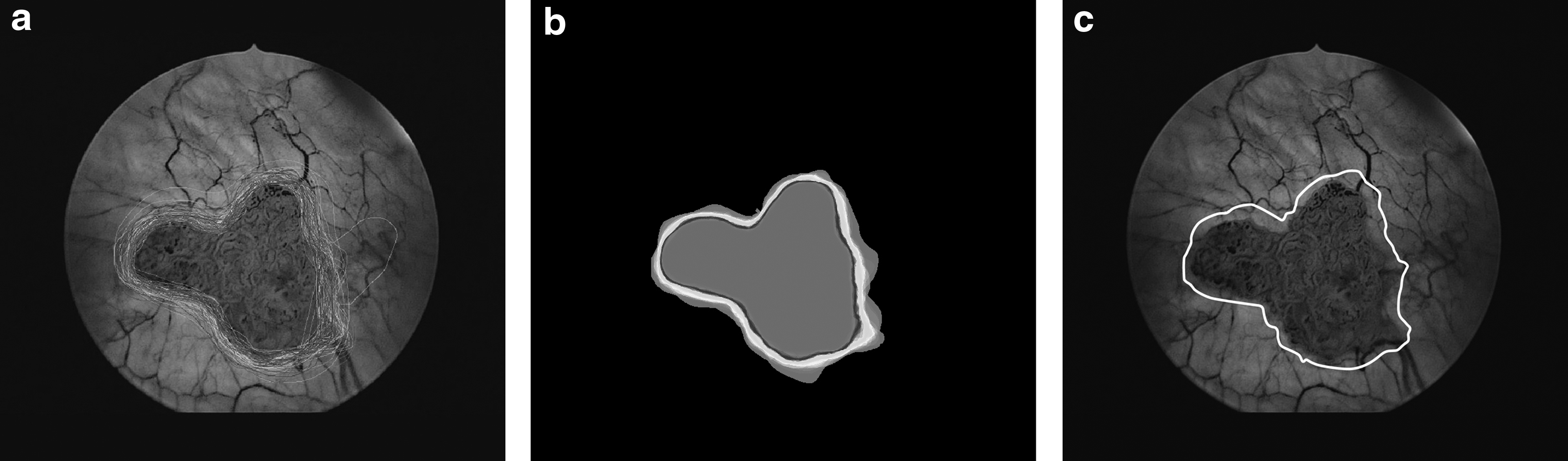
All participants were asked to delineate the suspected lesion as accurately as possible. This resulted in a multitude of drawn lines around the lesion.
The project was supported by an unrestricted educational grant of Storz. We would like to thank all urologists who participated in this study for their valuable contribution. We are looking forward to presenting the results at the upcoming World Congress of Endourology in Taipei.