
Abstract
Purpose:
We sought to evaluate the effect of holmium:yttrium–aluminum–garnet (Ho:YAG) laser exposure on ex vivo pig eyes and to test the protective action of different glasses in preventing eye lesions in case of accident.
Materials and Methods:
We pointed the tip of a Ho:YAG laser fiber from different distances (0, 3, 5, 8, 10, and 20 cm, respectively) toward the center of the pupil of the pig eye. The Ho:YAG laser was activated for 1 or 5 seconds at three different settings (0.5 J-20 Hz, 1 J-10 Hz, and 2 J-10 Hz, respectively). The experiment was repeated using laser safety glasses and eyeglasses. A total of 78 pig eyes were used. The effects of the Ho:YAG laser on pig eyes were assessed by histopathology. Comparable laser emission experiments were performed on thermal paper at different distances using different pulse energies.
Results:
Ho:YAG laser-induced corneal lesions were observed in unprotected eyes, ranging from superficial burning lesions to full-thickness necrotic areas, and were directly related to pulse energy and time of exposure and inversely related to the distance from the eye. When the laser was placed 5 cm or more, no corneal damage was observed regardless of the laser setting and the time of exposure. Similar distance/energy level relationships were observed on thermal paper. No damage was observed to the lens or the retina in any of the Ho-YAG laser-treated eyes or in any of the eyes protected by laser safety and eyeglasses.
Conclusions:
Ho:YAG lasers can cause damage when set to high energy, but only to the cornea, from close distances (0–5 cm) and in the absence of eye protection. Eyeglasses are equally effective in preventing laser damage as laser safety glasses.
Introduction
O
In the context of the upper urinary tract, the use of the Ho:YAG laser is actually recommended for several pathologies such as intracorporeal lithotripsy during flexible ureteroscopy in patients with kidney stones, 2 conservative treatment for upper urinary tract tumors, 3 and the endoscopic incision of strictures of the ureter or the ureteropelvic junction. 1
Despite its effectiveness, some concerns exist regarding laser safety. In particular, Ho:YAG laser manufacturers 4 –6 and the guidelines of European Association of Urology on laser and technologies 1 recommend all intraoperative personnel to wear proper eye protection to avoid any corneal or retinal damage in case of unintentional laser exposure.
A recent study reporting adverse events resulting from lasers used in urology in the United States showed that out of 433 total adverse events, 46% resulted from generator failure or fiber tip breakage and 37.8% were eye injuries associated with the use of neodymium:yttrium–aluminum–garnet (Nd:YAG), potassium titanyl phosphate (KTP), and diode lasers with improper eye protection. Interestingly, no eye injuries were reported with the use of Ho:YAG lasers. 7
To clarify the potential harmful effects of a Ho:YAG laser when the laser beam accidentally hits the surface of the eyes, we conducted an ex vivo animal study on pig eyes to evaluate the effect of a Ho:YAG laser, set at the most commonly used parameters and at different distances, on different eye structures.
In addition, we determined the real protection offered by laser safety glasses and eyeglasses in preventing eye lesions, and we assessed the photothermal effect of the Ho:YAG laser transmitted in air on thermal paper at different distances to implement the results obtained in the first part of the study.
Materials and Methods
All experiments included in the current study were conducted in the laboratory of the “Ecole Nationale Supérieure d'Arts et Métiers ParisTech” (Paris, France) in July 2014 using a 30-W Ho:YAG laser (Rocamed™-MH 01-ROCA FTS 30 [Serial No. LHT0661-0813, Monaco] and a 272-μm-core-diameter laser fiber (Rocamed™-MF272STs).
Evaluation of Ho:YAG laser effect on pig eyes
Overall, 78 pig eyes were used. The potential detrimental effect of the Ho:YAG laser on the different eye structures was evaluated by pointing the laser fiber toward the center of the pupil of the pig eyes. We decided to use such an ex vivo animal model because pig eyes share many similarities with human eyes not only in terms of size but also for some specific histologic and physiologic features. 8
Three different laser settings were examined, which approximately represent the ideal conditions in which clinicians should work to (1) obtain small stone fragments—the so-called “dusting effect”—(i.e., 0.5 J at 20 Hz, long pulse), 9,10 (2) reduce the stone burden when numerous large fragments are present—the so-called “popcorn effect”—(i.e., 1 J at 10 Hz, short pulse), 11,12 and (3) achieve a higher fragmentation volume by increasing the pulse energy (i.e., 2 J at 10 Hz, short pulse). 12,13
Because we sought to reproduce the condition of a beam-related adverse event that could occur in the daily clinical practice during any endourologic procedure requiring the use of a Ho:YAG laser, we assumed that if the tip of the laser fiber was accidentally pointed toward the eye of any person present in the operating theater, the exposure to the eye should last no more than some fraction of a second considering the blink reflex. Therefore, we established that activating the laser fiber in the direction of the pig eyes for 1 second would make the experiments reproducible and reliable, thus reflecting an unfavorable condition that would simulate an accidental injury.
We, in addition, tested a fourth laser lithotripter setting to evaluate an extreme condition by activating the laser fiber set at the highest energy (2 J at 10 Hz, short pulse) for 5 seconds.
The tip of the laser fiber was placed and activated in contact (0 cm) and at different distances from the eyes (3, 5, 8, 10, and 20 cm, respectively) (Fig. 1). Each eye was used to evaluate the effect of each laser setting at each different distance.

Two hundred seventy-two-micrometer-diameter laser fiber (Rocamed™-MF272STs) pointed toward a pig eye and placed in contact with the cornea
The experiment was repeated using laser safety glasses, eyeglasses, and without any eye protection. Six eyes served as nontreated controls. Eyes were fixed in 10% formol, after fixation embedded in paraffin and stained by hematein–eosin–saffron (HES). Cuts were 3 μm thick. The analysis was conducted by two independent pathologists who focused their attention on the cornea, lens, retina, and optic nerve of each eye. In the case of discordant findings, consensus lecture was undertaken.
Evaluation of Ho:YAG laser effect on thermal paper
The photothermal effect of the Ho:YAG laser was evaluated by using a thermal paper (Thermal Print Media, UPP-210HD; Sony Corporation, Tokyo, Japan). The laser fiber tip was positioned perpendicularly to the surface of the thermal paper at different distances for each test (1, 1.5, 5, 10, and 20 cm). Pulse energies included 0.5, 1, 1.5, and 2 J using the traditional short-pulse mode. For each distance/pulse-energy combination, laser emission was sustained for 3 seconds at 5 Hz. After laser emission, the darkened stains produced on the thermal paper were visually compared.
Results
Pathologic findings from the pig eyes exposed to Ho:YAG laser emission in the different situations are shown in Tables 1 –3. Compared to the nontreated controls (Fig. 2A), when the laser was set either at 0.5 J-20 Hz or 1 J-10 Hz, a superficial burning lesion with a loss of surface epithelium was observed on the HES on the cornea of the unprotected eye in contact with the laser fiber (Fig. 2B), whereas no effect was observed when the eye was placed at a distance greater than 3 cm from the laser fiber (Tables 1 and 2). When the laser was set at 2 J-10 Hz, eyes in contact or within 3 cm of the laser without any glasses showed an increase in corneal thickness, with necrotic areas in the anterior region and a complete loss of corneal epithelium (Fig. 2C). A superficial burning lesion was observed at 5 cm, and no relevant effect was observed from 8 cm onward. No damage was detected on the lens and on the retina in any of the Ho:YAG laser-treated eyes and in any of the eyes protected by laser safety and eyeglasses, regardless of the laser setting and the distance from the laser fiber (Tables 1 –3). Nevertheless, significant damage directly related to the pulse energy and inversely related to the distance from the laser tip was detected on both laser safety glasses and eyeglasses (Fig. 3).
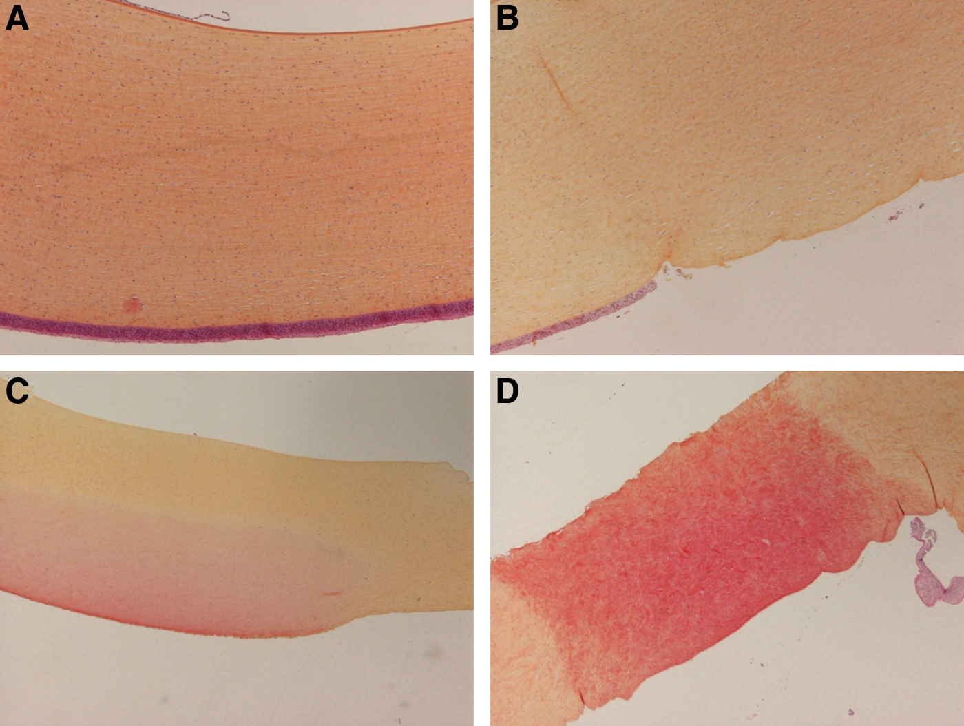
Hematein–eosin–saffron section of the cornea:

Ho:YAG = holmium:yttrium–aluminum–garnet; LSE = loss of surface epithelium; MP = melanophages; SBL = superficial burning lesion.
Ho:YAG = holmium:yttrium–aluminum–garnet; LSE = loss of surface epithelium; MP = melanophages; SBL = superficial burning lesion.
Ho:YAG = holmium:yttrium–aluminum–garnet; LSE = loss of surface epithelium; MP = melanophages; SBL = superficial burning lesion; ICT = increased corneal thickness; NA = necrotic areas.
In the extreme condition, a 2 J-10 Hz activation of the laser fiber for 5 seconds at contact (0 cm) and at 3 cm from the eyes caused full-thickness necrotic areas on the cornea, a complete loss of superficial epithelium, and an increase in corneal thickness (Fig. 2D), with no lesion on the lens, the retina, or the optic nerve. A superficial burning lesion was observed on the cornea at 5 cm, and no significant effect was found from 8 cm onward. No changes were observed in any of the eyes protected by either laser safety or eyeglasses (Table 4).
Ho:YAG = holmium:yttrium–aluminum–garnet; LSE = loss of surface epithelium; MP = melanophages; SBL = superficial burning lesion; ICT = increased corneal thickness; NA = necrotic areas.
Both the eyes exposed to the Ho:YAG laser and the nontreated control eyes showed the presence of melanophages on the optic nerve, compatible with chronic non-Ho:YAG laser exposure-related events. No other changes were documented in the control group (Tables 1 –4).
Concerning the evaluation of Ho:YAG laser effect on thermal paper, we observed that both pulse-energy level and distance from the paper were determinants of a considerable increase in the darkening effect or burn damage on the thermal paper.
Thermal paper was not affected by laser emission at a distance of 10 or 20 cm, regardless of the energy level used (Fig. 4).
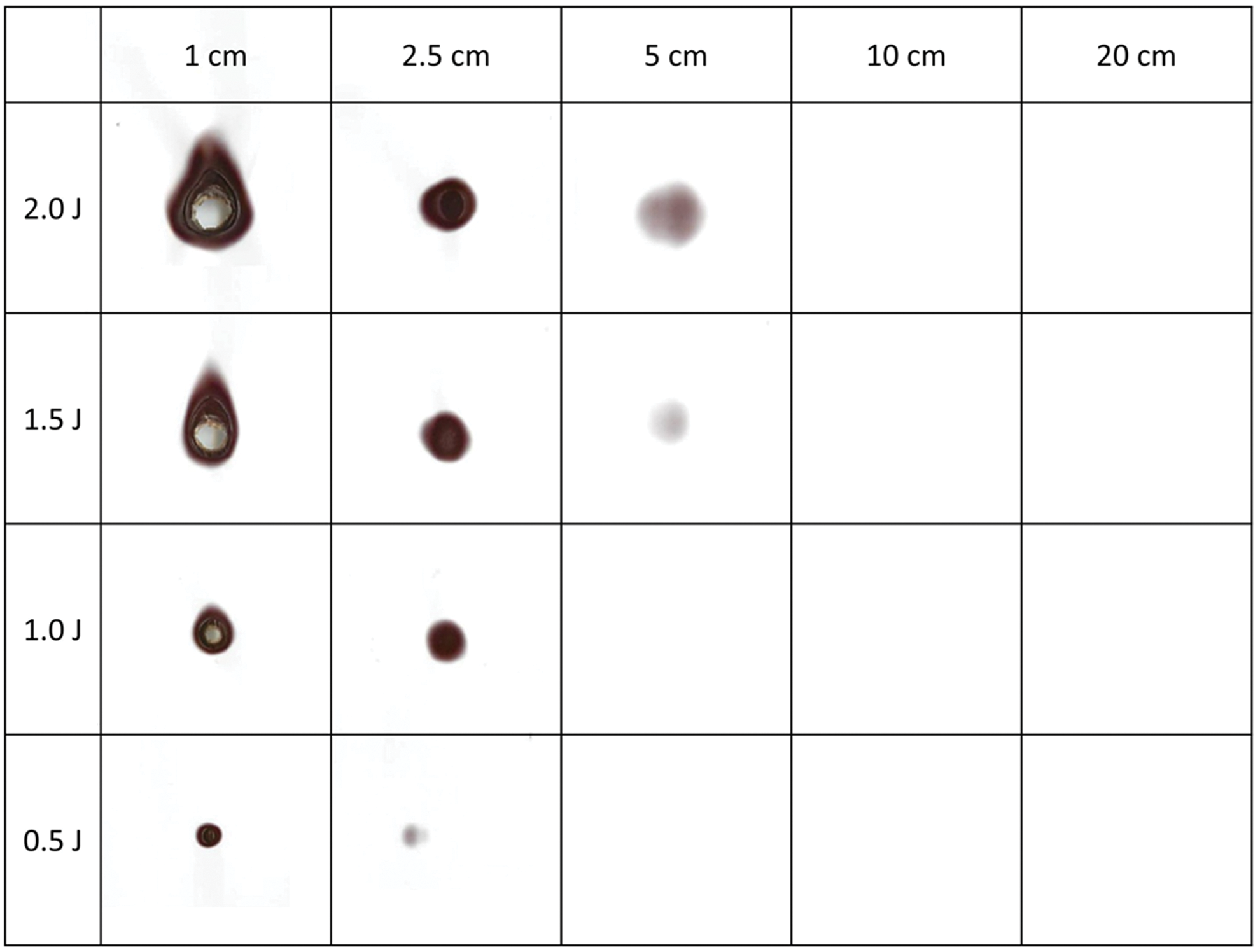
Effect of Ho:YAG laser exposure on a thermal paper (Thermal Print Media, UPP-210HD; Sony Corporation) according to the distance from the laser fiber tip (1–20 cm) and the pulse-energy setting (0.5–2 J). Color images available online at
Effects of laser emission began to be observed when the distance from the fiber tip to the paper surface was reduced to 5 cm or less: at 5 cm, only high pulse-energy levels of 1.5 and 2 J were able to darken the paper, whereas at shorter distances of 2.5 or 1 cm, all energy levels (0.5 up to 2 J) darkened the thermal paper. Moreover, at 1 cm distance, all pulse levels caused a significant burning of the thermal paper in an energy-dependent manner.
Discussion
Therapeutic applications of Ho:YAG laser in ophthalmology for the correction of hyperopia are well known. 14,15 Wearing proper laser eye protection is strongly recommended for all persons present in the operating theater when lasers are used for any kind of surgery, because there are case reports in the literature describing eye injury with significant loss of visual acuity due to unfavorable accidents after laser exposure. 16 However, a recent study investigating the rate of laser-induced adverse events showed that all eye injuries occurred when ND:YAG, KTP, and diode lasers were used, whereas Ho:YAG lasers caused only minor skin burns. 7 Our study analyzed for the first time in an ex vivo animal model the potential impairments of different eye structures following Ho:YAG laser exposure. We found that that the cornea of the pig eyes was the only layer damaged by laser exposure when no protective devices were used. In this context, the greater the pulse energy, moving from 0.5 to 2 J, the more severe the corneal injuries, ranging from superficial burning lesions with a loss of surface epithelium to full-thickness necrotic areas with increased corneal thickness and a loss of normal microarchitecture, when the laser tip was in contact or 3 cm from the eyes (Fig. 2B–D and Tables 1 –3). Similarly, Schmitz-Valckenberg et al. used an animal model to investigate the effect of Nd:YAG laser exposure on rat eyes. 17 They detected in vivo retinal cells undergoing apoptosis in a dose-dependent manner, with greater retinal burns for longer exposures (500 mS vs 300 mS) and at higher power levels (500 mW vs 300 mW). Conversely, our findings showed that neither the lens nor the retina was impaired regardless of the laser settings adopted. This difference seems to be explained by the intrinsic characteristics of the Ho:YAG laser, which has a wavelength of 2140 nm, is absorbed by water, and has a depth of tissue penetration of 0.4 mm; in contrast, Nd:YAG is absorbed by oxyhemoglobin, has a shorter wavelength (1064 nm), and has a higher depth of tissue penetration (4–18 mm). 1,18
A further interesting finding concerns the concept of distance: when the laser tip was more than 5 cm away from the cornea, Ho:YAG laser emission impaired neither the deeper eye structures nor more superficial ones; indeed, no superficial burning lesion or loss of surface epithelium was observed on the cornea, independent of the laser setting (low- or high-power energy), thus resulting in normal pathologic findings comparable to the nontreated controls.
The key role of power energy and distance in affecting the potential detrimental effect of the Ho:YAG laser in the case of accident has been confirmed by the experiments on thermal paper. Acute thermal injury is known to occur at temperatures of 65°C and above, even for brief periods of time. 19 The thermal sensitivity threshold of thermal paper is similar, starting at 60°C to 70°C. 20,21 Any significant thermal variation above that threshold would show up as darkened stains on the paper, such as in our experiments. Our results showed that unintentional Ho:YAG laser emission in air over distances of more than 5 cm may not be harmful, even if high pulse energies such as 2 J are used and for accidental emission periods of up to 3 seconds.
The distance to the terminal laser source, that is, the laser fiber tip, and power of the laser lithotripter, including pulse energy, are known factors that crucially influence the risk of laser injury. 22 The findings of the current study support this evidence, demonstrating that the damage caused by laser emission correlates directly with the pulse-energy level and inversely with the distance to the laser fiber tip. Of note, using high pulse energy (i.e., 2 J) is often not necessary and could be counterproductive, because it increases stone retropulsion, produces larger stone fragments, and induces greater fiber tip degradation. 12,23,24
In addition, we showed that laser safety glasses protect the eye's surface against all Ho:YAG laser exposure, even in the worst case scenario (i.e., small distance, high pulse energy, and long exposure duration—Table 4). Laser safety glasses for Ho:YAG lasers are certainly effective and do not cause any distortion of color perception, 25 thus not affecting the quality of view and the performance of the surgeon. Interestingly, regular eyeglasses act equally well in counteracting the potentially harmful effects of Ho:YAG laser exposure to the cornea. Taken together, the current data show that Ho:YAG laser emission can have detrimental effects on eyes only at small distances (no more than 5 cm from the laser source) and that regular eyeglasses are just as effective as laser safety glasses at protecting the eyes from these effects. However, because the current study systematically investigates for the first time the interaction between the Ho:YAG laser and eye structures in an ex vivo animal model, further in vivo studies are necessary to overcome the inability to evaluate the response of living tissues in terms of inflammatory events or late apoptosis occurring after Ho:YAG laser exposure, which could not be taken into consideration in the current study.
Moreover, it must be carefully considered that the human eye is highly radiosensitive, and prolonged radiation exposure increases the risk of developing cataracts. Taylor et al. 26 showed that the mean radiation dose received during 28 endourologic procedures was 0.208 mSv. Considering a threshold of 2500 mSv as the minimum fractionated long-term dose required to initiate cataract formation and an average of 20 endourologic cases per month, they stated that it takes about 50 years to reach that threshold and to start developing cataracts.
Overall, the benefit of wearing lead-lined glasses should be considered for young urologists and high-volume surgeons and compared with the usefulness of laser safety glasses, the protective effect of which against radiation exposure has never been proven.
Therefore, we may translationally suggest that wearing laser safety rather than lead-lined glasses during all endourologic procedures requiring the use of Ho:YAG lasers and X-ray should be a personal decision of the surgeon and the intraoperative personnel.
Conclusions
In the event of an accident during endourologic procedures, the Ho:YAG laser does not provoke injury to the lens or the retina of the eye. It can cause corneal lesions, but only in the absence of eye protection and only when set to high energy (2 J) and experienced from a short distance (0–5 cm). Eyeglasses are equally effective in counteracting harmful laser effects to the cornea as laser safety glasses. Although the risk of eye injury is minimal, the use of laser safety glasses or at least eyeglasses during endourologic procedures requiring Ho:YAG laser should be recommended to all intraoperative personnel who are close to the laser source.
Footnotes
Author Disclosure Statement
No competing financial interests exist.