
Abstract
Purpose:
Various cleave techniques have recently been shown to significantly impact initial laser fiber power output during holmium laser lithotripsy. The impact of cleave technique on long-term power output has not been well characterized. The purpose of this study was to determine the effect of laser cleave technique on power output over time.
Materials and Methods:
In this randomized single-blinded study, five cleave techniques were tested on two holmium laser fiber diameters (200, 365 μm) over 15 minutes of laser lithotripsy with calcium oxalate monohydrate stones. Comparisons between cleave techniques and fiber diameters were performed using independent samples Mann–Whitney U, Kruskal–Wallis, and homogeneity of variance tests with a significance of p < 0.05.
Results:
The 365-μm fiber was more durable and less affected by burnback degradation than the 200-μm fiber (p < 0.05). While initial power output varied between cleave techniques, all significance disappeared by 3 minutes. Power output decreased rapidly by a mean of 0.62 W over 4 minutes (p < 0.05), following which there was no significant change.
Conclusion:
These findings confirm that initial laser fiber power output is significantly influenced by cleave technique, and the ceramic scissor is the optimal tool for cleaving between procedures. However, because of rapid fiber tip degradation and power loss, this study argues against routine cleaving to improve procedural efficiency in lengthy ureteroscopy cases.
Introduction
T
The holmium:yttrium-aluminum-garnet (Ho:YAG) laser is the lithotrite of choice in many centers. The efficacy of this vaporization mechanism depends heavily on maintaining high power output at the laser fiber tip, ideally with a Gaussian beam profile for uniform energy impulses. 2 –5 The silicon dioxide optical fibers are small caliber and fragile, limited by the ureteroscope working channel to ∼200–400 μm diameter. During lithotripsy, the fiber tips are thermomechanically damaged, causing a reduction in pulse energy. 2,6 –8 Surgeons restore maximal energy output by routinely cleaving and stripping the fiber between procedures (for reusable models) or intraoperatively. Preceding work by Vassantachart and colleagues showed that cleave technique significantly impacts initial fiber power output, 9 however, the long-term effects of cleave technique are unknown. In this randomized single-blinded study, the change in power output that occurs through fiber degradation over 15 minutes of laser lithotripsy is characterized and quantified.
Materials and Methods
To simulate ureteroscopic laser lithotripsy (ULL), 330 calcium oxalate monohydrate (COM) stones of at least 70% purity were obtained from a stone reference laboratory and randomized (average maximum diameter 5.48 mm). A ureteral model was constructed using plastic tubing to constrict stone movement. A 1 × 1 mm wire mesh was affixed to one end of the tube for debris clearance and the setup was submerged in saline. Figure 1 shows the experimental setup.
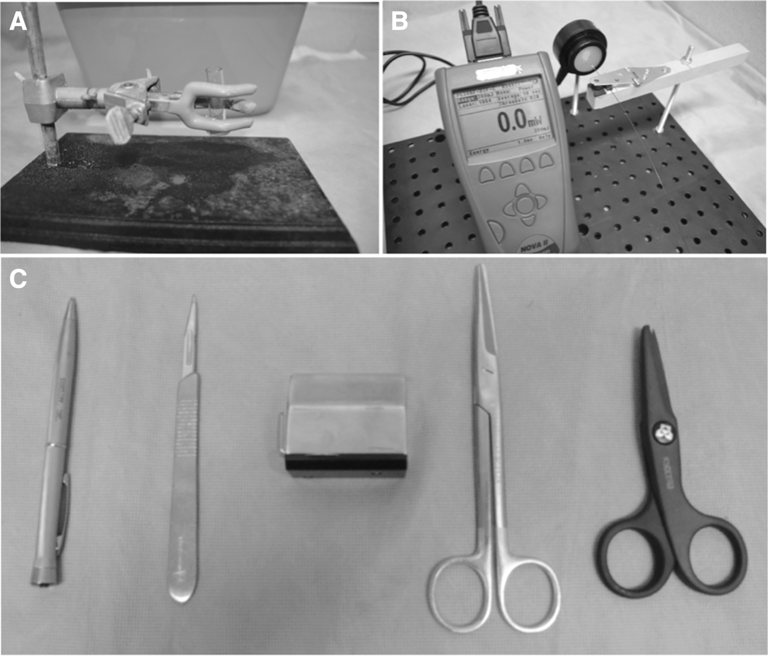
New Lumenis Slimline reusable fibers with 200 and 365 μm diameters were tested. Five cleave tools were utilized: a straight Mayo suture scissor (Aesculap, Center Valley, PA), a scribe pen cleaving tool (Sancliff, Inc., Worcester, MA), a diamond cleaving wheel (Cook Medical, Bloomington, IN), a number 11 blade scalpel (Aspen Surgical, Caledonia, MI), and a ceramic scissor (Lumenis, San Jose, CA). Tools are shown in Figure 1. The suture and ceramic scissors were used with the fiber held perpendicularly in the middle portion of the blades. The diamond wheel was placed on a flat table and used to score the fiber perpendicularly by pulling it along the full half-circumference of the wheel, following which the tip was removed by pulling along the fiber length. For the scribe pen and scalpel, the fiber was laid on a metal surface similar to an operating room table and scored perpendicularly, following which the tip was removed by pulling along the fiber length.
A Dornier Medilas H20 Ho:YAG pulsed laser (Dornier MedTech, Wessling, Germany) was used at a pulse frequency and energy of 8 Hz and 800 mJ (6.4 W). These settings are widely acknowledged as being within the range of most efficient settings for ULL. 3,10 –12 An Ophir PE50BF-DIF high-energy pyroelectric sensor (Ophir, North Logan, UT) was used to obtain power measurements with the fiber tip at a fixed distance of 1.5 cm from and perpendicular to the center of the 3.5-cm diameter sensor face to ensure that the entire beam output was collected. Laser beam spot size analysis demonstrated a beam profile of 7–8 mm with all tools using this setup. Data were collected with a Nova II pulse-triggered laser power meter using Starlab 2.0 software.
All COM stones were uniformly hydrated by submersion in physiologic saline at room temperature (0.9% NaCl, ∼68° F) in the ureteral model. All fibers were cleaved by one of two urology residents 1.0 cm from the tip, and the fiber jacket was stripped 8 mm using a fiber stripper (Micro Electronics, Inc., Seekonk, MA). Initial postcleave power output was recorded by a second investigator, who was blinded to all cleave techniques. The experimental model and power measurement setup are shown in Figure 1.
After measuring the initial cleaved power output, the fiber was held by hand in contact with the COM stone for 15 minutes of lithotripsy. Power measurements were taken from the fiber tip after each 60-second lasing period. At 8 Hz and 800 mJ, a total of 384 J was delivered to the fiber each minute (5760 J over 15 minutes). A new uncleaved fiber was measured for baseline power output and compared to subsequently cleaved fibers. The five cleave techniques were performed 10 times for each fiber diameter.
Data were analyzed using independent samples Mann–Whitney U, Kruskal–Wallis, and homogeneity of variance tests with a significance of p < 0.05. Cleave techniques and fiber diameters were analyzed with respect to postcleave initial power output and change over 15 minutes.
A VEGA LSH scanning electron microscope (SEM) was used to image fiber tips following lithotripsy (Tescan USA, Cranberry Township, PA). Newly cleaved tips were also imaged before use. Tips were mounted upright onto aluminum Pin Stub Mounts with PELCO Tabs™ Carbon Conductive Tabs (Ted Pella, Inc., Redding, CA) and plated with a Cressington 108 Auto Sputter Coater (Cressington Scientific Instruments Ltd., Watford, United Kingdom) for three 15-second periods using gold-palladium. Images were taken at 400× magnification with an accelerating voltage of 10 kV.
Results
Three hundred twenty-two of the original 330 stones were fragmented, which constituted the destruction of one 5.48-mm diameter stone per 5.31 minutes (2039 J) of lasing. For the 200-μm fiber, the ranked order of average initial power output before lithotripsy (time 0 minutes) was as follows: control (6.47 W); ceramic scissor (5.94 W, 91.8%); scribe pen (5.89 W, 91.1%); scalpel (5.72 W, 88.5%); suture scissor (5.19 W, 80.3%); and diamond wheel (4.74 W, 73.3%). For the 365-μm fiber, the ranked order of average initial power output before lithotripsy was as follows: control (6.60 W); ceramic scissor (6.46 W, 97.8%); scribe pen (6.01 W, 91.1%); scalpel (5.63 W, 85.3%); suture scissor (5.58 W, 84.5%); and diamond wheel (4.74 W, 71.8%). Figures 2 and 3 show the 15-minute power output trends for each cleave tool on the 200- and 365-μm fibers, respectively.
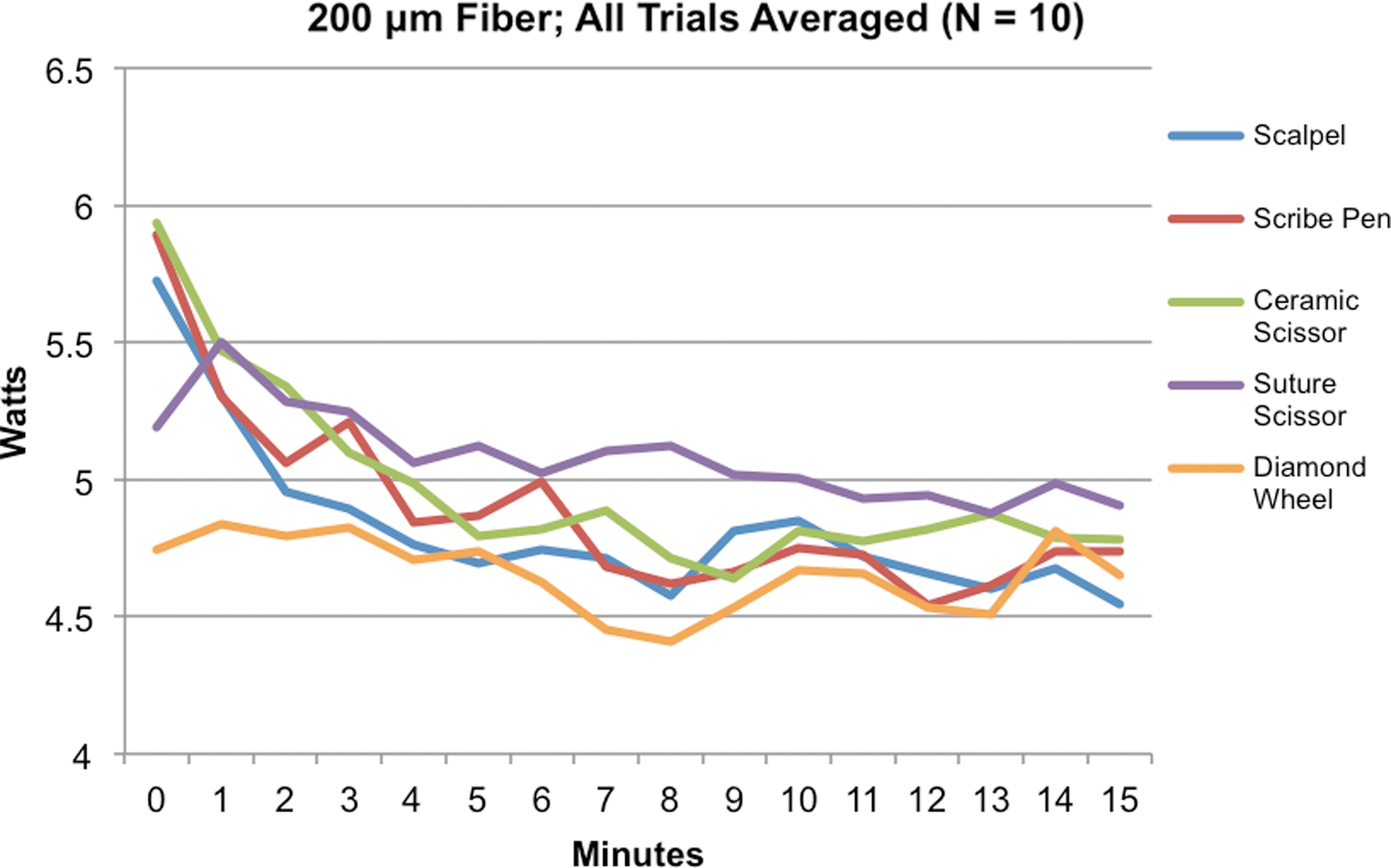
Data averaged for each tool over N = 10 trials on the 200-μm-diameter fiber. Power output at the fiber tip was taken every minute over a 15-minute lithotripsy trial. The ranked order of initial power output before irradiation was as follows: ceramic scissor, scribe pen, scalpel, suture scissor, diamond wheel.
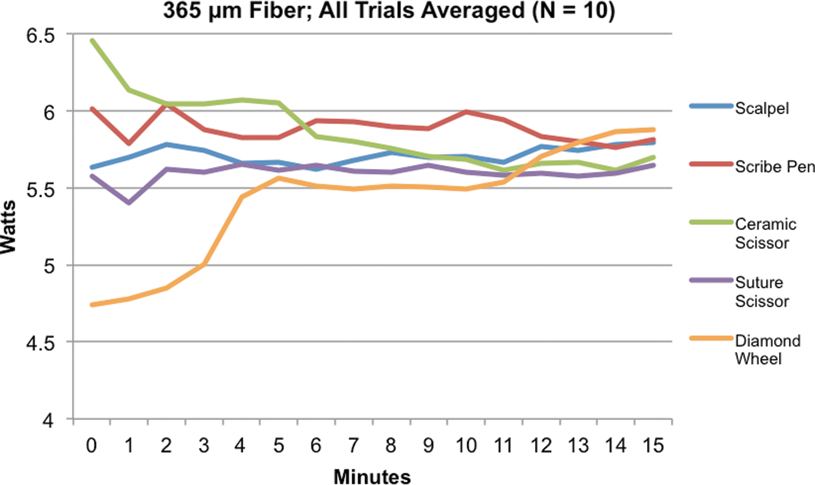
Data averaged for each tool over N = 10 trials on the 365-μm-diameter fiber. Power output at the fiber tip was taken every minute over a 15-minute lithotripsy trial. The ranked order of initial power output before irradiation was as follows: ceramic scissor, scribe pen, scalpel, suture scissor, diamond wheel.
With all cleave techniques combined, there was no significant difference in initial postcleave power output between the 200- and 365-μm fibers (p = 0.414). After 1 minute, the 200 μm power output was 0.27 W lower than the 365-μm fiber (p = 0.018). This difference increased to an average of 0.85 W and persisted over 15 minutes (p < 0.01). Figure 4 shows the fiber diameter power output comparison. In comparing the cleave techniques between fiber sizes, only the ceramic scissor tool showed an initial significant difference in power between the 200- and 365-μm fibers at 0 minutes, with the 365-μm fiber higher by 0.52 W (p = 0.019). The 365-μm fiber power output was significantly higher than the 200-μm fiber after 2 minutes for the scribe pen and scalpel tools, after 4 minutes for the diamond wheel, and after 6 minutes for the suture scissor (p < 0.05). Table 1 shows the difference in power output between fiber diameters analyzed by cleave tool.
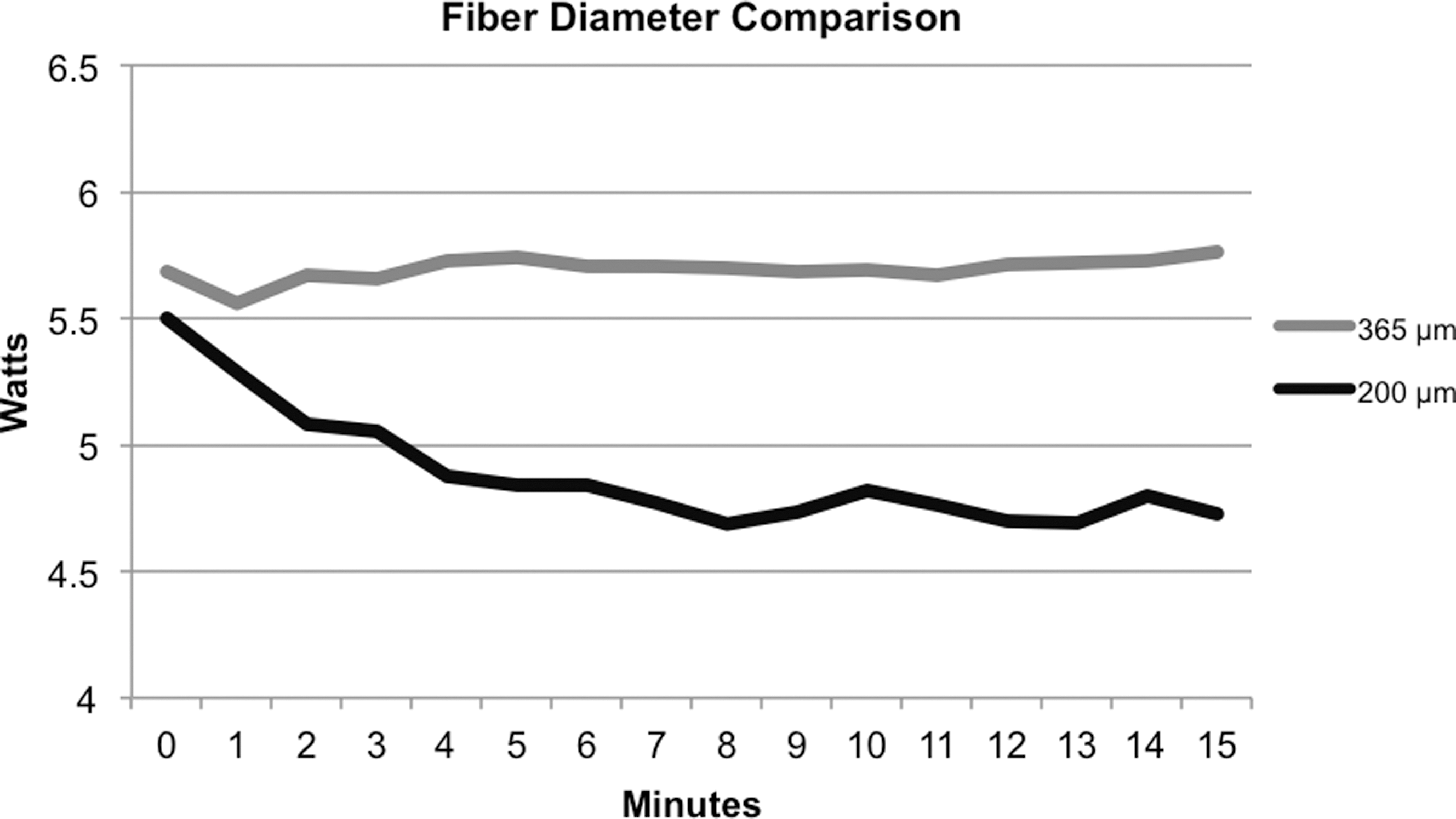
Comparison of fiber power output over time between the 200- and 365-μm fibers with all cleave techniques combined. There was no significant difference at minute 0 (p = 0.414). At minute 1, the 200-μm fiber was 0.27 W lower than the 365-μm fiber (p = 0.018). This difference increased to an average of 0.85 W and was maintained over 15 minutes (p < 0.01).
The composite comparison showed a significant drop in power in the 200-μm fiber at minute 1. This difference occurred at minute 0 for the ceramic scissor, at minute 2 for the scalpel and scribe pen, at minute 4 for the diamond wheel, and at minute 6 for the suture scissor. “Diff” represents the power output in watts of the 200-μm fiber subtracted from the 365-μm fiber power output.
The bold and shaded values represent those values that were statistically significant.
The mean power output of all cleave techniques combined in the 200-μm fiber decreased significantly by 0.73 W (13.25%) over the first 7 minutes (p < 0.05), following which there was no further change. Most of this power loss (0.62 W, 11.34%) occurred over the first 4 minutes. The 365-μm fiber showed no significant decrease in power output with all cleave techniques combined (p = 0.908). The ceramic scissor alone with the 365-μm fiber showed a significant decrease of 0.76 W (11.73%) over the first 9 minutes, with no change afterward (p < 0.05).
Within each fiber diameter, the power outputs of all cleave tools were compared in a pairwise manner at each minute (0–15). The 200-μm fiber showed no statistical differences between any cleave technique at any time point. The 365-μm fiber revealed statistical significance in the ranked trend of ceramic scissor, scribe pen, scalpel, suture scissor, and diamond wheel through minute 3 (p < 0.05), following which there were no statistical differences between cleave tools.
SEM images before and after lithotripsy revealed a distinct change in tip morphology. Before burnback, tips cleaved by the ceramic scissor and scribe pen in general showed a smooth appearance similar to the new fiber, while the scalpel, diamond wheel, and suture scissor produced jagged surfaces with many microfacets. For all fiber diameters and cleave tools, postburnback tip imaging revealed a roughened surface with many diffuse craters, often with central concavity or chipped edges. Figures 5 and 6 show SEM images for the 200- and 365-μm fibers, respectively.
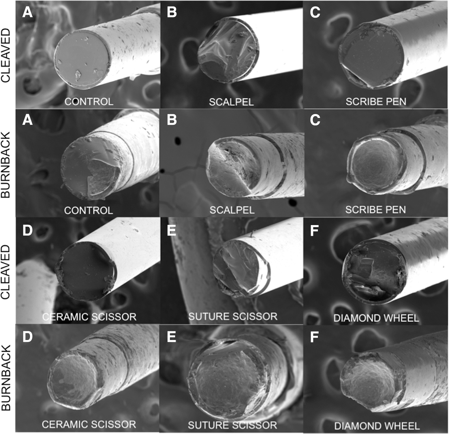
Scanning electron microscopy images of Lumenis Slimline 200-μm-diameter reusable laser fibers. Rows 1 and 3: Newly cleaved fiber tips before stone irradiation. Rows 2 and 4: Fiber tip burnback after 15-minute trials of stone irradiation. Degradation and a melted appearance of the fiber tips are evident.
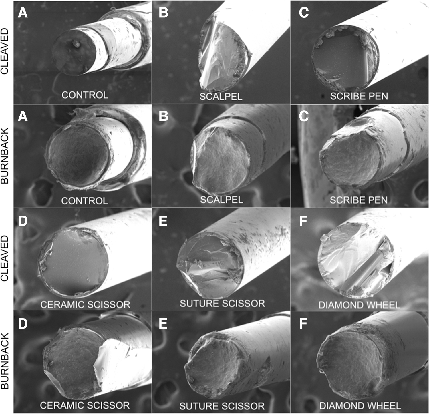
Scanning electron microscopy images of Lumenis Slimline 365-μm-diameter reusable laser fibers. Rows 1 and 3: Newly cleaved fiber tips before stone irradiation. Rows 2 and 4: Fiber tip burnback after 15-minute trials of stone irradiation. Degradation and a melted appearance of the fiber tips are evident.
Discussion
Several conclusions from this study may significantly impact ULL efficiency. First, the 365-μm fiber demonstrated higher durability over 15 minutes of lithotripsy. In 2009, Mues and colleagues studied the burnback and energy loss in several fiber brands and diameters, also showing that fibers in the 200 μm range were more susceptible to degradation. 6 This study used artificial stone material, one cleave technique, and a lasing duration of 3 minutes (2160 J total). Our data showed a difference in power output of 0.85 W between fiber diameters after 4 minutes. Given the well-documented advantage of higher power for lithotripsy efficiency, this quantified superiority in the 365-μm fiber argues for its preferential use over the 200 μm diameter whenever possible. Larger diameter fibers show more efficient lithotripsy in ex vivo experiments at the cost of diminished ureteroscope deflection and irrigation. 3,7,11,13,14
A previous study from our institution showed a statistically significant ranked order of fiber tip power output in the sequence of scribe pen, scalpel, diamond wheel, and suture scissor with a Lumenis Slimline 200-μm reusable fiber. 9 This data replicated the ranked order of initial postcleave power output, with the newly introduced ceramic scissor being superior to all other tools. However, in this study, after only 3 minutes of lithotripsy, there was no significant difference in power output between any tool. The recent work of Kronenberg and Traxer studied lithotripsy efficiency of stripped and nonstripped fibers cleaved with a ceramic or metal scissor on synthetic stones. 15 Their results corroborated the superiority of the ceramic scissor on stripped fibers only, however, lithotripsy duration was limited to 30 seconds (300 J total), and the authors described the need for a long-term study of fiber tip performance.
Across all cleave tools, the power output of poor cleaves increased for 1–3 minutes until equilibrating with trials having superior initial cleaves. The averaged diamond wheel trend in the 365-μm fiber displays this effect in particular (Fig. 3). This is the first study to show that such cleaves with poor initial power output may increase in power rapidly to equal better cleaves.
These results contribute to an argument against routine intraprocedural cleaving. The time to remove the fiber, cleave, strip, and reinsert during ULL may gain less than 4 minutes of an additional 0.62 W (11.34%) with the 200-μm fiber and may achieve no improvement with the 365-μm fiber. A rapid decline from initial power output argues against intraprocedural fiber cleaving for the purpose of increasing power output alone. These findings corroborate trends observed by Lee and colleagues in 2003, 2 whose work with Lumenis 200- and 365-μm fibers showed a rapid initial loss of power, which appeared to stabilize afterward. This study used a lasing duration under 10 minutes (2100 J total), employed a potentially softer stone composition (struvite), did not test different cleave techniques, and did not present statistical analysis.
Previous work by Grant and colleagues demonstrated burnback power loss in 600-μm fibers over 5 minutes at high powers (10–25 W), however, soft tissues were used for ablation and no statistical analysis was presented. 7 Spore and colleagues demonstrated significant differences in power loss and fragmentation efficiency in a 365-μm fiber used on various human stone compositions, however, the lasing duration was limited to 20 seconds (200 J total), and cleave techniques were not tested. 12 Molina and colleagues performed a recent retrospective study on 100 ULL cases with similar stone characteristics, laser fibers, and energy settings, in which no single case required more than 15 minutes, and only one case required more than 5760 J. 4 To our knowledge, this is the first study using real COM stones to characterize the effect of cleave techniques on power output over 15 minutes of lithotripsy, replicating surgical lasing time. These findings may benefit surgeons when selecting fiber diameters and considering cleaving, depending on stone burden and the anticipated lithotripsy time.
Another novel finding of this study is SEM characterization of the fiber tip morphological change following cleaving and lasing of COM stones. The best cleave tools with respect to power output also showed the most similar SEM morphology to the new uncleaved control fiber. Rough surfaces produced with poor cleaving or burnback are less ideal due to beam scattering and a non-Gaussian profile. 2 These SEM images depict a combined thermomechanical degradation of fiber tips, with jagged mechanical chips from stone fragments as well as a thermally seared appearance.
The practice of fiber cleaving in the urology community is inconsistent, and drawing conclusions on how to most efficiently perform ULL remains complex. Clarifying the fiber cleaving and burnback process may help surgeons make simple and effective decisions with respect to ULL protocol. For single and multiuse fibers, improved cleaving methodology can increase procedural efficiency, prolong fiber lifetime, and save thousands of dollars annually. 16,17 Compared to single use fibers, reusable fibers reduce ULL costs by as much as $142 per procedure, and extending fiber lifetime can further reduce expense. 18
Also to consider, each fiber withdrawal for cleaving risks ureteroscope perforation upon reinsertion, particularly with the jagged edges of poorly cleaved fibers. 16 The integrity of fiber cladding may be compromised with multiple reinsertions, allowing laser energy delivery to the ureteroscope or surrounding tissue. Such perforations may cause procedural complications, injure the patient, and add extensive costs, as much as $5900 19 for major ureteroscope repair and $1800 17 for minor repairs.
There are several limitations to this study. Only Lumenis holmium fibers were tested, however, Lumenis has been employed widely in other studies and is one of the most commonly used manufacturers. 6,11,13,14,18,20 A second limitation was the use of only one stone composition. COM was utilized because it is the most common stone. 21,22 In addition, COM represents one of the most challenging compositions for fragmentation as it holds the highest ablation threshold at the 2.1 μm laser wavelength, creating the greatest thermal stress on the fiber tip during lithotripsy. 10,12,20,23 A third limitation was the use of a bench top model. While unable to completely replicate all physical factors in vivo, this model employed real COM stones to study the effect of cleave technique on laser fiber output over time in a realistic manner not previously achieved. The handheld stone contact technique also realistically modeled fiber stabilization in the ureteroscope working channel. Because the model allowed for continuous contact between the stone and fiber tip, these results may overestimate the rate of power degradation in the clinical setting, where stone retropulsion may prevent constant stone contact. Another limitation of this article is that the volume of stone fragmented during each trial was not directly measured. Repeat studies with tip polishing, varying energy levels, fiber diameters, fiber brands, and stone compositions would augment these results.
Conclusion
The ceramic scissor is the optimal tool for cleaving reusable fibers between ULL cases. The 365 μm is more durable than the 200-μm fiber and should be used whenever possible. There is no significant difference between any cleave tool after ∼3 minutes of lithotripsy, and power loss due to fiber tip degradation occurs in the first few minutes. For these reasons, we do not recommend routine lithotripsy interruption for the sole purpose of intraprocedural cleaving to improve power output. The time required to cleave will likely outweigh the power output benefit and the overall procedural efficiency will decrease. However if circumstances require fiber withdrawal from the ureteroscope for other reasons, cleaving with the ceramic scissor is likely to increase efficiency by temporarily improving power output.
Footnotes
Acknowledgment
We would like to acknowledge and thank Dr. Michael Kirby in the LLU Perinatal Neurobiology Research Department for his generous provision of experimental space.
Author Disclosure Statement
D.D.B. is a lecturer for Cook Medical, a consultant for Boston Scientific, and holds a research grant from Teleflex Medical. For the remaining authors, no competing financial interests exist.