




Abstract
Introduction:
Over the past 20 years, the use of fluoroscopy to guide urologic surgical interventions has been constantly growing. Thus, in their daily practice, urologists and other operating room (OR) staff are exposed to X-radiation increasingly frequently. This raises questions as to the risks they encounter and the actions needed to reduce them.
Objective:
Evaluate X-ray dose exposure in the members of the surgical team and determine urologist radioprotection knowledge and practices.
Materials and Methods:
A prospective bicenter study was conducted within AFUF (French urology resident association) and in association with The French Nuclear Safety Authority/The Institute for Radiological Protection and Nuclear Safety (ASN/IRSN). Radiation exposure was measured on 12 operators using dosimeters (seven per operator), in staff-occupied locations in the OR using ionization chambers, and on anthropomorphic phantoms. A survey was used to gather information on radiation knowledge and safety practices of the AFUF members.
Results:
Annual whole-body radiation doses were low (0.1–0.8 millisieverts [mSv], mostly at around 0.3 mSv), and equivalent doses were low for the fingers (0.7–15 mSv, mostly at around 2.5 mSv), and low for the lens of the eye (0.3–2.3 mSv, mostly at around 0.7 mSv). In percutaneous nephrolithotomy, extremity doses were lower when the patient was placed in dorsal decubitus compared with ventral decubitus. Pulsed fluoroscopy reduced radiation dose exposure by a factor of 3 compared with continuous fluoroscopy with no image quality loss. Radiation safety practices were poor: only 15% of urologists wore dosimeters and only 5% had been trained in the handling of X-ray generators.
Conclusion:
In the present study, radiation exposure for urologists was low, but so was knowledge of radiation safety and optimization practices. This absence of training for radiation safety and reduction, teamed with novel techniques involving long fluoroscopy-guided interventions, could result in unnecessarily high exposure for patients and OR personnel.
Introduction
T
Thus, within the framework of AFUF (French association for urology residents), we wished to assess radiation doses received by OR personnel during radio-guided interventions. Toward this, we developed a collaborative effort with The Institute for Radiological Protection and Nuclear Safety (IRSN) and The French Nuclear Safety Authority (ASN) to undertake a dosimetry study with the objective of assessing radiation doses and identifying good practices. In parallel, we performed a survey of urologists to assess differences between real and perceived risks, and between radiation safety practices and recommendations.
Materials and Methods
OR dosimetry study
A prospective study was performed over a 1-month period involving two university hospitals (Tenon Hospital, Paris, France and Lyon Sud Hospital, Pierre-Bénite, France) and three urologic fluoroscopy-guided interventions: retrograde placement of Double-J stents (RDJs), ureterorenoscopy (URS), and percutaneous nephrolithotomy (PCNL).
The IRSN was brought in upstream of study launch so that they could understand the procedures and define technical elements. One center used four Siemens Siremobil Compact L devices (Siemens Healthcare, Erlangen, Germany) and the other a Stenoscop 2 series 6000 and a OEC 7700 (General Electric Medical System). Tube potential and current were, respectively, 88 kV and 2.9 mA in fluoroscopy and 88 kV and 9 mA in digital radiography with a 23 cm field. The IRSN verified that the X-ray emissions reported by the device were accurate.
Measurement methods
Measurements were taken during interventions directly on the urologists (personal dosimeters) and at different locations and distances in the OR (ionization chambers). In the absence of patients, measurements were taken using CIRS ATOM (701–706:CIRS) anthropomorphic phantoms.
The operational quantities retained for the study were Hp(10) for personal dose equivalent, Hp(3) for the lens, and Hp(0.07) for the extremities. Detection limits were estimated at 10 μSv for Hp(10), 19 μSv for Hp(3), and 24 μSv for Hp(0.07).
The urologists wore seven calibrated dosimeters (Figs. 1 and 2). These included two radiophotoluminescence (RPL) dosimeters, one placed at chest level under the lead apron and the other at the neck in alignment with the eye, and five thermoluminescent dosimeter (TLD) chips, one on the forehead and four on the hands (at the tips of the first and second digits).
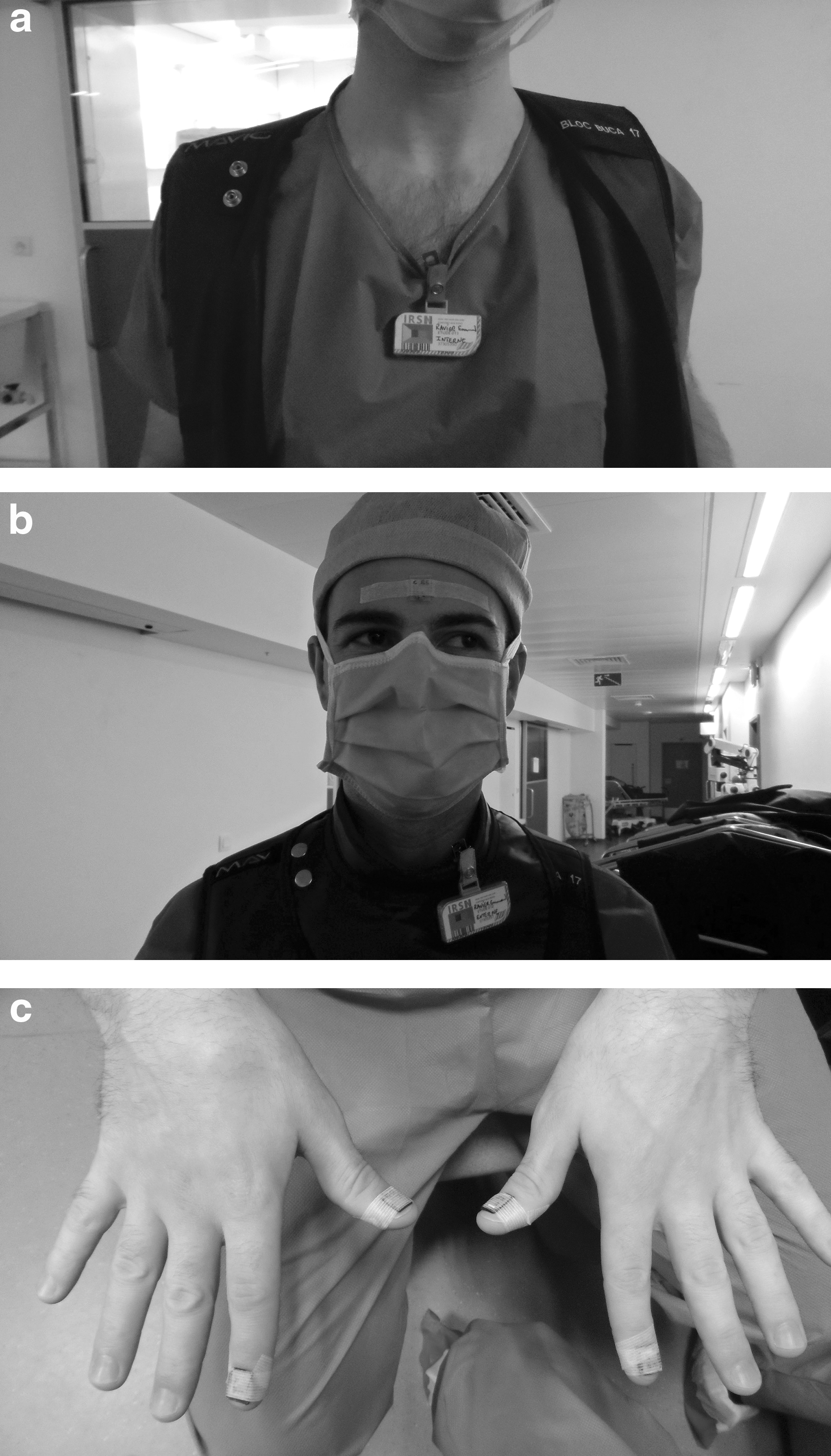
RPL dosimeters under the apron
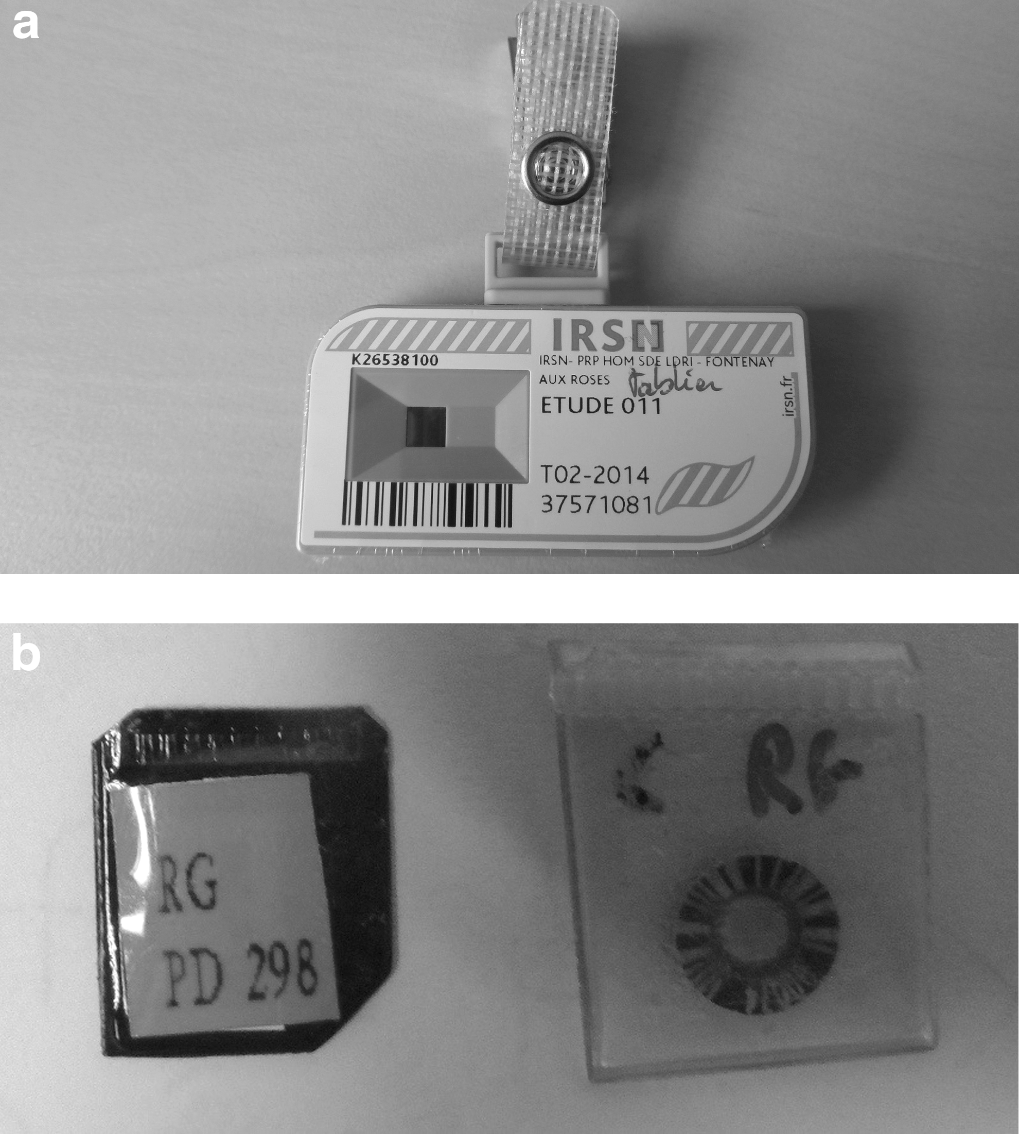
The 3 mm TLD chips were enclosed in impermeable plastic pouches marked with the user's initials.
The anthropomorphic phantoms permitted prolonged exposures (6 minutes) to strengthen results. “Hand phantoms” were used to verify extremity doses.
Patients were placed most frequently in variants of dorsal decubitus (lithotomy position, posterior oblique). Ventral and dorsal decubitus were compared in simulated PCNL using phantoms (Figs. 3 and 4).
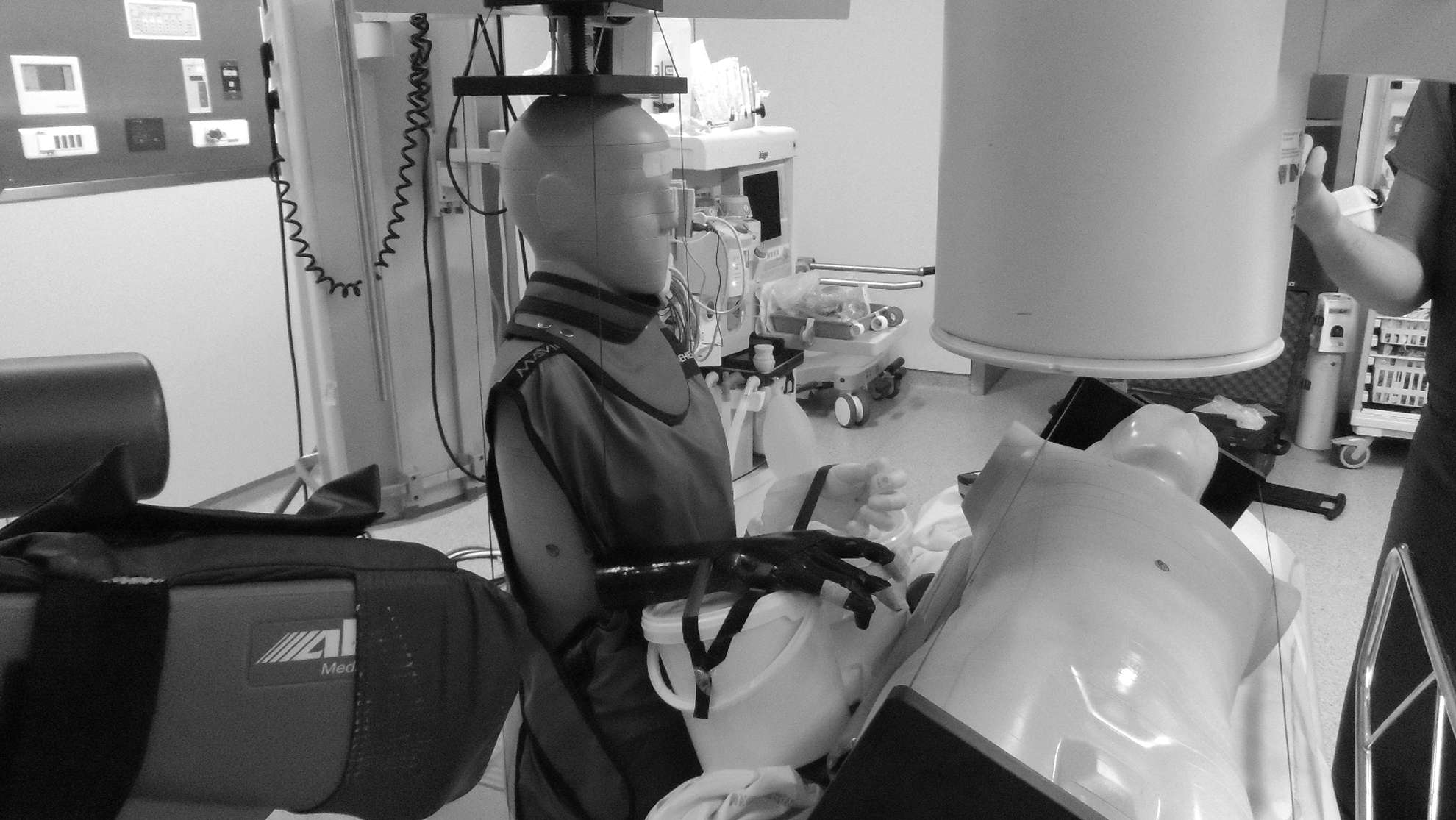
Measurements on CIRS ATOM phantoms. PCNL with patient in the left posterior oblique position. PCNL = percutaneous nephrolithotomy.
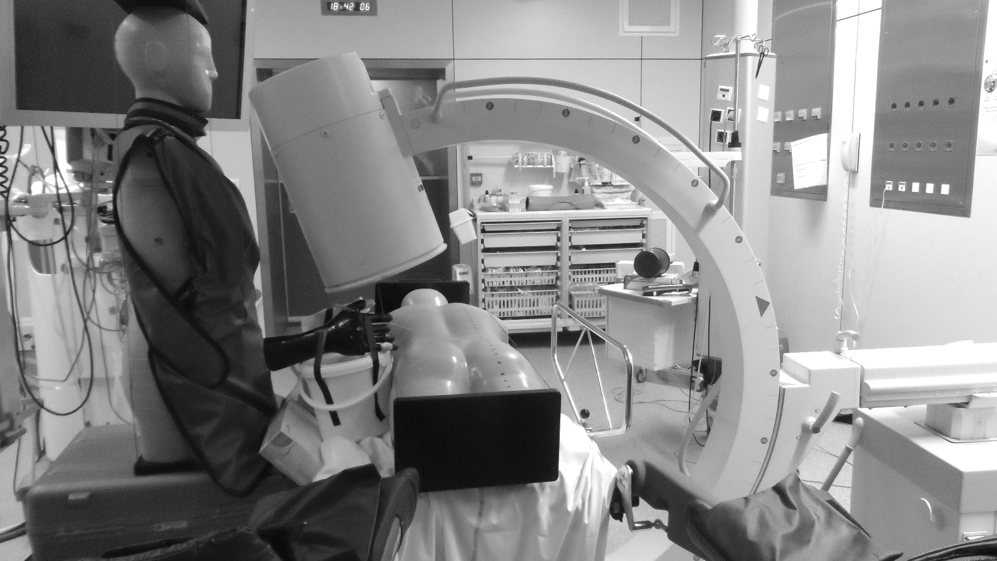
Measurements on CIRS ATOM phantoms. PCNL with patient in ventral decubitus.
Extruded polymethyl methacrylate (PMMA) plates were used as diffusers to evaluate image quality. Doses absorbed through scattered radiation were measured using two ionization chambers, one of which was large volume (1800 cm3). A Radical device (RADCAL) was used to verify calibration of the high voltage generator. Image quality (spatial resolution and low-contrast sensitivity) was evaluated with a test object FL-18.
The precision of measurements included the calculation of relative uncertainty with a 95% confidence interval.
Estimation of operator annual radiation dose
Interventions were defined in terms of “URS equivalent” doses as follows: 3 RDJ = 1 URS = 0.5 PCNL. Yearly estimations were calculated from the URS equivalents, considering the Historical department activity of Tenon hospital, which is an expert center, and an equal workload for surgeons (86 URS, 8 PCNL, and 28 RDJ per operator per year). For the extremities, the maximum absorbed doses were taken into account.
Estimation of radiation doses for other OR personnel
Radiation exposure for other OR staff (perioperative nurse [PN], nurse anesthetist [NA], surgical assistant) was estimated by placing ionization chambers in the zones normally occupied by these personnel and taking measurements using various settings and acquisition modes and various collective protection equipments (Fig. 4).
Assessment of operator knowledge and attitudes
A knowledge, attitude, belief, and practice (KABP) survey was proposed to the 321 AFUF members to assess these aspects in the setting of urology radiation risks and prevention. The questionnaire, comprising 20 items, was made available to the AFUF members through the association's website. Only simple descriptive statistical analyses were done for the responses.
Results
Dosimetry in interventional urology
The study was performed over 15 days in each center. In all, 12 volunteers performed 35 URS. Eleven operators wore their dosimeters correctly, one wore none, and two TLD chips were lost.
Urologist dose exposure during a single URS
It was not possible to distinguish dose exposures between the surgeon and surgical assistant as the attending and resident surgeons alternated in these roles. Table 1 summarizes dosimetry results for a single URS.
Doses expressed in μSv for one intervention. Time expressed in seconds [Hp(10)].
95% CI = 95% confidence interval; <DL = below detection limit; NE = not exploitable; NW = not wear; URS = ureterorenoscopy.
Whole-body doses (measured under the lead apron) were all below the detection limit (10 μSv). Eye lens doses were undetectable for 8 of the 11 urologists wearing their dosimeters; none wore lead glasses. Extremity doses (for the most-exposed finger) ranged from 15 to 59 μSv. The index received more radiation than the thumb on the same hand. There were no significant differences between the two hands.
The wide variations in exposure were attributable to the role played by the subject (surgeon or assistant) injury location, concomitant retrograde stent placement, patient morphology, and operator experience (resident or attending).
Anthropomorphic phantom measurements
The measurements made on phantoms confirmed those made on urologists for whole body and eye lens exposure. Extremity doses for phantoms were lower than those recorded by the finger TLD dosimeters (absence of hand movement).
The measurements on phantoms permitted a comparison of two patient positions during PCNL: extremity doses were ∼1.5 times higher in ventral decubitus compared with dorsal decubitus (Table 2).
Doses expressed in μSv for one intervention.
PCNL = percutaneous nephrolithotomy.
Annual radiation exposure (dosimetric workplace study)
AFUF urologists
Potential effective annual whole body doses ranged from 100 to 800 μSv according to the operator. Yearly exposure in millisieverts (mSv) was most frequently (for 9 of 10 operators) at or around 0.3 mSv, that is, 60 times less than the regulatory limit of 20 mSv.
Eye lens dose equivalents without protective eyewear ranged from 0.31 to 2.3 mSv. Seven of 10 urologists had yearly doses at or about 0.7 mSv, that is, 200 times less than the regulatory limit of 150 mSv.
Extremity dose equivalents ranged from 0.7 μSv to 15 mSv. Nine of 10 urologists had yearly doses at or about 2.5 mSv, that is, 200 times less than the regulatory limit of 500 mSv.
The results of potential exposure to radiation over a year depending on the number for URS practice in 1 year are summarized in Table 3.
Doses expressed in μSv for one intervention.
Nursing staff (exposure from urologic interventions)
As they were positioned at least 2 m from the X-ray source, the nursing staff had low radiation exposure levels (Table 4).
Doses for PNs were divided between time in movement (90%) and time seated at the workstation (10%) (Table 5).
On the basis of 1000 “URS equivalents” per year (activity of one of the centers) and 12 PNs and 24 NAs called upon for urologic interventions, the potential annual effective doses due to urologic interventions were 0.4 mSv (0.323–0.395) for any one PN and 0.06 mSv (0.058–0.071) for any one NA, in the absence of protective aprons or screens.
Dose reduction optimization factors
The influences of different settings were studied.
No differences in low-contrast resolution and only inconsequential differences in spatial resolution were observed between images obtained by continuous fluoroscopy and those obtained by pulsed (15/second) fluoroscopy (Table 5). In contrast, pulsed fluoroscopy reduced air kerma by a factor of 2.9 (ambient dose equivalent rate of 45 μGy/s vs 130 μGy/s for continuous fluoroscopy).
Influence of collimation and fluoroscopy mode on scattered radiation
The use of pulsed fluoroscopy with collimation (10 cm) reduced scattered radiation by a factor of 5 (Table 6).
PMMA = polymethyl methacrylate.
Influence of table shields
The use of a 0.5 mm lead equivalent table shield (under-table X-ray tube) reduced operator leg radiation by a factor of 30 and chest radiation by a factor of 4.3.
Knowledge and attitude survey
The rate of participation for the KABP survey was 37% (119/321). Only one questionnaire was not exploitable. The responses suggested that the knowledge and the use of radiation protection measures were poor. Furthermore, training appeared largely insufficient: only 10% of the responders reported receiving patient protection training and 17% staff protection training.
Training for C-arm manipulation was reported by only 5% of the responders. Furthermore, 12% could not differentiate between the X-ray tube and the detector, 46% used the fluoroscope with the X-ray tube above the table or there where they found it, and 78% did not know the difference between continuous and pulsed fluoroscopy.
In contrast, shielding (lead aprons, 91%) and distance (96%) were better identified as radioprotective measures. Nonetheless, 16% of the responders reported not using a lead apron systematically. The absence of collective protective equipment (barriers, table shields, etc.) in the OR was reported by 90% of responders.
The responding urologists who reported using a passive dosimeter “almost always or always” during X-ray interventions were 14%. The use of ring dosimeters was rare (1%).
Of note, 95% of the responders stated that radiation protection in the OR was mainly their responsibility and that they were important factors in radiation safety. Furthermore, and importantly, 80% knew that other personnel in the room could also be exposed. Thus, the results of the survey suggest more so a lack of training than a lack of responsibility.
Finally, the urologists perceived risks as being greater than what was objectively encountered, but their use of recommended daily radiation protection methods was poor.
Discussion
In the present study, we calculated yearly radiation doses taking into account the substantial activity of an expert center hospital. Although indicative, these doses cannot cover the gamut of situations that may be encountered in radio-guided interventions. Residents spent more time with the fluoroscope than attending urologists did. The annual estimations reported in this study may best be thought of as superior limits for attending urologists in other structures rather than average levels. Precisely, estimating annual whole-body doses for PNs and NAs would require adding all doses received in all of the interventions (not only urologic interventions) in which they participate.
Our study was performed in two university hospitals. Had we had more sites, we could have identified differences and tied them to practices, equipment, or training. However, our use of anthropomorphic phantoms expanded the scope of our measurements.
Other centers may use more powerful fluoroscopes than ours. However, all mobile fluoroscopes undergo the same quality controls, and the settings used for our study were not particularly optimized.
To a greater extent than in other similar studies, 5,6 our results suggest poor radiation prevention knowledge, a near total absence of specific training, and poor practices among the urologists who responded to our survey.
None of our urologists used radiation eyewear. Taylor and colleagues reported that the threshold for a risk of cataract formation was 2500 mSv (eye lens cumulative equivalent dose). 7 Those authors reported that it would take 50 years of normal urologic surgery to reach that threshold, and we could imagine 100 years with our lens dose measurements. Thus, the utility of lead glasses appears open to debate.
The extremity doses observed in our study were 28 times greater than those of Hellawell and colleagues. 2 However, in their study, dosimeters were worn on the fifth finger and their kV and mA parameters were lower than ours.
Considering these results on urology radiation risks, department chiefs could decide to not implement specific complementary (lens, extremities) dosimetric follow-up, as long as they carefully verify local general occupational exposure aspects in coherency with studies.
Few of our urologists had received specific fluoroscope training, despite the fact that the optimization of settings and C-arm use can reduce patient radiation exposure 10-fold. These optimization elements include the use of pulsed radiation, lower pulse rates and doses, image holding/fluoroscopy store, minimized zooming, correctly collimated large fields of view, complementary filtration, under-table X-ray tubes as far away as possible, detectors as close as possible, and limited use of left anterior oblique position >20°.
Conclusion
In the present dosimetry study performed conjointly by AFUF, IRSN, and ASN, we found that urology residents were exposed to only low annual levels of radiation. However, these same residents had major shortcomings in their knowledge and practice of radiation reduction measures, likely due to the absence of radioprotection training. This absence of training, teamed with the arrival of novel techniques involving long fluoroscopy-guided interventions, could result in unnecessary high exposure for patients, urologists, and other OR personnel. Specific training courses, focused particularly on the practical aspects of fluoroscope settings and manipulation, need to be developed.
Footnotes
Author Disclosure Statement
No competing financial interests exist.