




Abstract
Objective:
To compare the retropulsion of stones with the use of holmium: yttrium aluminum garnet (Ho: YAG) laser and thulium: yttrium aluminum garnet (Tm: YAG) laser in settings that could be used in clinical practice.
Methods:
The experimental configuration included a glass tube set in a water bath filled with physiologic saline. Plaster of Paris stones were inserted in the tube. Tm: YAG and Ho: YAG laser systems were used along with a high-speed slow-motion camera. The lasers were activated with different settings. The displacement of the stone was measured according to a custom-made algorithm.
Results:
Ho: YAG: the retropulsion of stones was the lowest with the energy setting of 0.5 J and the frequency of 20 Hz with long pulse duration. The highest retropulsion was observed in the case of 3 J, 5 Hz, and short pulse. Tm: YAG: the retropulsion of stones was the lowest with the energy setting of 1 J and the frequency of 10 Hz with either long or short pulse duration. Practically, there was no retropulsion at all. The highest retropulsion was observed in the case of 8 J, 5 Hz, and short pulse.
Conclusion:
Ho: YAG laser has a linear increase in stone retropulsion with increased pulse energy. On the other hand, the retropulsion rate was kept to the minimum with Tm: YAG as much as the energy level of 8 J. The activation of lasers with short pulse resulted in further displacement of the stone. Lower frequency with the same power setting seemed to result in further stone retropulsion. Higher power with the same frequency setting resulted in further displacement of the stone.
Introduction
D
Methods
Experimental configuration
A glass tube (5 mm in diameter) was set in a water bath filled with physiologic saline. Plaster of Paris stones weighing 0.07 g were used for the experiment. An Optilite 273 μm (COOK Medical, Limerick, Ireland) and a 271 μm Laser fiber (Lisa laser products OHG, Katlenburg-Lindau, Germany) were used for the Rhapsody H-30 Holmium Laser system and Revolix 200 devices, respectively. These laser fibers were appropriate for flexible ureteroscopy and were placed in contact with the stone. A high-speed camera (Olympus i-speed 2; Olympus Corp., Tokyo, Japan) was set close to the glass tube to track the movement of the stone. The laser was activated with a variety of settings with both long and short pulse durations (Fig. 1).
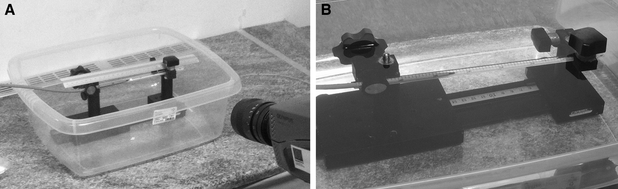
Measurements and equipment settings
The laser devices were set to different energy, frequency, and pulse duration settings within the specifications of the manufacturer for the fibers that were used. The settings were chosen to reflect the clinical practice as well as the worst-case scenarios. The camera was set to obtain 300 frames/sec. The available illumination was provided by two 1000 W projector lamps and the camera image was meticulously focused. A total energy of 100 J was applied to the stone. For each laser setting, at least three measurements were obtained. The videos were evaluated during the experiment to be sure that the laser fiber was touching the stone. Otherwise, the experiment with this particular setting was repeated.
The acquisition of images for each operation mode of the laser (power and frequency) was carried out by the means of a high-speed camera in a relative medium acquisition mode (300 frames/sec). The camera was close to the object so that an acceptable illumination was available and simultaneously the image was perfectly focused. The experimental procedure followed for the estimation of the position of the stone was to facilitate the algorithm to work only on the region of movement. Since the specific region of the image containing the stone is much smaller than the total image acquired, this specific region was extracted from the frames of each file as a separate image sequence. Accordingly, by working on a much smaller region, which contains the stone, the algorithm performed faster and the movement detection procedure was concentrated only on the region of interest. This way, false detection of the position was avoided. The position detection procedure was based on finding a specific part of the stone, which was clearly appearing in the image, by matching it with any similar part of all images in the same sequence. The algorithm for the image processing was custom-made for the current experiment. The method was based on cross-correlation, which is a well-known signal and image processing technique. 7,8 The position of the specific part of the stone was exactly determined in each image with high accuracy of less than a pixel. Under the current conditions, the resolution of the obtained images was 0.01613 cm/pixel.
Results
Tables 1 and 2 present the laser settings and the retropulsion measurements of the Ho: YAG and Tm: YAG lasers, respectively.
Ho: YAG: The retropulsion of stones was the lowest with the energy setting of 0.5 J and the frequency of 20 Hz with long pulse duration. The highest retropulsion was observed in the case of 3 J, 5 Hz, and short pulse. When the retropulsion was measured for the same energy settings and pulse duration, the higher frequencies resulted in shorter retropulsion (Fig. 2). For the same energy and frequency, the long duration of pulse resulted in shorter retropulsion (Fig. 3). The latter effect was noted for the same frequency and pulse duration when the energy was set in lower values (Figs. 2A, 3B).
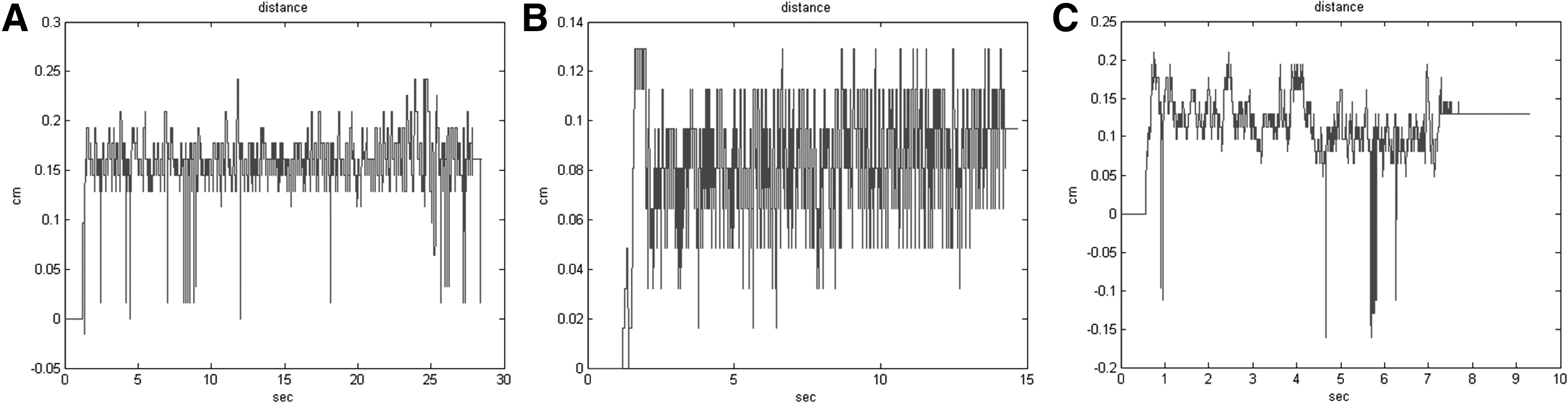
Displacement values with different Ho: YAG laser settings are presented. Graph
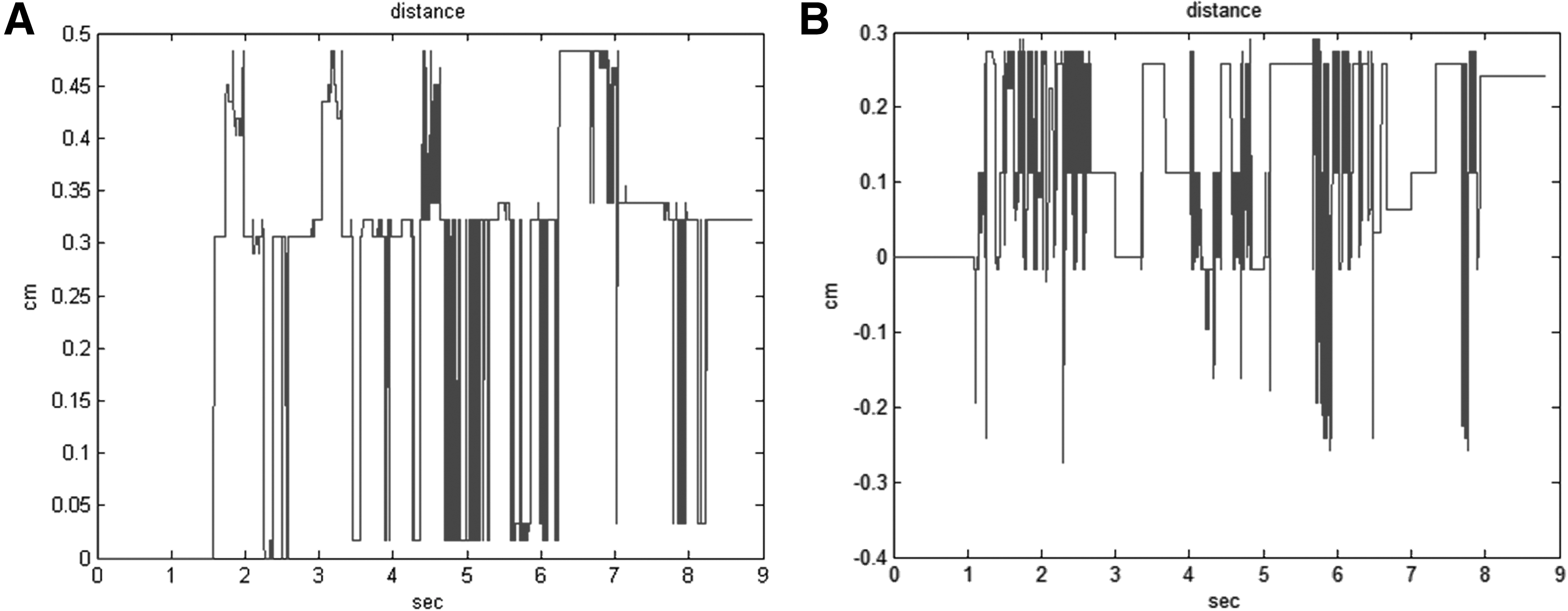
Displacement values with different Ho: YAG laser settings are presented. Graphs
Tm: YAG: The retropulsion of stones was the lowest with the energy setting of 1 J and the frequency of 10 Hz with either long pulse duration. Practically, there was no retropulsion at all at these settings. The highest retropulsion was observed in the case of 8 J, 5 Hz, and short pulse. Any changes in the laser settings (energy or frequency or pulse duration) exhibited similar change in the retropulsion with those described for the Ho: YAG above (Fig. 4). Nevertheless, the Tm: YAG showed a lower retropulsion profile in comparison with the Ho: YAG, especially when the considering that the energy applied to the stone was higher in the case of the Tm: YAG. The Tm: YAG had the potential for continuous mode, which was evaluated and showed no retropulsion.

Displacement values with different Tm: YAG laser settings are presented. Graphs
Discussion
Over the last two decades, Ho: YAG is considered the gold standard lithotripter. Nonetheless, an interest among investigators has been generated in Tm: YAG as an alternative in stone management. 9 –12 Investigators suggested that the Tm: YAG characteristics may have more advantages than Ho: YAG. Specifically, Tm: YAG has an excellent spatial beam profile that yields coupling of higher laser power into smaller fibers. This character is of great clinical importance, which allows for increased irrigation rate through the working channel within the ureteroscope and hence better visibility. The smaller fiber also allows greater flexibility when using a flexible ureteroscope. In addition, the wavelength of Tm: YAG (λ = 1908 nm) matches more closely a high-temperature water absorption peak in the tissue than the wavelength of Ho: YAG (λ = 2120), which may lead to improved stone ablation. Although the Ho: YAG laser has the capability of operating at high pulse energies, its efficiency is limited to relatively low pulse repetition rates (10–20 Hz) during lithotripsy. Moreover, the Ho: YAG is also associated with noticeable retropulsion. On the other hand, the Tm: YAG is capable of operating efficiently at high pulse rates (as much as 1000 Hz). 12 In an ex vivo study, Blackmon and colleagues compared Tm: YAG with Ho: YAG and showed that the Tm: YAG had 5 to 10 times higher vaporization rate than holmium laser. 5 In an in vitro study, Hardy and colleagues demonstrated that the Tm: YAG rapidly fragmented stones with reduced retropulsion. 6 Our group evaluated the raise in temperature of the irrigation fluid and the safety of Tm: YAG in the urinary tract in vitro and in vivo studies and concluded that temperature increases in the irrigation fluid during Tm: YAG activation did not represent a risk for the renal tissue during the upper urinary tract (UUT) endoscopic surgery. 13,14 Nonetheless, current literature is lacking on evidence regarding the clinical efficacy and safety of Tm: YAG in the management of urinary calculi. To our knowledge, there are no publications regarding the clinical use of Tm: YAG for UUT calculi.
Considering the aforementioned promising experimental use of the Tm: YAG in the UUT, we evaluated the stone retropulsion of the Tm: YAG in comparison with the Ho: YAG in different power settings in an in vitro setting. The use of the Ho: YAG laser was related to an increase in stone retropulsion with increased pulse energy. Nonetheless, the retropulsion distance was kept to the minimum with Tm: YAG with energy levels as much as 8 J. Despite the higher energy and low-frequency settings, the Tm: YAG exhibited shorter retropulsion and the effect of pulse duration showed some significance only in the higher energy settings. If we consider the power applied to the stone, the Tm: YAG activation resulted in shorter retropulsion with a power of 40 W in comparison with the Ho: YAG, which was evaluated in power settings as much as 15 W. In fact, the lack of average displacement in Table 2 for some of the Tm: YAG measurements is associated with minimal displacement of the stone during laser activation and the return of the stone to its initial position after receiving 100 J of energy. Moreover, activation of the laser with short pulse resulted in further displacement of the stone, while the lower frequency with the same power setting seemed to result in further stone retropulsion. Higher power with the same frequency setting resulted in further displacement of the stone. These findings were similar for both lasers and they confirm the currently available evidence in the literature. 15 Nevertheless, the current study provides a detailed evaluation of the retropulsion effect of two lasers with emphasis on the settings that could be used in clinical practice.
Limitation of the current study is the lack of measurements with exactly the same power settings for both lasers, which was not possible due to the manufacturer's specifications for the two laser devices and the respective fibers. The investigation of the ablative potential of the two lasers with settings used in the current experimental configuration would have been interesting. Nevertheless, ablation effectiveness was an aim of the current investigation. An additional limitation of the study is that only the retropulsion with a specific stone composition was investigated. The selection of standardized stone composition and size aimed to eliminate any variations of results, which might be related to the stone composition and size. Thus, the retropulsion effect of each laser could not be extrapolated to other stone compositions.
Conclusion
Ho: YAG laser has a linear increase in stone retropulsion with increased pulse energy. On the other hand, the retropulsion rate was kept to the minimum with Tm: YAG as much as the energy level of 8 J. The activation of either of the lasers with short pulse resulted in further displacement of the stone. Lower frequency with the same power setting seemed to result in further stone retropulsion. Higher power with the same frequency setting resulted in further displacement of the stone.
Footnotes
Author Disclosure Statement
No competing financial interests exist.