
Abstract
Purpose:
Transurethral surgery has been traditionally done using the nonelectrolyte, isotonic 1.5% glycine solution as irrigation fluid. The emergence of modern technologies, which can be applied with electrolyte solutions, such as bipolar resection and LASER evaporation, as well as the worry of transurethral resection (TUR) syndrome have driven urologists away from glycine toward the use of physiologic solution. Differences in the transparencies of these fluids have not been studied.
Materials and Methods:
The ability to resolve two bars at 1 mm apart using a 30° cystoscope lens immersed in different solutions was studied. Physiologic solution, distilled water (DW), and 1.5% glycine solutions containing increasing concentrations of blood, from 0.5% to 2%, were tested. Solutions containing 2% blood were inspected with magnification and microscopy.
Results:
One-millimeter resolution was reached in as much as 2% blood in 1.5% glycine solution and as much as 1% blood in DW, but in none of the blood–saline solutions. Magnified and microscopic views of 2% blood solutions showed an even distribution of red blood cells (RBCs) in physiologic solution, clumps of RBCs in 1.5% glycine, and an almost complete hemolysis in DW.
Conclusions:
Glycine solution increases the transparency compared to physiologic solution or DW owing to the clumping of RBCs. When the risk of TUR syndrome is low, as in resection of bladder tumors or small prostates, we propose that 1.5% glycine solution should be preferred over saline, owing to its improved visibility.
Introduction
F
The frequency of TUR syndrome is often cited as occurring in 2% of the cases. 3 TUR syndrome is rare when resection time is less than 90 minutes and gland size is less than 45 g. 3 In contemporary series, the rate of TUR syndrome is low. In a randomized trial comparing monopolar to bipolar prostatectomy, TUR syndrome occurred in 0.7% of the patients in the monopolar arm vs 0 in the bipolar arm (p = 0.495). 4 In fact, many experienced urologists may find it difficult to recall their last case of TUR syndrome.
On the other hand, there are clinical observations favoring the use of glycine solution. During surgery, when switching from saline prostatectomy (e.g., with LASER) to a 1.5% glycine solution (to achieve monopolar hemostasis at the end of surgery), it is often noted that visibility immediately improves. Conversely, when completing a monopolar surgery with relatively clear glycine irrigations and switching to physiologic solution in the recovery room, irrigation fluids often turn cloudy red. These well-recognized phenomena were not studied systematically.
We examined the transparency of 1.5% glycine, physiologic solution, and DW mixed with increasing concentrations (0.5%–2%) of blood, and examined these solutions under the microscope to elucidate the differences in their clarity.
Materials and Methods
Phantom
To simulate the visibility conditions during a transurethral surgery, a phantom was designed. An automatic retractor (Nathanson retractor) connected to a camera was fixed to a table, holding a 30° optical transurethral lens (Karl Storz, Germany) (Fig. 1). The lens was submerged in a glass canister containing 100 mL of the different fluids and fixed 1 cm away from the canister walls. A red millimetric paper was attached to the canister on the outside. Fresh whole blood drawn from five healthy individuals was used for the tests. The ability to discriminate two bars at 1 mm apart was tested with increasing concentrations of blood, from 0.5% to 2%. Test solutions included 1.5% glycine (Teva Medical), physiologic solution (Teva Medical), or DW (Baxter), all kept at room temperature to resemble surgery conditions. Subsequently, blood was added to each canister, gentle stirring was applied, and a picture was taken. The screenshots were presented to a forum of four surgeons for comments. The analysis of images was unanimous.
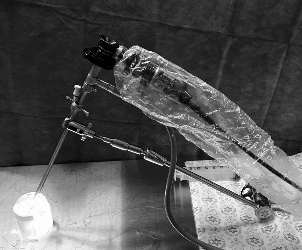
A phantom built from an automatic retractor holding a 30° optical lens attached to a camera and submerged in a glass canister containing 100 mL of the different liquids in front of a red millimetric paper.
Microscopy
Solutions of 2% blood in 1.5% glycine, physiologic solution, or DW were dropped on glass slides and examined under magnifying loupes ( ×4 magnification) and under a microscope (Olympus) ( ×200 magnification) connected to a camera.
Images analysis
Images taken from the cystoscope and microscope are presented without applying any filters or graphical adjustments.
The test was done by two surgeons; then, after finalizing the images, they were presented to a forum of four surgeons for comments. The analysis of images was a consensus.
Results
Representative phantom screenshots are presented in Figure 2. The 1 mm resolution was preserved in solutions containing 2% blood in 1.5% glycine and 1% blood in DW, yet adjacent bars could not be discriminated in none of the blood–saline mixtures. Mixtures of 2% blood in physiologic solution, DW, and 1.5% glycine examined under × 4 magnification are presented in Figure 3. The physiologic solution and DW mixtures presented as homogenous red opacity, whereas the 1.5% glycine mixture appeared a clearer, yet grainy appearance. Microscopic examinations ( × 200 magnification) of these mixtures are presented in Figure 4, revealing an even distribution of the red blood cells (RBCs) in physiologic solution, whereas clumps of RBCs were present in 1.5% glycine. Complete hemolysis of the RBCs occurred in the DW.
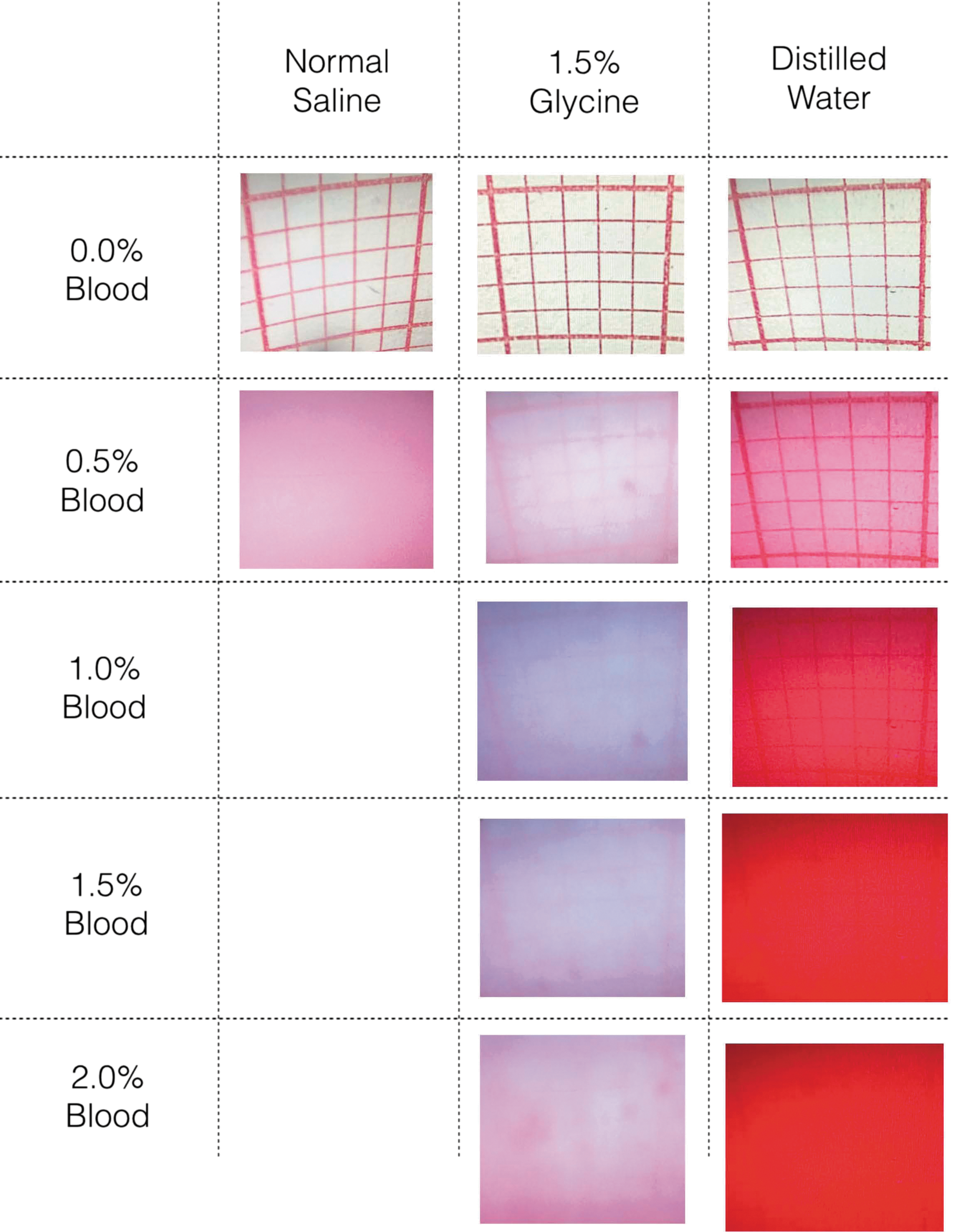
Screenshots taken with the camera. The 1 mm resolution is lost in 0.5% blood in physiologic solution and in 1% blood in DW, yet it is maintained at 2% blood in 1.5% glycine solution. DW, distilled water.
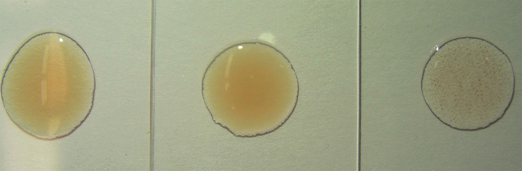
Drops of 2% blood in DW (left), physiologic solution (middle), and 1.5% glycine (right) examined under × 4 magnification. Physiologic solution and DW show homogenous red opacity and the 1.5% glycine shows a clearer, but grainy appearance.
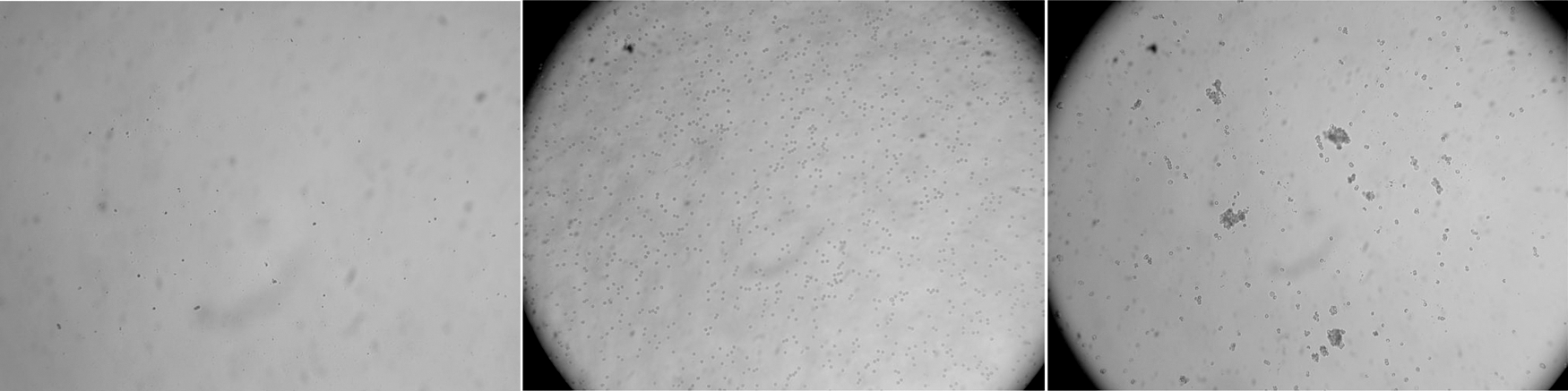
Microscope examinations ( ×200 magnification) of 2% blood in DW (left), physiologic solution (middle), and 1.5% glycine (right). RBCs are evenly distributed in physiologic solution, completely hemolyzed in DW, and form clumps in 1.5% glycine. RBCs, red blood cells.
Discussion
This study shows that a mixture of 2% blood in 1.5% glycine is clearer compared with blood in saline. Although 1 mm resolution was possible at as much as 2% blood in glycine, the view in physiologic solution was obscured already at 0.5% of blood. In DW, the 1 mm resolution was maintained at as much as 1% of blood. Surgeries using DW could be appropriate for resections of small bladder tumors, but will not be discussed further due to its hemolytic effect. 5
In addition, RBCs form clumps when mixed with 1.5% glycine, whereas in physiologic solution, they remain individually distributed. The explanation to these phenomena lies in the different electrical behaviors of RBCs in different solutions.
RBCs are coated with sialic acid, which confers their surface a negative charge. In electrolyte solutions, such as blood or physiologic solution, cations like Na+ are attracted to the negative surface of the RBCs as counter-ions. The closest layer to the interface is tightly bound and is composed only of cations (the Stern layer). The next layer is more diffusely bound and is composed of a mixture of cations and anions, with predominance of cations. These two layers are termed the “diffuse double layer.” Beyond this layer, the liquid is electroneutral. The potential difference between the tightly bound layer and the electroneutral region is defined as the zeta potential of the particle. The zeta potential and the thickness of the diffuse double layer determine the magnitude of the overall electrical repulsion between the RBCs, which is high in plasma. When the zeta potential is reduced to a certain level or when the thickness of the diffuse layer decreases, the attraction forces between the RBCs (monolayer bridging by plasma macromolecules) overcome the repulsive forces and the RBCs clump (flocculate). 6 The zeta potential decreases when counter-ion concentration is markedly increased, when the valence of the counter-ion is increased, or when the sialic acid is removed from the RBCs with neuraminidase. 7 Physiologic solution does not affect the zeta potential and only moderately decreases the thickness of the diffuse layer, although not to an extent that will produce flocculation. Conversely, 1.5% glycine (which is a zwitterion) is an electroneutral solution that markedly diminishes the thickness of the diffuse double layer around the RBCs, resulting in their partial flocculation. The result is a decrease in the number of particles per volume and greater transparency.
The enhanced transparency of glycine was demonstrated using a phantom (Fig. 1). The drawback of this model is the lack of fluid motion typical to transurethral surgery; so, in fact, it simulates more closely the situation when the inflow stream is turned off. This maneuver is often used by surgeons toward the end of the surgery for corroborating the absence of active bleeding. We used room temperature fluids to resemble real-life conditions; we cannot predict the effect of other conditions on the visibility. We did not check other fluids such as sorbitol and mannitol, thus we cannot predict their effect and it might be the subject for further research. Since the use of glycine can cause transurethral resection syndrome, we recommend its use instead of NS when the risk is low. Nevertheless, better view might shorten the time of surgery, thus reducing the risk of transurethral resection syndrome.
Conclusions
This study showed that the transparency of blood containing glycine solution is greater compared with physiologic solution. This observation is completely expected from the physicochemical properties of RBCs. To improve visibility, we propose that in patients with low risk of TUR syndrome, 1.5% glycine solution should be preferred over physiologic solution, even when bipolar or LASER systems are used. This conclusion could also apply to other disciplines that employ endoscopic surgery, such as gynecology.
Footnotes
Acknowledgement
We are grateful to Prof. Simon Benita for elucidating the physicochemical aspects of the findings in this research.
Author Disclosure Statement
No competing financial interests exist.