




Abstract
Objectives:
The aim of this study was to investigate “caliceal” fluid temperature changes during holmium laser activation/lithotripsy using settings up to 40 W power output with different irrigation flow rates.
Materials and Methods:
The experimental system consisted of a glass test tube (diameter 10 mm/length 75 mm) filled with deionized water, to mimic a calix. Real-time temperature was recorded using a thermocouple (Physitemp, NJ) positioned 5 mm from the bottom of the tube. A 200 μm laser fiber (Flexiva; Boston Scientific, MA) was introduced through the working channel of a disposable ureteroscope (LithoVue; Boston Scientific) and the laser fiber tip was positioned 15 mm above the bottom of the test tube. Deionized water irrigation (room temperature) through the working channel of the ureteroscope was delivered at flow rates of 0, 7–8, 14–15, and 38–40 mL/minute. A 120-W holmium laser (pulse 120; Lumenis, CA) was used. The following settings were explored: 0.5 J × 10 Hz, 1.0 J × 10 Hz, 0.5 J × 20 Hz, 1.0 J × 20 Hz, 0.5 J × 40 Hz, 1.0 J × 40 Hz, and 0.5 J × 80 Hz. During each experiment, the laser was activated continuously for 60 seconds.
Results:
Temperature increased with increasing laser power output and decreasing irrigation flow rate. The highest temperature, 70.3°C (standard deviation 2.7), occurred with laser setting of 1.0 J × 40 Hz and no irrigation after 60 seconds of continuous laser firing. None of the tested laser settings and irrigation parameters produced temperature exceeding 51°C when activated for only 10 seconds of continuous laser firing.
Conclusion:
High-power holmium settings fired in long bursts with low irrigation flow rates can generate high fluid temperatures in a laboratory “caliceal” model. Awareness of this risk allows urologist to implement a variety of techniques (higher irrigation flow rates, intermittent laser activation, and potentially cooled irrigation fluid) to control and mitigate thermal effects during holmium laser lithotripsy.
Introduction
T
Materials and Methods
The experimental apparatus consisted of a 13 L water bath maintained at 37°C using an immersion heater (Ulanet, CT). A glass test tube (inner diameter 10 mm/length 75 mm) filled with deionized water simulated a renal calix and was positioned in the water bath with its opening 1 cm above the surface of the water. Temperature of the fluid in the test tube was recorded once per second using a thermocouple (Physitemp, NJ) positioned 5 mm from the bottom of the tube. A second thermocouple recorded temperatures in the water bath 10 mm distance from the test tube. A disposable ureteroscope (LithoVue; Boston Scientific, MA) was inserted into the test tube with distal tip secured 20 mm above the bottom of the test tube. A 200 μm laser fiber (Flexiva; Boston Scientific) was introduced through the working channel of the ureteroscope with the laser fiber tip positioned 15 mm above the bottom of the test tube (Fig. 1).
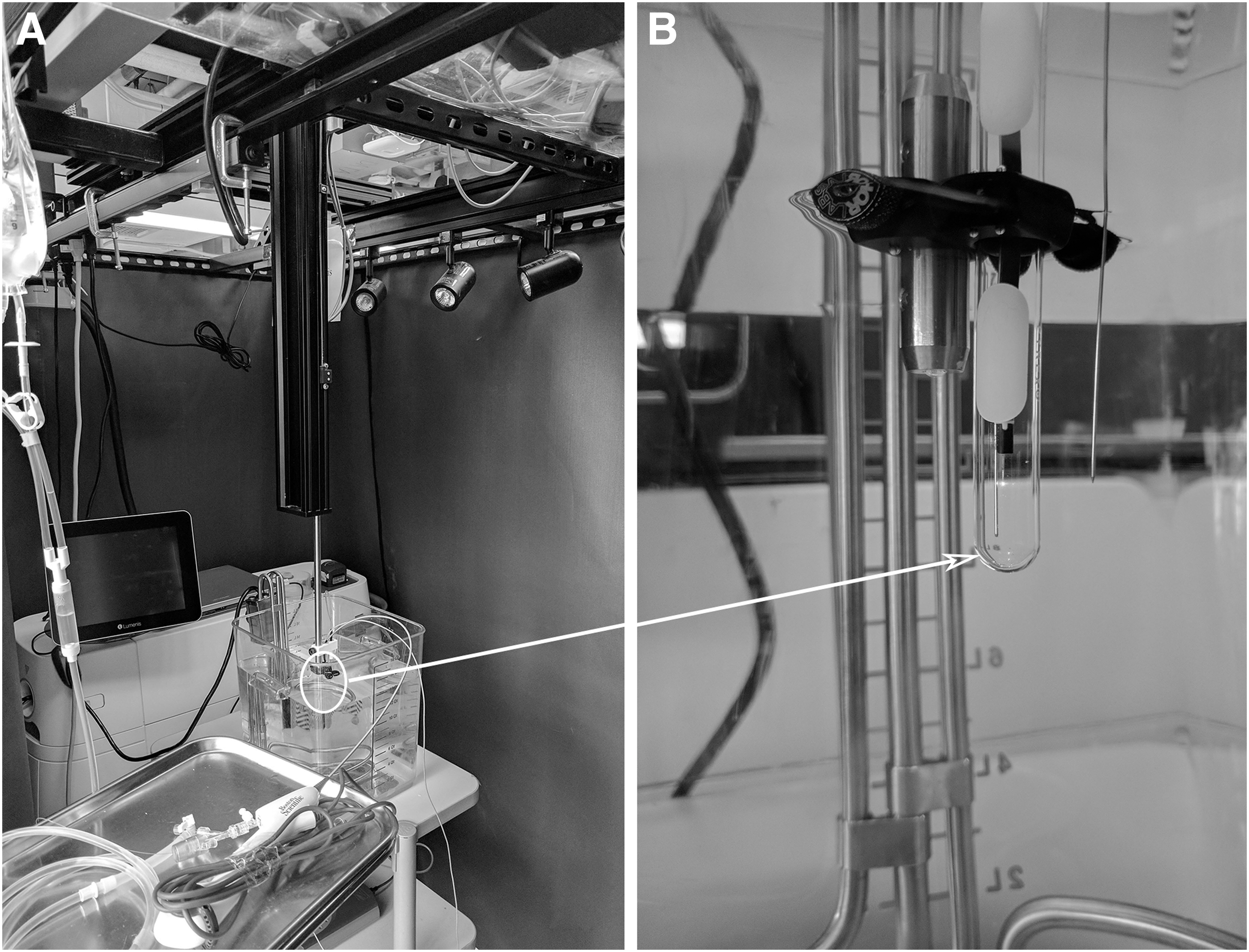
Deionized water irrigation (room temperature; 23°C) through the working channel of the ureteroscope was delivered at flow rates of 0 mL/minute, 7–8 mL/minute (60 cm H2O—irrigation bag 60 cm above the tip of the ureteroscope), 14–15 mL/minute (100 cm H2O), and 38–40 mL/minute (304 cm H2O, 100 cm irrigation height and 150 mmHg pressure irrigation).
Each trial was conducted for 120 seconds during which the temperature was monitored continuously. Baseline temperature was recorded for the first 20 seconds of the experiment (time 0–20 seconds). At 20 seconds, irrigation was started at the prescribed rate. At 40 seconds, laser energy was delivered continuously until 100 seconds. Thermocouple measurements were continued for 20 additional seconds. Each trial was stopped at 120 seconds.
A 120-W holmium laser (pulse 120; Lumenis, CA) was used in this study. The following settings were explored: 0.5 J × 10 Hz (5 W), 1.0 J × 10 Hz (10 W), 0.5 J × 20 Hz (10 W), 1.0 J × 20 Hz (20 W), 0.5 J × 40 Hz (20 W), 1.0 J × 40 Hz (40 W), and 0.5 J × 80 Hz (40 W). All experiments were conducted utilizing short pulse mode. The laser fiber was stripped and cleaved before each experiment. Each experiment was repeated five times.
Data are presented as mean and standard deviation (SD). The mean temperatures recorded during the 60 seconds of laser activation at each setting were used to calculate the thermal dose based on Dewey and Sapareto T43 equivalence calculations. 4 Safe exposure was defined by Dewey and Sapareto to be 120 minutes at 43°C. Statistical analysis was performed using Microsoft Excel (Redmond, WA).
Results
The highest temperature, 70.3°C (SD 2.7), occurred with laser setting of 1.0 J × 40 Hz and no irrigation. Temperatures exceeded 55°C for 20 W and higher settings. Table 1 presents the temperature data measured at the end of 60 seconds of continuous laser firing. Figure 2 shows representative tracings of temperature during trials with select laser settings and 14–15 mL/minute irrigation rate. In these experiments, the temperatures rapidly rise and then reach a plateau after the first 20 seconds. Figure 3 depicts temperature tracings for trials with laser settings of 1.0 J and 40 Hz at four different irrigation rates. Increasing irrigation flow rate (active cooling) decreased the 1-minute (plateau) temperature. Importantly, after the first 10 seconds of continuous laser firing, none of the tested laser setting irrigation produced temperatures exceeding 51°C (Table 2).
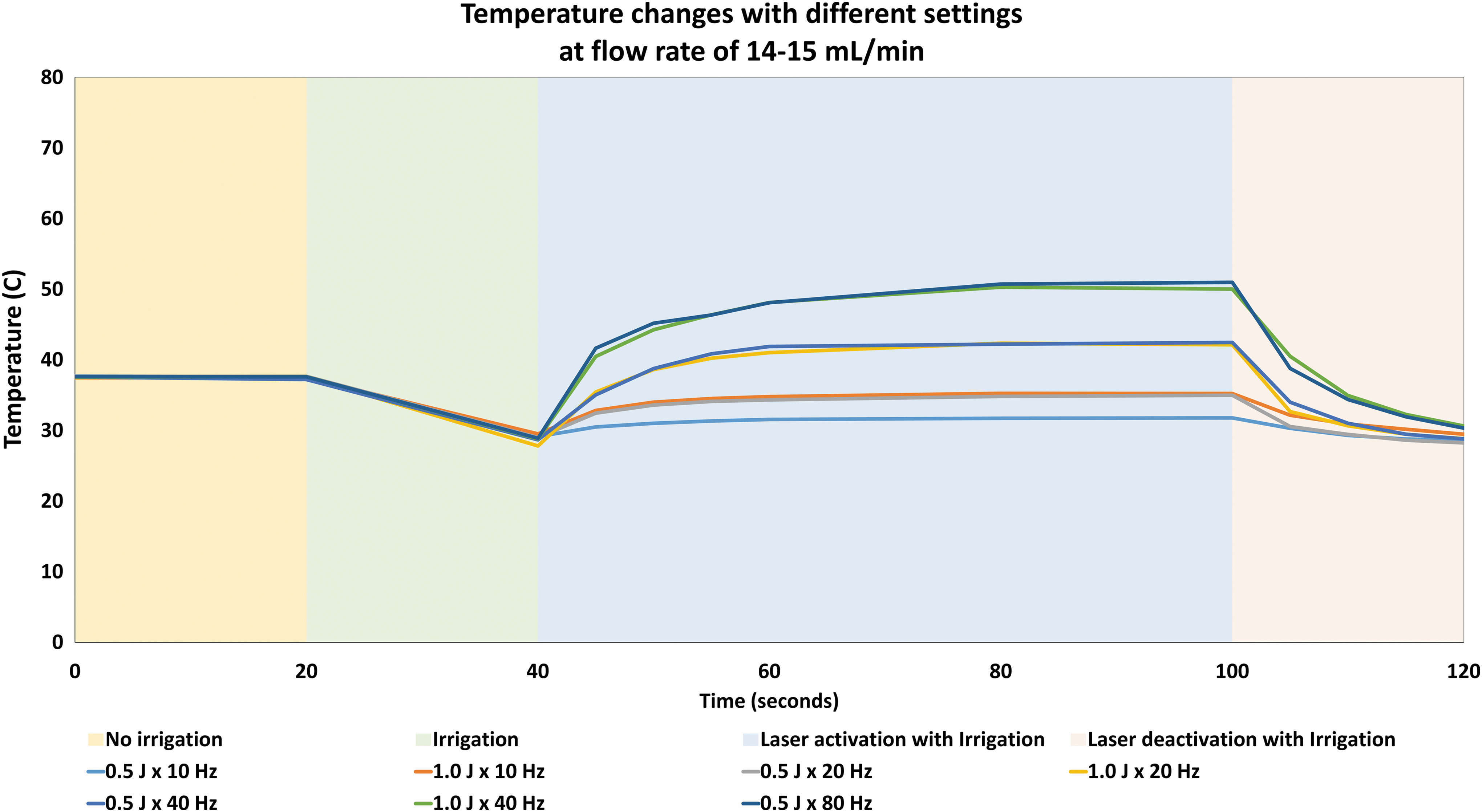
Temperature changes during holmium laser activation utilizing different settings on a short pulse mode at irrigant flow rate of 14–15 mL/minute.
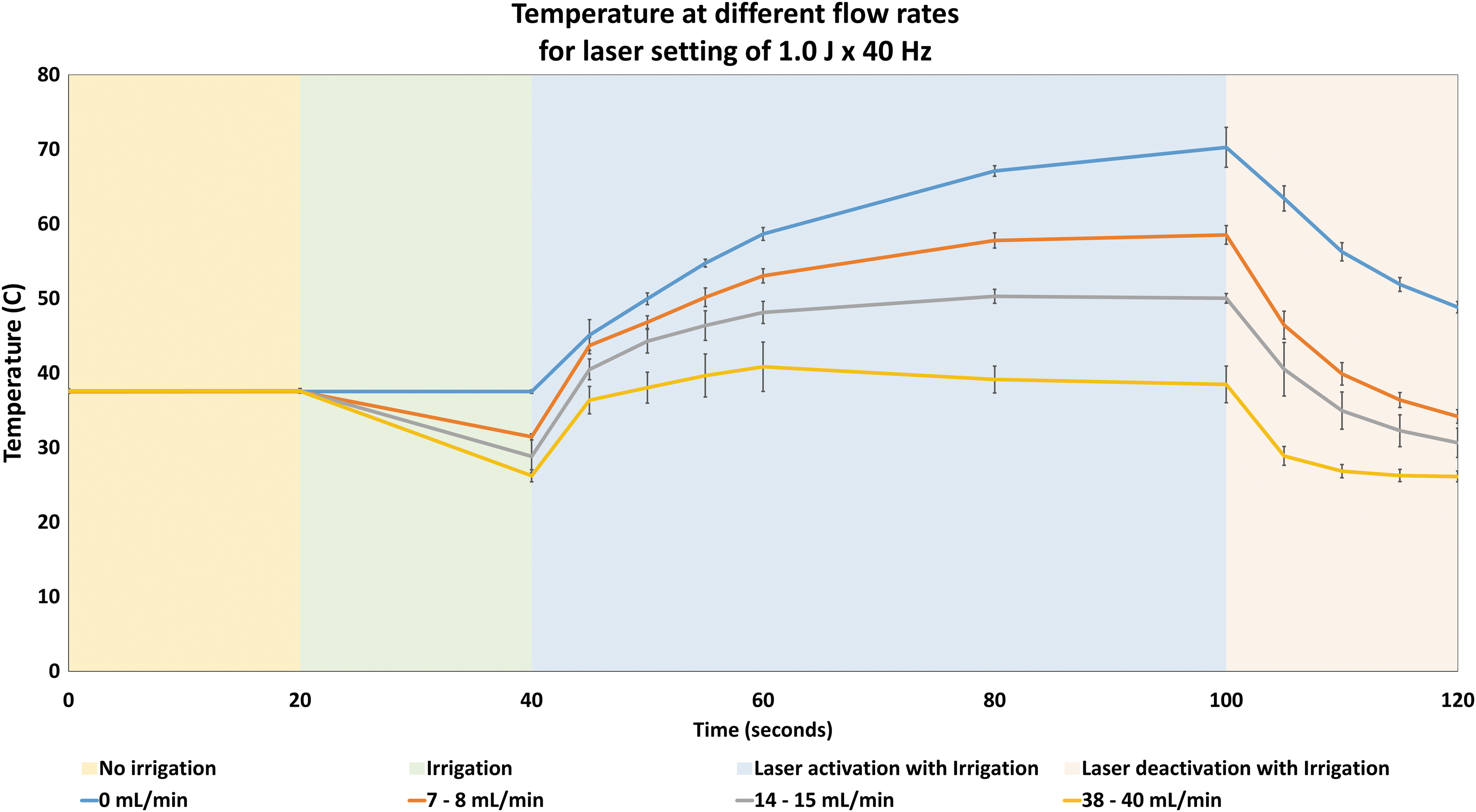
Temperature changes during holmium laser activation utilizing 1.0 J × 40 Hz setting on a short pulse mode at different irrigant flow rates.
SD = standard deviation.
The thermal dose calculated from 60 seconds of laser activation at each setting is presented in Table 3. The safe exposure limit (T43 equivalent of 120 minutes) was exceeded when low irrigation and high-power outputs were used. When high irrigation flow rates 38–40 mL/minute) were used, none of the settings exceeded the safety threshold.
Discussion
Holmium laser systems have been in clinical use for almost 30 years. The first generation systems had limited parameter settings and were only capable of producing power output up to 15 W. 5 However, recent advances in laser technology have resulted in the availability of systems with higher power output up to 120 W. Ureteroscopy laser techniques are increasingly utilizing high-power settings with increased thermal deposition.
It is known from the thermal tissue ablation literature that both magnitude of temperature elevation and time maintained at elevated temperatures are factors contributing to cell death. 4 This time/temperature relationship has been quantified by Dewey and Saparato. 4 A temperature of 43°C for 120 minutes was found to be lethal for normal tissue. Using this as a reference point, Dewey and Sapareto proposed a formula to calculate T43 equivalence (the time that is required to maintain tissue at a specified temperature to produce cell death). Using this formula, T43 equivalence can be calculated for specific temperatures and used to determine the maximal safe exposure time for tissue at each temperature. For example, thermal injury and cell death will occur if tissue is maintained at 51°C for more than 28.1 seconds or at 53°C for more than 7.1 seconds.
Two prior studies have examined heat generation during holmium laser lithotripsy. Molina et al. recorded temperature changes in the ureter during holmium laser lithotripsy in an ex vivo study. 3 Using a laser setting of 1.0 J × 10 Hz, they found the ureteral wall temperature reached 112.4°C without irrigation. However, irrigation from a saline bag at 3 feet reduced the temperature to 49.7°C. Butticè et al. evaluated temperature changes in an in vitro kidney model, “K-Box” ureteroscopy simulator, during holmium laser lithotripsy with 50 cm H2O irrigation pressure. 6 Laser settings up to 20 W, 0.5 J × 20 Hz, 1 J × 10 Hz, and 2–4 J × 5 Hz were utilized. Temperature exceeded 45°C without irrigation but was controlled below this level with continuous irrigation. 6
This current study explores thermal effects of newer laser settings with greater maximum power output up to 40 W. Several of these high-power laser settings with continuous firing for 60 seconds and no irrigation or low-flow irrigation resulted in temperatures that could cause thermal injury based on the Dewey and Sapareto T43 equivalence calculations.
The “heat-sink” effect of vascular perfusion in renal tissue is not accounted for in this model and may mitigate the temperature elevation by constantly moving heated blood away from the kidney. 7 However, the rapid rise in temperature at the initiation of laser lithotripsy could still expose directly adjacent tissue to harmful temperatures. 8 While these findings are relevant in the renal collecting system, they may in fact be of greater importance in the ureter, where heat-sink effects are minimal due to less perfusion from smaller blood vessels than in the kidney. 9
Active measures to reduce heat production and limit cellular damage include intermittent laser firing during the procedure. In this study, continuous laser firing of 10 seconds did not reach damaging temperatures with any of the tested laser parameters. Typically, laser activation is interspersed with pauses to allow stone debris to clear from the visual field and provide an assessment of the remaining stone burden. This pause is also beneficial to allow cooling of the fluid. The optimal length of pauses needs to be further studied, although insight can be gained from the temperature decay curves following laser deactivation (Figs. 2 and 3). Another consideration when using high-power laser setting is providing adequate irrigation during ureteroscopy. This can be achieved with pressure irrigation and facilitated by using an access sheath to improve irrigant outflow and decrease the intrarenal pressure. However, the use of pressure irrigation must be carefully regulated to minimize pyelovenous back flow, fluid extravasation, and the concomitant risk of systemic inflammatory response syndrome and sepsis. 10,11 Additional considerations include using room temperature or chilled saline irrigant rather than body temperature irrigant to mitigate temperature increase.
There are several limitations to this study. First, our experimental model does not fully capture the active heat-sink effect of tissue perfusion. However, this is partially compensated for by the relatively larger surface area of the test tube (∼24 cm3 compared to a renal calix) in contact with the water bath. Second, the glass test tube might be less conductive to heat than human tissue. Using a transparent glass test tube in this study was necessary to keep the ureteroscope, thermocouple, and laser fiber distance the same for all the experiments. Finally, our experimental model does not include laser treatment of a stone, rather laser firing into a fluid filled test tube as part of the experimental methods. However, no difference was noted in temperature measurements when this was trialed with and without a stone.
Future studies to follow up on this study include exploring the effect of using chilled irrigation with in vitro studies. Additional animal studies are also needed to confirm these findings in vivo and to investigate the extent to which vascular perfusion acts as a heat sink in vivo with the rapid temperature rise seen with high-power laser lithotripsy.
In conclusion, high-power laser lithotripsy settings fired in long bursts with low irrigation flow rates can generate high fluid temperatures in a laboratory caliceal model. Awareness of this risk allows the urologist to implement a variety of techniques (higher irrigation flow rates, intermittent laser activation, and potentially cooled irrigation fluid) to control and mitigate thermal effects. In addition, this research is important as laser techniques continue to evolve with perhaps even higher power settings and greater emphasis on low irrigation flow rates to prevent high renal pelvic pressures.
Footnotes
Acknowledgment
Funding for this project was provided through a scientific grant from Boston Scientific.
Author Disclosure Statement
K.R.G. is a consultant for Lumenis and Boston Scientific.