
Abstract
Background:
Laparoscopic donor nephrectomy (LDN) converted a retroperitoneal (RP) procedure into a transperitoneal (TP) operation with reports of bowel and solid organ injuries leading to mortality in occasional cases. Laparoscopic RP donor nephrectomy can reduce these risks but never became popular because of the muscle cutting approach. Lumbotomy incision can be used to approach retroperitoneum by incising fascial planes, eliminating disadvantages of the RP approach. This report compares the outcomes of the standard multiport TP LDN with translumbar laparoendoscopic single-site donor nephrectomy (LESS-DN).
Methods:
Between January 2016 and June 2017, 50 voluntary kidney donors out of 267 donors were randomized to undergo LESS-DN vs LDN. Donors with body mass index ≥30 kg/m2, multiple renal arteries, and right-sided nephrectomy were excluded from the study. Postoperative pain, duration of surgery, length of graft vessels and ureter, warm ischemia time, intraoperative blood loss, incision length, convalescence period, duration of hospital stay, and recipients' creatinine at discharge were compared among both the groups. Pain assessment was done using visual analogue scale (VAS).
Results:
The RP group experienced lesser pain (VAS score 0.3 ± 0.3 vs 1.1 ± 0.0, p = 0.000), lesser analgesic requirement (186 ± 51.07 mg vs 254 ± 62.7 mg, p = 0.000), and faster convalescence (7.0 ± 3.0 days vs 10.7 ± 3.3 days, p = 0.00) related to smaller cumulative incision (7.8 ± 0.8 cm vs12.4 ± 2.0 cm, p = 0.00), and had reduced operative time (142 ± 26.2 minutes vs 170.8 ± 34.75 minutes, p = 0.001) and blood loss. Other recorded parameters were similar in both the groups.
Conclusions:
The single port RP approach significantly reduced postoperative pain and hastened recovery when compared with the TP approach. Converting to a RP approach presents an opportunity for surgeons to further reduce morbidity associated with the donor nephrectomy.
Introduction
T
A recent systematic review and meta-analysis have shown that the laparoscopic RP approach had significantly less complications than its TP counterpart (odds ratio [OR] = 0.5, 95% confidence interval [CI] 0.2–1.1) with all intra-abdominal injuries reported in the TP group. 7 But the RP approach has not gained popularity given the disadvantage of a muscle cutting flank incision through which the kidney is extracted. Both TP and RP nephrectomies have been traditionally performed through multiple ports. Recently, single port nephrectomies have been reported either through transumbilical or flank incision, further reducing postoperative pain and convalescence. 8,9 A single port laparoscopic RP procedure that can use the same incision for kidney retrieval and avoid cutting of abdominal muscles would be an ideal approach for donor nephrectomy. Lumbotomy incision wherein the retroperitoneum is entered through a fascial incision has been previously used in urologic surgeries. 10 In this context, a single port procedure using the lumbotomy incision can fulfill most of the criteria for an ideal operation for donor nephrectomy, provided it meets the safety and ease of laparoscopic TP nephrectomy. LDN through lumbotomy incision was first performed and published by our center in 2016. 11 This report compares the outcomes of translumbar laparoendoscopic single-site donor nephrectomy (LESS-DN) with those of standard multiport TP LDN performed at our center.
Aim
To compare duration of surgery, perioperative pain and recovery, hospital stay, recipient allograft function, and surgical complications in patients undergoing RP single port donor nephrectomy with those of TP multiport donor nephrectomy.
Materials and Methods
This was a prospective randomized controlled study in 50 voluntary kidney donors (VKDs), operated in the Department of Renal Transplant Surgery, Post Graduate Institute of Medical Education and Research, Chandigarh, between January 2016 and June 2017. The trial was registered at
Fifty donors were recruited from a total of 267 VKDs who underwent donor nephrectomy at our center during this period and were followed up for 6 months. Donors with body mass index ≥30 (23), multiple renal arteries (63), right-sided nephrectomy (17) and those who did not consent (114) were excluded. The procedures were performed by one of three surgeons in the team each having an experience of >500 LDNs. Fifty VKDs were randomized into two groups:
Group A, RP LESS-DN; Group B, TP LDN. A random sequence of 50 numbers was generated by a computer. Numbers 1–25 were allocated to Group A and numbers 26–50 were allocated to Group B. On the day of surgery, a patient was allocated to a particular group on the basis of the random sequence generated. The surgeon was informed of the randomization on the day of the procedure to eliminate selection bias.
The following parameters were recorded and compared in the two groups.
Intraoperative
Duration of surgery, warm ischemia time, length of vessels, ureter, and incision were documented. Lengths of individual ports were added to the length of retrieval incision for the TP group. Any complications during surgery were recorded.
Postoperative
Pain assessment was done at 8-hour intervals on postoperative day (POD) 0 and 12 hourly thereafter till POD 5 using the comfort score (CS) 12 and visual analogue scale (VAS). 13 If the CS was less than 6, then pain assessment was done using VAS and patient response was graded as follows: no pain (0–4 mm), mild pain (5–44 mm), moderate pain (45–74 mm), and severe pain (75–100 mm). Inj. tramadol 50 mg i.v. 8 hourly was given as baseline analgesia. Inj. morphine 3 mg i.v. stat was used as a rescue drug if VAS was >44 and amount of morphine given was recorded. Patient's convalescence was noted as number of days taken to resume daily activities to preoperative level without any pain. Renal allograft function in the recipient was also assessed by creatinine at discharge and the number of days to achieve the baseline creatinine.
Statistical analyses
Quantitative variables were estimated using mean, median, and standard deviation. Mann–Whitney test was used to compare two groups. Qualitative variables were described as frequencies and proportions. Proportions were compared using chi-square or Fisher's exact test whichever was applicable. All statistical tests were two sided and were performed at a significance level of ≤0.05. All data were analyzed with SPSS 24 software.
Operative technique: RP laparoscopic single-site donor nephrectomy
Patients were placed in lateral position and retroperitoneum was entered through fascia cutting incision 14 (Fig. 1). After entering the retroperitoneum, Gerota's fascia was opened and lower pole of kidney was identified. Dissection was carried out anterior to psoas till ureter and gonadal vein were identified. Alexis wound retractor was applied to the incision with a sterile latex-free surgical glove rolled over the inner ring of the retractor so that the fingers project out of the outer ring, creating airtight RP compartment. Three fingers of the glove were used to insert one 10 mm camera port, two working ports (10 mm and 5 mm) and pneumoretroperitoneum was obtained 10 (Fig. 2).

Cutting the lumbodorsal fascia.
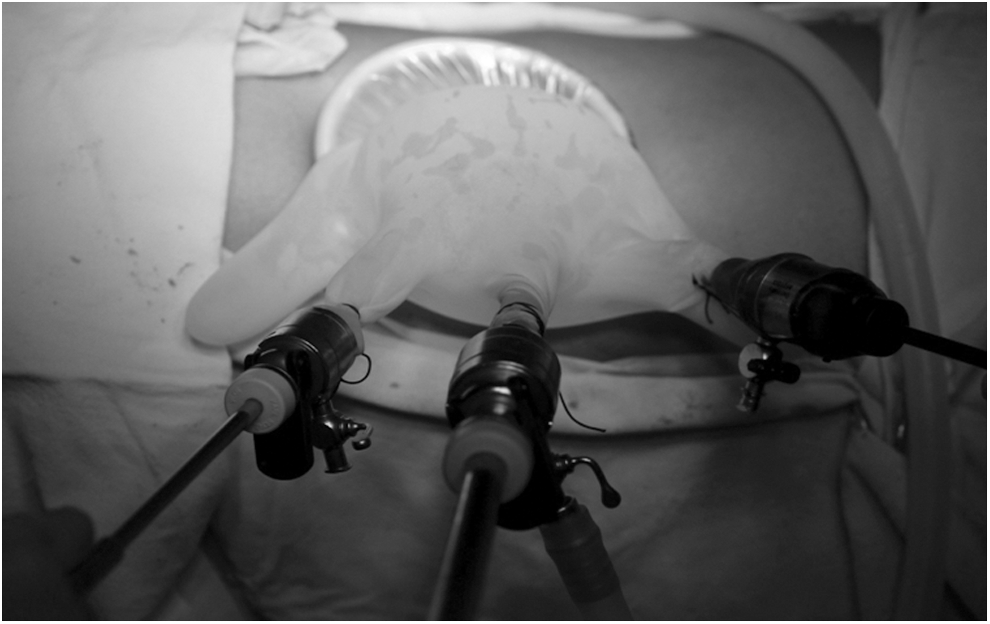
Final port assembly.
Further dissection was carried out laparoscopically. The ureter was lifted from the retroperitoneum and the gonadal vein was dissected till its drainage into the renal vein and was ligated. The gonadal artery, if encountered, was ligated. Pulsations of the renal artery were easily observed on division of the lumbar veins. The renal artery was dissected till its origin. Thereafter, the renal vein was dissected and the adrenal vein was identified. Once the vascular dissection was completed, the lower pole of the kidney was separated from the peritoneum, and the kidney that was hanging from the peritoneum so far became free, allowing dissection to be carried out on all surfaces of kidney to free it from the surrounding fat till the renal vein was seen anteriorly. The adrenal gland was separated from upper pole of the kidney and its vein was ligated. The ureter was divided once the kidney and renal vessels were free. The renal artery and renal vein were separately ligated with two Hemolok clips each (polymer clips have been issued a black box warning by U.S. Food and Drug Administration but its use is in practice in most centers in India) and cut with scissors. The kidney was retrieved into the Alexis wound retractor and taken out along with the retractor (Fig. 3).
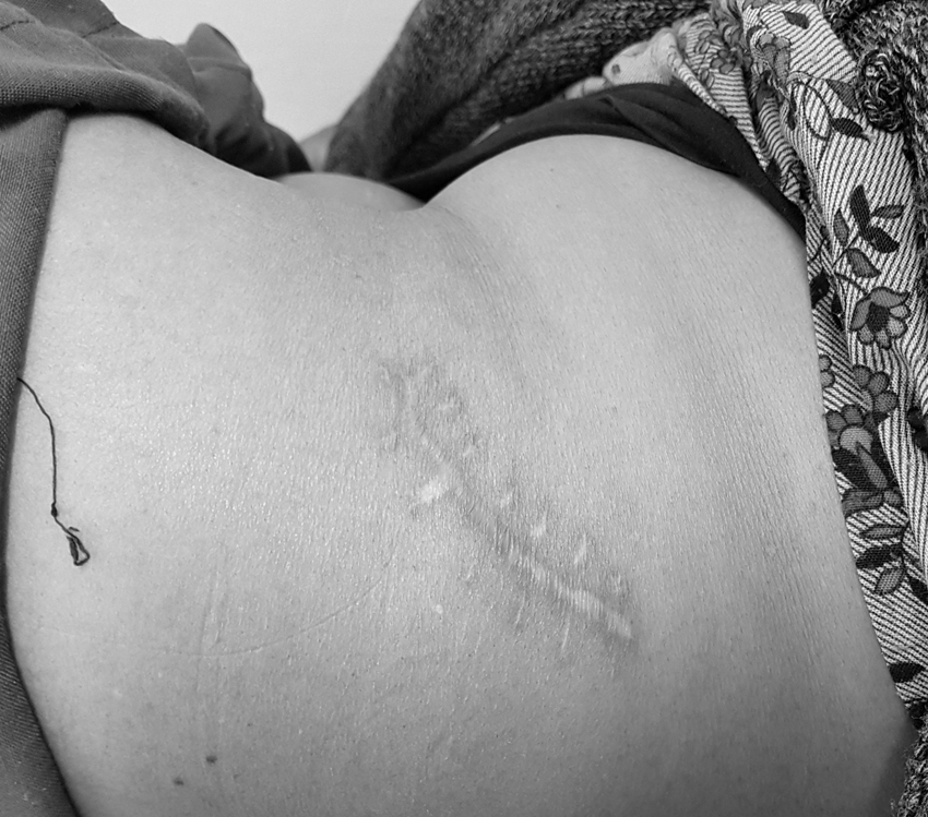
Lumbotomy postoperative incision.
Transperitoneal LDN
Pneumoperitoneum was created by Veress needle, which was inserted into left lower quadrant. Two 10 mm and one 5 mm port were used. An additional 5 mm subcostal port was used if required.
The left colon was mobilized by dividing the line of Toldt, reflecting the colonic mesentery upto observation of gonadal vein. Remaining steps were same as in the RP approach. After completing vascular dissection, the kidney was freed along its lateral border. Renal vessels and ureter were ligated using polymer clips and divided, and kidney was retrieved through a Pfannenstiel incision.
Results
The demographics are given in Table 1. The mean age of patients in Group A was more than that in Group B (46.4 ± 11.9 vs 38.5 ± 10.0 years, p = 0.014), but on multivariate analysis, there was no impact of this age difference on pain scores. The RP procedure was completed with an average duration of 28 minutes less than that of the TP procedure (142 ± 26.2 vs 170.8 ± 34.75 minutes, p = 0.001). Mean estimated blood loss was significantly lower in the RP group. Intraoperative complications in the form of bleeding were seen in two donors in the TP group. One donor was converted to open surgery because of bleeding from mesenteric vessels, whereas in the other case, hemostasis was secured laparoscopically from a bleeding adrenal vein. No blood transfusion was required in any of the patients.
BMI = body mass index; RP = retroperitoneal; TP = transperitoneal; UTI = urinary tract infection; WIT = warm ischemia time.
Mean incision length in the RP group was significantly smaller than that in the TP group (7.8 ± 0.9 cm vs 12.4 ± 2.0 cm, p = 0.00). Patients experienced more pain during initial 12 hours postsurgery, being significantly more in the TP group. Average pain scores for first 2 PODs were higher in the TP group and difference was statistically significant (Table 2). Mean analgesic requirement was more in the TP group and the difference was statistically highly significant (186 ± 51.07 mg vs 254 ± 62.7 mg, p = 0.000). Three donors in the TP group required rescue drugs on POD 0 but none in the RP group. The duration of hospital stay was similar in the two groups, although patients were clearly more comfortable in the RP group. Patients in the RP group recovered significantly faster and were back to their daily routine on an average 3 days earlier than the TP group.
POD = postoperative day.
Constipation was more in the TP group (16% vs 4%) possibly because of bowel handling during intraperitoneal dissection. One donor in the TP group had urinary tract infection and serous discharge from the Pfannenstiel incision site, which was managed conservatively. No wound infection was seen in any patient.
The mean creatinine and days taken to reach the baseline creatinine were similar in both the groups. No vascular or ureteral complications were seen in any of the 50 recipients.
Discussion
LDN has replaced the conventional open donor nephrectomy after 1995 because of lesser pain and early convalescence. 15 To further minimize discomfort, single port nephrectomy was performed by Raman et al. 16 and many reports of living donor nephrectomies using TP LESS approach were published subsequently. 8,9,16,17 Although the first effective retroperitoneoscopic donor nephrectomy was reported in 2000 by Gill and colleagues, 18 it never gained popularity. However, a recent systematic review and meta-analysis have shown that the RP approach had significantly less complications than its TP counterpart (OR = 0.5, 95% CI 0.2–1.1) with all intra-abdominal injuries reported in the TP group. 7 However, TP nephrectomy remains the preferred procedure as muscle cutting incision associated with RP approach negates the advantages of minimally invasive procedure. Therefore, it was thought that the use of lumbotomy incision to approach retropertitoneum would be advantageous to the donors and would have significant impact on postoperative pain and convalescence.
In this study, it was observed that pain was significantly less in the RP group. Patients in both groups received similar baseline analgesia and pain assessment was done using CS 12 and VAS. 13 The intensity of pain was maximum during the first 12 hours postsurgery in both the groups, which was, however, significantly more in the TP group (p = 0.004). Higher pain in the TP group could be attributed to longer cumulative length of the incision (7.8 ± 0.9 cm vs 12.4 ± 2.0 cm, p = 0.00) or because of the TP approach. It is difficult to attribute the reduction in pain to the single-site approach or an RP approach. Both factors likely played an important role. Although some studies in the literature have shown that postoperative pain is less with the RP approach, 19,20 there have been equivalent outcomes in others wherein nephrectomy has been performed for indications other than donor surgery. 21,22 The peritoneal irritation by CO2 insufflation is thought to be responsible for greater pain in the TP approach. Similarly, single-site surgery for donor nephrectomy has also been shown to reduce the pain associated with the procedure. 23 It can be expected that single-site lumbotomy incision would add the benefits of both and would be associated with the least pain and morbidity associated with donor nephrectomy. 10
The study also demonstrated a significant improvement in recovery after surgery with the RP approach. The mean convalescence period was significantly shorter in the RP group because of lesser postoperative pain, lesser analgesic requirement, and hence early return to daily routine activities.
Intraoperative complications (hemorrhage) were seen only in the TP group, requiring conversion to open approach in one donor. There were no intraoperative bowel/solid organ injuries in both the groups. The differences in late complications of the surgery such as incisional hernia or adhesive obstruction require a longer follow-up and were not the primary objective of this study.
One of the reasons for low acceptance of the RP route is the limited working space, which increases difficulty of performing surgery. However, in our experience, larger working space can be created if one remains in the plane outside the Gerota's fascia rather than dissecting on the surface of the kidney and allowing the fat to remain on the peritoneum. The space hence created is good enough to comfortably proceed with the surgery. This was also reflected in this study wherein the RP donor nephrectomy could be completed in a shorter time than the TP procedure. This was despite the fact that the RP approach was done with a single port, which traditionally has longer operative time. 7 Studies in patients undergoing partial and radical nephrectomy have also shown that the RP route has shorter operative time. 22,24 The RP single port approach eliminates steps of colon mobilization and placing/closing multiple ports, which reduces operative time.
Most single port surgeries require specialized angulated instruments, which are expensive. However, all the procedures in this study were completed comfortably with regular instruments. The kidney during the RP surgery remains attached to the peritoneum while the vascular dissection takes place, this reduces the need of retraction and manipulation. The home-made single port device used a full incision as compared with single ports such as single incision laparoscopic surgery, hence allowing a greater freedom of movement of individual instruments, obviating the need of angulated instruments. 25 This home-made single port device reduces cost and has been used for other surgeries. 26
Longer renal vein and ureter were retrieved in the RP group. This difference can be explained because of the direct access to the retroperitoneum and is advantageous in the recipient surgery. There was brisk diuresis intraoperatively in all the cases. The mean PODs to achieve baseline creatinine and mean creatinine at discharge in a recipient were comparable in both the groups. However, meta-analysis has shown that the RP approach is better in terms of fewer complications and lesser delayed graft function in the recipients receiving these kidneys. 27 Increased intra-abdominal pressure during TP nephrectomy adversely affects the ventilatory and hemodynamic parameters, 28 leading to decreased urine output. 29 These intraoperative changes can also affect the recovery of residual kidney function in the donor and a larger initial decline in renal function may happen with the laparoscopic approach in the donors when compared with open or RP donor nephrectomy, and these effects can persist till 1 year. 15
Limitations
The study had a short follow-up period as it focused on immediate postoperative recovery and excluded patients with right-sided nephrectomy as well as donors with multiple renal arteries. The study could not be blinded for obvious reasons as both the medical staff and patients knew the type of surgery in the postoperative period, which could have influenced the results. The study was underpowered to know the difference in complications as the incidence of these is currently low. Switching to an RP technique would also involve a learning curve for the new approach for about 10–30 cases as it is not as widely practiced. 30
Conclusions
The single port RP approach significantly reduced postoperative pain and hastened recovery when compared with the TP approach in renal donors undergoing laparoscopic nephrectomy. Further study is required to know the applicability of this technique in donors with complex anatomy.
Footnotes
Authors' Contributions
N.C. performed study design and data collection. D.B.K. carried out study design and surgical intervention. N.S. was involved in study design, writing of article, and surgical intervention. S.S. carried out surgical intervention. A.S. performed study design, surgical intervention, and writing of the article. K.K. and S.K. wrote the article.
Author Disclosure Statement
No competing financial interests exist.