
Abstract
Purpose:
We came up with a dynamic anatomical study intended to validate the safety of intercostal approach used by our center to access the upper pole of the kidney during percutaneous surgery.
Materials and Methods:
A total of 101 patients presenting randomly to the radiology department for CT evaluation of the abdomen and superior pelvis were involved in this study. Deep inspiration and expiration sequences in the prone position were evaluated to establish the location of the parietal pleura in relation to different anatomical landmarks. Three-dimensional reconstruction was performed to simulate the access needle course through the retroperitoneum.
Results:
Our data show that the position of parietal pleura is invariably higher on the right side irrespective of anatomical relation or respiratory changes. Higher position of the parietal pleura was noted in all considered landmarks upon full expiratory sequences. Using the midclavicular line as a landmark, our data show that on the right side, the parietal pleura was higher than the 10th intercostal space (ICS) in 100% of patients. Going up to the level of the ninth and eighth ICS, the pleura is higher in 89.1% and 66.3% of patients, respectively. Moreover, on the left side, the level of the parietal pleura was higher than the 11th ICS in 100% of patients. Reaching the 10th ICS, the parietal pleura still is higher in 92.07% of cases. Going up to the ninth ICS reduces the margin to 64.35% and using the eighth ICS would convey a margin of 24.7%.
Conclusions:
Supracostal access for percutaneous nephrolithotomy carries a risk of pulmonary complications, limiting its use worldwide. We have shown in this study that using the differences in inspiration and expiration along with the right anatomical landmarks could substantially lower the risk of complications. However, regardless of the side or landmark used, supracostal access is safe in >90% of cases.
Introduction
P
The supracostal approach, although providing high success rates, has been associated with various complications, including pneumothorax, hydrothorax, bleeding, and parenchymal injury. 2 We have shown in our department that adopting full expiration during access creation has reduced the risk of hemo/pneumothorax to nil. 3
We proposed a radiologic model to assess and compare the levels of diaphragm and parietal pleura during full inspiration and full expiration to try to set a safety anatomical landmark that can be used for supracostal access during PCNL.
Materials and Methods
This study was reviewed and approved by an institutional review board.
A total of 101 patients presenting for CT evaluation of the abdomen and superior pelvis for different purposes were randomly included in this study. After taking informed consent, CT scan was done in the prone position on double bolsters thoracic support, which mimics the PCNL position used in our medical center. The patients' arms were placed in adduction, and deep inspiration and expiration sequences were taken.
The same technique was applied to all 101 patients in the prone position. The patients were selected randomly, ages between 17 and 83 years, mean age 46.17 years; 51 men and 50 women were selected with average body mass index of 26.7 kg/m2, ranging between 19 and 36 kg/m2 (Table 1).
BMI = body mass index.
The selected population was all previously healthy. Exclusion criteria included any chronic lung disease and severe anatomical abnormalities.
Three anatomical landmarks were studied. The first line was drawn vertically along the edge of both paraspinatus muscles.
The second line was drawn through the midclavicles, whereas the third was taken midaxillary. Full expiratory and inspiratory CT sequences were evaluated by obtaining a 30° angle with the horizontal line at the lowest point of the pleura bilaterally, thus having the maximum threshold to prevent occurrence of a pneumothorax (Figs. 1 and 2). The intercostal space (ICS) below the lowermost edge of the pleura was then noted.
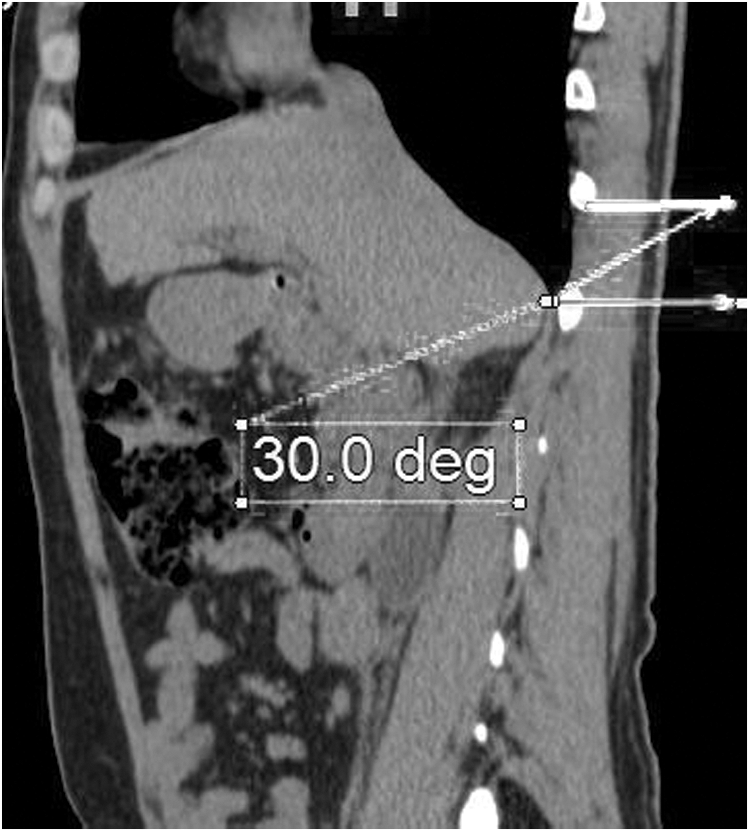
Needle course simulation under CT scan in percutaneous access to the kidney.

Needle angulation and point of entry under CT scan in percutaneous access to the kidney.
Acquisitions were taken using a General Electric high-resolution seventh-generation CT scan and three-dimensional reconstruction made by Magnum image rendering.
Results
Comparison was done in each anatomic landmark between inspiratory and expiratory sequences to accentuate the difference. Our data show that the position of parietal pleura is invariably higher on the right side irrespective of anatomical relation or respiratory changes.
Higher position of the parietal pleura was noted in all considered landmarks upon full expiration as compared with the inspiratory sequences (Tables 2 –5).
SC = subcostal.
Using the midclavicular line as landmark (the most commonly used landmark for percutaneous access in our center), our data show on the right side the following:
The parietal pleura lies above the 10th ICS in 100% of patients in full expiration (Tables 4 and 5).
Going up to the level of the ninth ICS, the margin decreases to 89.1% and 66.3% at the level of the eighth ICS.
Moreover, on the left side, upon full expiration, the parietal pleura sits above the 11th ICS (using the midclavicular line as landmark) in 100% of the cases. In 92.07% of cases, the pleura is above the 10th ICS; going up to the 9th ICS reduces the margin to 64.35%, and in 24.7% of the cases the pleura is above the 8th ICS (Tables 4 and 5).
Furthermore, using the paraspinatus muscle as a landmark, the parietal pleura rises above the 10th ICS in 88.2% of patients on expiration, whereas at the posterior midaxillary line, it rises beyond the 9th ICS in 85% of cases on expiration.
Discussion
The primary goal of treatment of renal calculi is complete stone clearance with minimal morbidity to the patient. 4 PCNL is a safe and effective approach to treatment of large kidney stones, and gaining percutaneous access to enter the appropriate calix is critical to the procedure. 5
Traditionally, supracostal renal access is avoided because of increased risk of pleural complication; however, it should not be excluded as a potential access point. 5,6
Hossain et al. considered the upper caliceal approach to be ideal for management of upper ureteral, upper caliceal, and staghorn stones. Although technically more demanding and challenging, entry through upper calix provided a straight access along with a better observation of upper and lower calices, renal pelvis, and ureteropelvic junction. 7
Among the complications related to this access, violation of the pleural space leading to pneumothorax or hydrothorax was the primary limiting factor to its widespread use. Our results showed that the risk of pleural injury is minimal upon upper pole access and can be further reduced upon full expiration. In a series done by our center, no pulmonary-related adverse event was noted regardless of access point. 3 These results were in concordance with this anatomical study, which is one of the first to employ an imaging technique showing the safety of intercostal access through simulation of needle trajectory while reproducing intraoperative settings.
According to our data, using the posterior midclavicular line as landmark, intercostal access in PCNL can be safely implemented whenever needed if coupled with full expiration.
On the right side, we can safely access the kidney up to the level of the 10th ICS with 100% safety; going up to the 9th ICS conveys an 89.1% margin, whereas the 8th ICS conveys a 66.3% safety margin.
On the left side, accessing the 11th ICS would be 100% safe, whereas the 10th ICS would carry a 92.07% safety margin.
Regardless of the landmark used, intercostal access is safe in >90% of cases.
Conclusion
Supracostal access for PCNL carries a risk of pulmonary complications, limiting its use worldwide. We have shown in this study that using the differences in inspiration and expiration along with the right anatomical landmarks could substantially lower the risk of complications.
The parietal pleura on the right side is invariably higher than on the left side.
However, regardless of the side or landmark used, supracostal access is safe in >90% of cases.
Footnotes
Author Disclosure Statement
No competing financial interests exist.