




Abstract
Objective:
The aim of the study was to assess the feasibility of laparoscopic nephrectomy (LN) in the treatment of patients with xanthogranulomatous pyelonephritis (XGP).
Methods:
Retrospective review of medical records of 17 patients (mean age 60.0 ± 13.3 years) who underwent LN by a single surgeon from 2010 to 2018. Sociodemographic and clinical data including diagnosis, presenting clinical features, surgical management, and postoperative course were analyzed.
Results:
LN was successfully performed in 15 (88.2%) patients. Two (12.5%) patients with disseminated disease were electively converted to open nephrectomy (ON) due to failure to progress. Two (11.8%) patients experienced intraoperative complications (grades 3b and 4b). Among patients in whom LN was successfully performed (n = 15), the mean operative time was 198.0 ± 107.1 min and was shorter when no intraoperative complications occurred (169.0 ± 48.1 min). Three (20%) of these patients required transfusions and nine (60.0%) required postoperative pelvic drainage (PD). Six (40%) patients experienced postoperative complications: one grade 1, four grade 2, and one grade 5. Mean hospital stay was 4.4 ± 4.3 days, and 3.4 ± 2.2 for those experiencing manageable or no complications. Among patients without postoperative complications (n = 6), mean hospital stay was shorter when no PD was placed (1.6 vs 2.6 days).
Conclusion:
LN is a feasible surgical option in patients with XGP although given the nature of XGP, it is associated with complications—nearly all manageable—which makes it a challenging surgical procedure. Advanced laparoscopic skills and experiences are needed. Dissemination of the disease is associated with the occurrence of more severe complications and conversion to ON. PD placement seems associated with shorter hospital stay.
Introduction
X
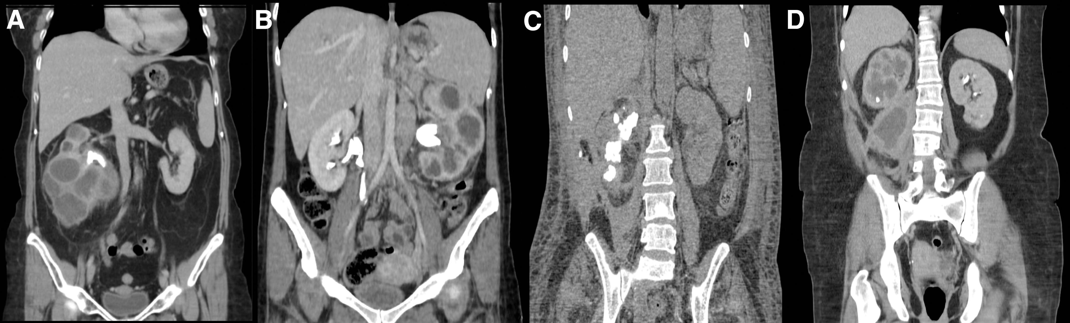
Xanthogranulomatous pyelonephritis.
Open nephrectomy (ON) is the treatment of choice for XGP. However, surgery can be challenging given the inflammatory involvement of the renal pelvis, hilum, and adjacent structures, which leads to obliteration of the tissue planes. 1,2,4 Since first reported, 5 laparoscopic nephrectomy (LN) has proven to be a less invasive alternative to ON for several renal indications and offers a number of benefits, including less blood loss, better cosmesis, decreased postoperative pain, quicker recovery, and shorter hospital stay. 3,6 –11 However, the first reports of its use in the management of XGP were discouraging, with high rates of conversion to ON and postoperative complications. 12 –16 More recent reports—as experience and skills with LN have improved—showed better outcomes, with rates of effective completion >80%, at least in appropriately selected patients. 2 –4,17 –22
More experience with the LN technique is still needed to prove feasibility, reduce morbidity, and for the adoption of this technique into routine surgical procedure for patients with XGP. We report on our experience with LN for the treatment of XGP performed by a single surgeon (A.J.) in a contemporary series of patients. Two aspects of the surgical procedure—the insertion of percutaneous preoperative nephrostomy (PPN) and placement of postoperative pelvic drainage (PD)—which have been the subject of controversy for the past decade, are further discussed here.
Methods
We retrospectively reviewed the medical records of all patients who had undergone LN between April 2010 and February 2018 at our institution (n = 17). Sociodemographic and clinical information and data on the diagnosis, presenting clinical features, surgical management, and postoperative course and complications were analyzed. The cases were classified according to the extent of involvement of the adjacent tissue using the staging system proposed by Malek and Elder (M&E). 23 Complications were graded according to the Clavien–Dindo classification. 24
Laparoscopic procedures were performed using a conventional transperitoneal four-port approach. A combined technique that uses both retroperitoneoscopic and transperitoneal approaches was chosen in cases where the xanthogranulomatous mass exceeded 15–20 cm, or when there was major involvement of the vascular pedicle caused by large adenopathy. In these cases, the retroperitoneoscopic approach allows control of the renal artery from the beginning, and then the surgery can be completed using the transperitoneal access, which in turn allows a larger working space. For the transperitoneal approach, the patient is placed in lateral decubitus position. An umbilical optical port is inserted through a small cut. The other three ports are placed under direct vision in the subcostal region and iliac fossa at the anterior axillary line for adequate triangulation of the vascular pedicle. Retroperitoneoscopic access and freeing of the perirenal adhesions are performed through the avascular line of Toldt (Fig. 2). When large renal masses are detected, the common iliac artery is located and dissected. The ureter is then found at the level of the iliac vessels. Ascending dissection is continued to reach the major vessels (aortic artery and vena cava, depending on the side). After these vessels are completely dissected, the exit of the artery or the renal vein is reached and tied up at this level by using Hem-o-Lok™ clips. LigaSure™ Maryland is used for sealing, which allows fine dissection with precise electrocoagulation and cutting of the structures to be performed. This strategy improves visualization of major vessels and facilitates laparoscopic repair in the event of a vascular accident. Dissection of the renal parenchyma is performed extracapsularly to avoid damage to the surrounding tissues. Finally, the mass is placed in a retrieval bag and extracted through a Gibson incision that connects the inferior ports of the iliac area. The decision to convert to ON is based on the surgeon's criteria. PPN is inserted only in cases where an active infection is present. PD is not routinely practiced unless renal parenchymal rupture or perinephric abscess occurs.

Placement of the four renal ports.
The safety protocol “Vascular Accident Code” was activated in all cases. This is a novel standardized work procedure developed by our center for all presurgical and intrasurgical actions in relation to the operating room's equipment and the role of each of the specialists who take part in the surgery in the event of massive vascular bleeding. 25
Statistical analysis
The analysis performed is descriptive, with continuous variables expressed as mean ± standard deviation, and categorical variables expressed as n (%). The occurrence of complications was analyzed according to M&E stages. The length of hospital stay was analyzed according to PD placement. The significance of the differences observed was not statistically analyzed, given the low number of patients and events. The SAS 9.4 statistical package was used.
Results
Patients
Mean age was 60.0 ± 13.3 years, with 58.8% (n = 10) being women. Table 1 summarizes the demographic and clinical characteristics. Fifteen (88.2%) patients presented with signs or symptoms suggestive of XGP, with fever being the most frequently reported symptom (46.7%). XGP was associated with urinary infection in 6 patients (50.0%), with Escherichia coli being the most common pathogen (66.7%), and/or renal calculi in 13 patients (76.5%). In three patients, XGP was found incidentally while performing LN for other renal diseases. Two of these patients (women aged 64 and 71 years) reported iliac fossa pain: one of them after a left ureteroscopy for ureteral fibrosis associated with ureteral stones, the other woman was referred from a private center for a LN for a suspected renal tumor. The third patient (a man aged 65 years) presented with hematuria and recurrent pyelonephritis. M&E classification was only available for the latter patient (stage II). Ten (71.4%) patients of the 14 where XGP was suspected had stage II or III according to the M&E classification. Both patients without signs or symptoms had renal calculi, and one of them also had a urinary infection and risk factors.
Valid percentages are given. M&E classification: Stage I (nephric), in which the disease is confined to renal parenchyma only; Stage II (nephric and perinephric), in which the disease process involves renal parenchyma along with perinephric fat; Stage III (nephric and perinephric), in which the disease is extending into adjacent structure or diffuse retroperitoneum. 23
Missing values: n = 2.
M&E = Malek and Elder; SD = standard deviation.
Six (40.0%) and 4 (26.7%) patients presented M&E stages II and III, respectively. In patients in whom LN was successfully performed (n = 15), only patients with M&E stage III (n = 2) presented grade 3 to 4 complications. In turn, all complications in these two patients were grades 3 to 4 and intraoperative. Patients with M&E stages I to II more often had no complications (Table 2).
Only in patients in whom LN was completed and did not experience a Grade-5 complication (n = 14). Valid percentages are given. Patients converted to open nephrectomy were excluded from this analysis. These patients were both M&E stage III, and only one had a pelvic drain placed. No nephrostomies were inserted into any of these patients. Complications of one patient were not collected. The other patient experienced abdominal collection and septic shock requiring intensive care management (CD grade 4).
Missing values = 1.
All XGPs were unilateral, with 53% (n = 9) cases being right sided.
Perioperative outcomes
LN was successfully performed in 15 patients (88.2%). Two cases (12.5%) were electively converted to ON due to failure to progress. One of these patients was a woman aged 55 years with M&E stage III disease in whom even ON could not be completed. Intraoperative or postoperative complications were not collected for this woman. It was necessary to insert a nephrostomy tube early in the postoperative period due to the occurrence of pyelonephrosis (grade 3a complication). The other patient was a paraplegic man aged 57 years who also had M&E stage III disease, who came to our office due to passage of urine through the sacral fistula, requiring emergency LN. The patient developed postoperative septic shock with respiratory insufficiency associated with abdominal collection requiring drainage and management in the intensive care unit (grade 4b complication). In both cases right-sided XGP was diagnosed by imaging.
PPN was inserted in seven (41.2%) patients. Two (11.8%) patients experienced intraoperative complications: one grade 3b and another grade 4b. Both complications were pneumothorax caused by accidental opening of the pleural cavity, the second one leading to septic shock requiring management in the intensive care unit (Table 3).
O
Valid percentages are given.
Only in patients in whom LN was completed (n = 15).
LN = laparoscopic nephrectomy.
Among patients in whom LN was successfully performed (n = 15), the mean operative time was 198.0 ± 107.1 minutes (range 100–520). Mean time was shorter in patients with no intraoperative complications 169.0 ± 48.1 minutes. Three (20%) patients were given transfusions. Nine (60.0%) patients required PD (Table 3).
Postoperative outcomes
Six (40%) patients of the 15 in whom LN was successfully performed experienced postoperative complications (Table 4). One complication was grade 1, and four were grade 2. One patient (a woman aged 71 years in whom the XGP was detected while performing LN and in whom dense adhesions were found in the vascular pedicle) experienced perisplenic abdominal collection requiring percutaneous drainage. A pancreatic fistula (grade 5 complication) due to accidental opening of the pancreatic tail mass was diagnosed, which was unsuccessfully treated with somatostatins and empiric antibiotic therapy. The patient died 18 days after the procedure due to septic shock.
Valid percentages are given. CD classification: grade 1 includes any deviation from the normal postoperative course, including wound infections opened at the bedside, without the need for pharmacological treatment (except antiemetics, antipyretics, analgesics, diuretics, and electrolytes) or surgical, endoscopic, and radiological intervention; grade 2 includes complications requiring pharmacological treatment with drugs other than such allowed for grade 1 complications, blood transfusion, or total parenteral nutrition; Grade 3 includes complications requiring surgical, endoscopic, or radiological intervention (3a under local/regional anesthesia, 3b under general anesthesia); Grade 4 includes life-threatening complication (including CNS complications) requiring intermediate care or intensive care unit management (4a single-organ dysfunction, 4b multiorgan dysfunction); Grade 5 includes death of the patient. 24
Only in patients in whom LN was performed (n = 15).
CD = Clavien–Dindo; ICU = intensive care unit.
Mean hospital stay of patients in whom LN was successfully performed was 4.4 ± 4.3 days, and 3.4 ± 2.2 days in those experiencing manageable complications (grades 1–4) or no complications (n = 14) (Table 3). Among patients with no postoperative complications (n = 6), the mean hospital stay was shorter in those in whom postoperative PD was not inserted (1.6 vs 2.6 days in patients with postoperative PD).
Discussion
Results from our retrospective study in a series of 17 consecutive patients with XGP undergoing LN from 2010 to 2018 add to the evidence on the feasibility of this surgical procedure in challenging patients in the contemporary era as recent reports show. 2 –4,17 –22 LNs were performed by a highly experienced surgeon and were able to be successfully accomplished in 15 (88.2%) patients. Moreover, the traditional benefits of laparoscopy, such as a shorter hospital stay, were available to these patients, with a mean of 4.4 ± 4.3 days for patients in whom LN was successfully performed, which was shorter (3.4 ± 2.2 days) for most patients (i.e., those experiencing manageable complications or no complications). The transfusion rate of patients in whom LN was successfully performed was 20%. Transfusions were classified as grade 2 complications.
Rates of intraoperative (11.2%) and postoperative complications (41.2%)—the latter only among patients in whom LN was successfully accomplished—are comparable with those described in other contemporary series (20%–50%). 2 –4,17 –22 One patient experienced a grade 5 complication and died within 18 days. This patient was a special case as the XGP was detected during the procedure and presented with dense adherences that highly complicated the removal. Aside from this, all other complications were effectively managed, providing further evidence on the low morbidity associated with LN. One patient experienced an intraoperative grade 4b complication, which led to an increased hospital stay. Another patient experienced an intraoperative grade 3a complication that mainly increased the operative time. In both cases, this complication was pneumothorax caused by accidental perforation of the pleural cavity.
Mean operative time in patients in whom LN was successfully performed (198.0 ± 107.1 minutes) was within the range of time reported in other contemporary series. 3,4,21 In two of these series, which compared ON and LN approaches, mean operative times were similar. 3,4 In our case, mean operative time was much greater in the two cases in which there were intraoperative complications (330 and 520 minutes) vs 169.0 ± 48.1 minutes in patients who did not experience an intraoperative complication. Mean operative time in patients requiring conversion to ON was 150 and 255 minutes, which are within the range of effective LNs in our series.
LN is the initial approach for all patients with XGP at our center, with the option of conversion to ON when severe inflammatory changes and extension of the disease preclude progression. LN offers an opportunity to better visualize the anatomic structures and safer dissection of the upper part of the kidney, which facilitates ON when this is needed. In fact, two patients were electively converted to ON. Both cases were M&E stage III and right sided, a risk factor for conversion to ON. 22 The low number of patients converted to ON in our series precludes any analysis of potentially predictive factors. It is worth noting that hand-assisted laparoscopic surgery, which has been shown to minimize the need for conversion to ON when difficulties arise, 18 is not used in our center, given its elevated cost and the low benefits it provides in surgery progression.
Some aspects related to the LN surgical procedure deserve a special consideration. The first is the use of a combined retroperitoneoscopic and transperitoneal laparoscopic technique in cases where large XGL masses were involved, or when there was major involvement of the vascular pedicle. This quite novel approach, which was first described in 2009 by Srivastava et al. 26 for large right-sided T2 renal tumors, has also been successfully employed in the laparoscopic surgical treatment of tumors with renal vein and inferior vena cava involvement. 27 We used this approach for better management of the renal artery when removing the large mass from the woman who was referred to our center with a suspected renal tumor.
The second is the insertion of a PPN. Contrary to other authors who consider PPN a facilitator for the surgical procedure, 28 PPNs are not routinely inserted in our center unless an active infection is present. PPN was inserted in seven (41.2%) patients, and LN was successfully performed in all of them. Information regarding PPN insertion before LN in patients with XGP is sparse. In a retrospective study on 17 patients with XGP who had undergone LN between 2001 and 2009, Shah et al. 4 reported PPN insertion in 10 patients. Length of hospital stay was longer for patients with nephrostomy (12.6 ± 4.6 vs 5.0 ± 4.7 for those without), which indicates the higher severity of patients selected for PPN.
The third is the placement of a postoperative PD. The so-called “tubeless” approach is the strategy of choice in our center unless renal parenchymal rupture or perinephric abscess occurs. Ten (58.2%) patients had a PD placed after the surgery. While PD placement increases postoperative pain and mobility difficulties, 29 the benefits of the “tubeless” strategy, such as early ambulation and narcotic avoidance, may contribute to early recovery by returning bowel function and preventing ileus. 30 This strategy also helps prevent the occurrence of rare but severe complications associated with PD placement or removal. 30 In our series, mean hospital stay in patients without postoperative complications (n = 6) was shorter when no postoperative PD was placed (1.6 vs 2.6 days), although the low number of patients analyzed precludes drawing conclusions.
Our report is among the largest contemporary single-surgeon series of XGP being managed with LN. Besides the inherent limitation of retrospective studies, when interpreting these results it should be noted that given the low incidence of XGP, our series of patients was small, which makes analysis sensitive to the occurrence of an event. This is a problem common to other contemporary reports. Also, the relative heterogeneity of the results may respond to the heterogeneity of the disease, ranging from an incidentally discovered renal mass (11.8% of our patients) to patients with a high degree of disease extension, who are likely to present adjacent organ involvement and concomitant infection.
Conclusion
LN is a feasible surgical option in patients with XGP although given the nature of XGP, it is inherently associated with complications—nearly all manageable—which makes it a challenging surgical procedure. Surgeons are encouraged to develop advanced laparoscopic skills and to gain broad experience. Dissemination of the disease is associated with the occurrence of more severe complications and conversion to ON. PD placement may be associated with a shorter hospital stay.
Footnotes
Acknowledgments
The authors thank Beatriz Viejo, PhD, for her assistance in the writing of the article and David Calbet for his statistical support.
Author Disclosure Statement
No competing financial interests exist.