
Abstract
Introduction:
The Moses technology for the Ho:YAG laser introduces a pulse-shape modulation that optimizes energy delivery through water and can be utilized for lithotripsy at a distance from the target. In light of this advance, we undertook an in vitro study to assess the effect of fiber tip to stone distance on fragmentation, incorporating the use of a variety of pulse modes.
Methods:
Experiments were conducted with a three-dimensional (3D) positioning system, a 30 mm flat plate BegoStone, and a 230 μm core laser fiber connected to a 120W holmium laser utilizing short pulse (SP), long pulse (LP), Moses contact (MC), and Moses distance (MD) modes. Ablation crater volume was measured by 3D confocal microscopy, after a single pulse (1.0 J) was activated with the fiber tip positioned at 0, 0.5, 1, 2, and 3 mm from the stone. Fragmentation efficiency (1 J × 10 Hz) was assessed with the fiber tip at 0 and 1 mm distance, programmed to fragment the stone over 3 minutes. Fragmentation was defined as difference in stone mass before and after each experiment.
Results:
For all tested pulse modes, ablation crater volume and fragmentation were greatest when the fiber tip was in contact with the stone. Ablation declined as the working distance increased with no ablation occurring at 3 mm. At 1 mm distance, the ablation crater volume using MD mode was significantly higher when compared with SP, LP, and MC modes (p < 0.05). Compared with all pulse modes tested, MD resulted in 28% and 39% greater fragmentation at both 0 and 1 mm working distances, respectively (p < 0.05).
Conclusion:
Holmium laser lithotripsy is significantly affected by fiber working distance with the greatest ablation obtained with the fiber in contact with the stone. At 0 and 1 mm distance, MD had the greatest fragmentation efficiency, suggesting this mode may have advantages during ureteroscopy.
Introduction
Ureteroscopy (URS) with Ho:YAG laser lithotripsy is the most common modality for urinary stone treatment in North America. 1 Holmium laser energy is highly absorbed in water and has a low penetration depth limiting the amount of energy reaching surrounding tissue. 2 Energy transmission when the laser is activated in saline is inversely proportional to the fiber tip to tissue target working distance and directly proportional to the pulse energy delivered. 2 However, studies assessing the relationship between fiber tip and stone working distance on ablation are limited. 3
The use of the term “Moses” in relation to laser ablation was first described when investigating the interaction between laser energy and fluid medium. 4 Isner et al. described the bubble that forms when laser energy is transmitted through fluid. The energy causes a “vapor tunnel [that] serves as a pathway permitting transmission of radiation between the parted seas of blood or water” hence the term the “Moses effect.” 4,5 In contrast, the “Moses technology” is a pulse modulation technique in which energy is delivered in two pulses, the first pulse to create the vapor channel and the second pulse to deliver the energy through the already formed channel. 6 This ensures that more energy is delivered to the stone compared with regular pulse mode. 7
Next-generation holmium lasers allow the surgeon to choose from multiple parameters for lithotripsy, including selection of variable pulse width and modulation modes. 8 The Moses platform has two settings, Moses contact (MC) mode—intended for operation at a close distance, and Moses distance (MD) mode—intended for lithotripsy at a distance of 2 mm. 7 Despite introduction of this new technology, it is unclear how pulse modulation changes and variable pulse width modes impact fragmentation when varying the working distance between the laser fiber and the stone. A better understanding of the role of fiber to stone working distance and selection of pulse parameters may help guide laser lithotripsy techniques during URS. Thus, the aim of this study was to assess the effect of fiber to stone working distance on ablation crater volume and fragmentation efficiency utilizing a variety of pulse modes.
Methods
Single-pulse experiments were performed to understand the relationship between pulse mode and crater ablation. Further experiments were performed with fragmentation over a period of time while maintaining a fixed working distance to understand how these parameters may be clinically relevant.
Experimental setup
Square BegoStones with a flat surface measuring 30 × 30 × 3 mm were specifically created as the stone model for this study. Laser lithotripsy was performed using a 120W holmium laser (Moses P120; Lumenis, San Jose, CA) and a 230 μm core laser fiber (Moses 200 D/F/L; Lumenis). The laser fiber was attached to a three-dimensional (3D) positioner system (Velmex, NY) using a fiber chuck to allow for precise fiber positioning during the experiments. Energy was delivered utilizing short pulse (SP), long pulse (LP), MC, and MD modes for all experiments.
Ablation crater
BegoStone composition was 15:3. Stones were hydrated in deionized water for 12 hours before each experiment. A single pulse of 1 J utilizing SP, LP, MC, and MD modes was delivered from a laser fiber placed at various distances (0, 0.5, 1, 2, and 3 mm) from the stone in a grid of locations. Five trials were performed for each fiber position and pulse mode. The fiber was stripped and cleaved with a ruby scribe before each trial. By using the lowest pulse frequency possible (5 Hz), and a quick tap on the foot switch, a single pulse was delivered. Single pulses were confirmed by high-speed imaging at 10,000 frames per second (Photron Fastcam SA1.1, Tokyo, Japan). Ablation crater volume, depth, and area at the stone surface were measured by a 3D laser confocal microscope (LEXT OLS 4000; Olympus, Tokyo, Japan).
Fragmentation efficiency
Since completely smooth and flat horizontal stones were important to maintain consistent laser fiber to stone working distance during fiber movement, BegoStone composition of 15:5 was used to allow the material to be poured in an injection mold. Dry weight of the stone was measured and then stones were hydrated in deionized water 12 hours before each experiment. Two laser fiber to stone distances were assessed, 0 and 1 mm. At each laser fiber position, fragmentation was assessed at 1 J × 10 Hz (10W) utilizing SP, LP, MC, and MD modes. The 3D positioner system was programmed using MATLAB (MathWorks, MA) to advance at a speed of 1 mm/s to make 10 lines. Each line was 20 mm long with 2 mm spaces in between; the fiber was checked and repositioned before each line to account for fiber tip degradation and maintain fiber to stone distance. This was done to simulate “painting” technique during laser lithotripsy. 9 During each experiment, a total energy of 2 kJ was delivered over 200 seconds. Stones were desiccated in a dry environment for 24 hours before being weighed. Five trials were performed for each fiber position and pulse mode. The fiber was stripped and cleaved with ruby scribe before each trial. Fragmentation was defined as difference in stone mass before and after each experiment.
Additional experiments
To demonstrate differences in vapor bubble shapes between SP, LP, MC, and MD modes, we performed high-speed imaging at 30,000 frames per second (Photron Fastcam SA1.1, Tokyo, Japan). In addition, power output was measured using a pyroelectric energy meter (FPE80BF, Ophir, UT) to determine the amount of energy delivered for each pulse mode during the experiment. Since the Moses modes deliver the energy over two pulses, we were interested in determining if the Moses modes deliver the same amount of energy as SP and LP modes. Furthermore, the optical pulse profile was assessed using a photodetector (DTE05D2, Thorlabs, NJ) to determine pulse duration for each pulse mode. MATLAB (MathWorks, MA) was used to reconstruct the optical pulse profiles to accurately determine the pulse duration.
Statistical analysis
Student's t-test was used to compare cohorts' mean (R statistical computing v3.4). The probability of a type I error was set at 0.05 and all testing was two sided.
Results
Ablation crater
Figure 1 presents mean ablation crater volume data for each pulse mode at different fiber to stone working distances. For all pulse modes tested, the crater volume decreased as the working distance increased, with no ablation occurring at 3 mm. At 0.5 mm, the ablation volume was significantly lower than at 0 mm distance (p < 0.0002). At 0 mm distance, MC mode resulted in significantly larger stone craters than LP and MD modes (p < 0.03), and nearly reached significance compared with SP mode (p = 0.06). At 0.5 mm and 1 mm distance, SP produced significantly smaller craters than all other pulse modes (p < 0.01). At 1 mm distance, the mean crater volume when using MD mode was significantly greater when compared with the other pulse modes (p < 0.01). At 2 mm distance, while LP and MD produced significantly larger craters, the differences were small from a clinical perspective (p < 0.002). While there were no significant differences in crater volume between LP and SP when the fiber was in contact with the stone (0.111 vs 0.116 mm 3 ; p = 0.52), LP was superior to SP at 0.5, 1, and 2 mm working distances (p < 0.01).
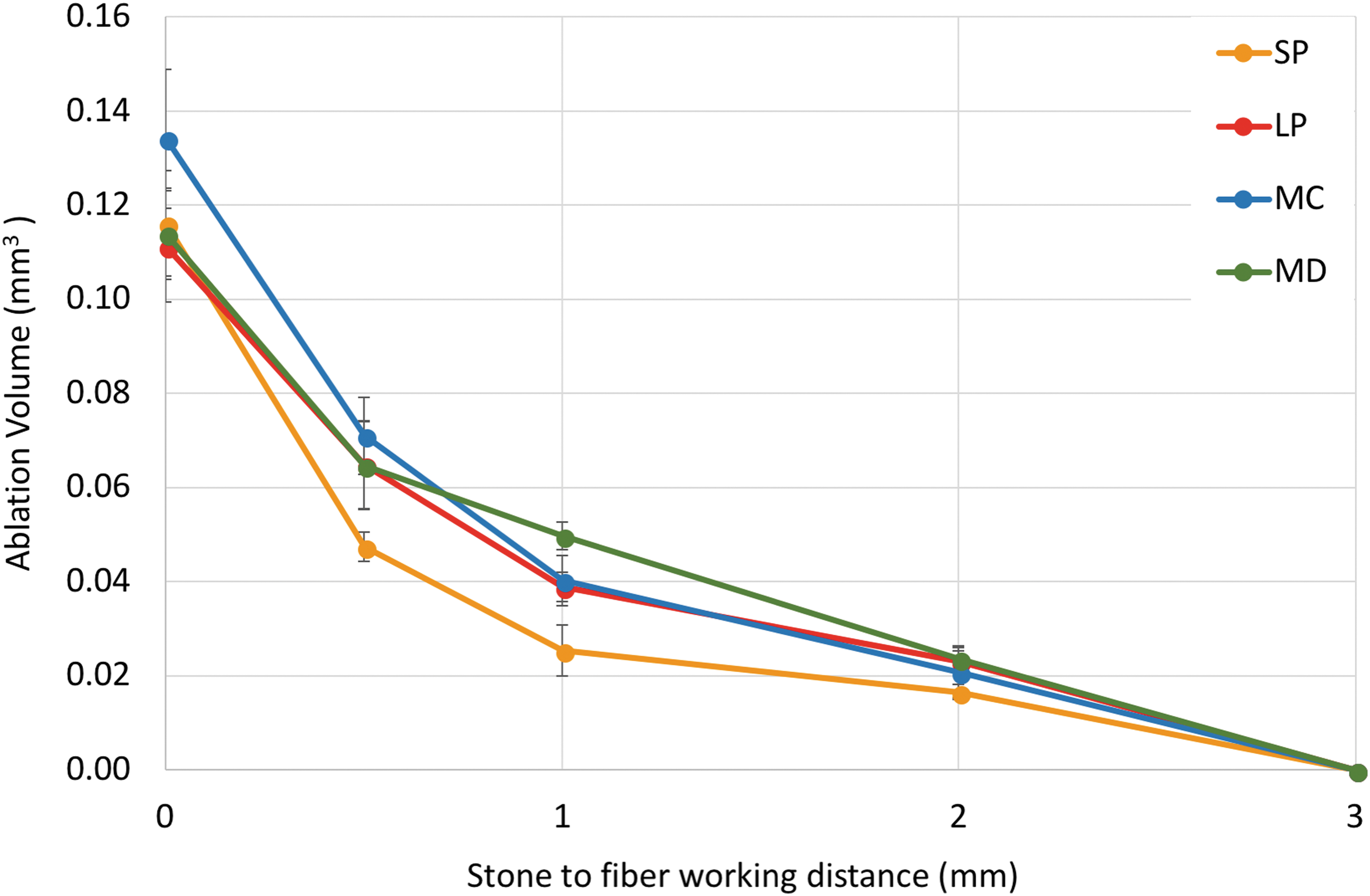
Mean crater volume following single pulse at 1.0 J at different laser fiber to stone distances utilizing SP, LP, MC, and MD modes. LP = long pulse; MC = Moses contact; MD = Moses distance; SP = short pulse.
Crater depth and crater area are presented in Tables 1 and 2, respectively. Crater depth decreased with distance for all pulse modes. The deepest crater when at 0 mm distance was demonstrated when using the MC mode, while at 1 mm fiber distance, MD mode had the deepest crater (Table 1). For crater area, SP mode had the largest area at 0 mm distance and the area decreased with increasing fiber to stone working distance. For MC mode, the area peaked at 0.5 mm distance and decreased as the working distance increased (Table 2).
Crater Depth Following Single Pulse (1.0 J) at Different Laser Fiber to Stone Working Distances Utilizing Short Pulse, Long Pulse, Moses Contact, and Moses Distance Modes
p < 0.05: SP vs MC, LP vs MC, MC vs MD.
p < 0.05: SP vs LP.
p < 0.05: SP vs LP, SP vs MC, SP vs MD.
p < 0.05: MD vs SP, MD vs MC.
LP = long pulse; MC = Moses contact; MD = Moses distance; SP = short pulse.
Crater Area Following Single Pulse (1.0 J) at Different Laser Fiber to Stone Working Distances Utilizing Short Pulse, Long Pulse, Moses Contact, and Moses Distance Modes
p < 0.05: SP vs LP, SP vs MC, SP vs MD.
p < 0.05: SP vs LP, SP vs MC, SP vs MD, LP vs MC, MC vs MD.
p < 0.05: SP vs MD.
p < 0.05: SP vs LP, LP vs MD.
Fragmentation efficiency
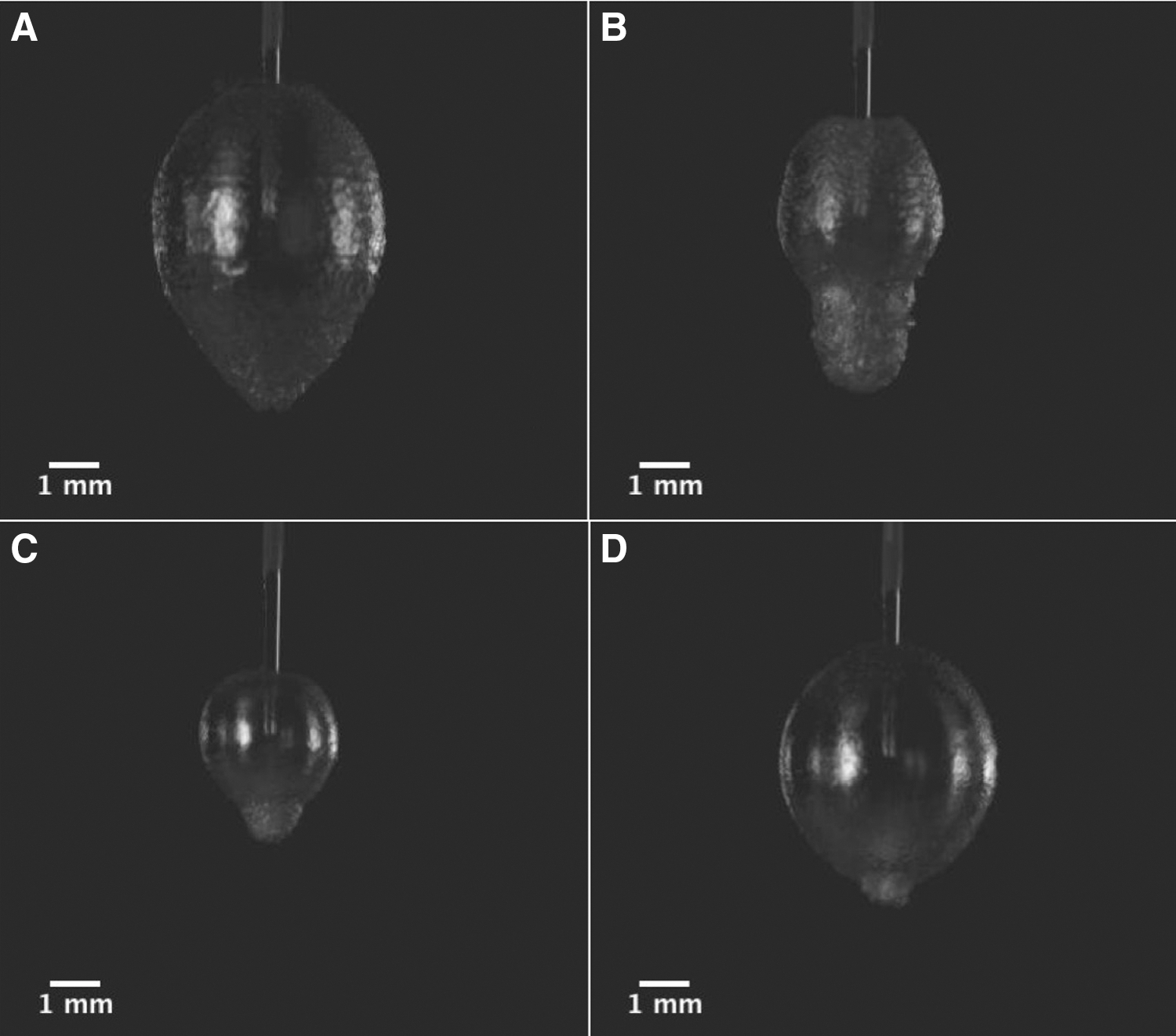
Initial stone mass was similar for all experiments (mean 5.0 g; range 4.7–5.1 g). Compared with 1 mm working distance, fragmentation was greater when the fiber tip was in contact with the stone (0 mm distance) (p < 0.0002; Fig. 2). When the working distance was 1 mm, fragmentation was reduced by 34% to 40% depending on the pulse mode. However, MD resulted in 28% and 39% higher fragmentation compared with all other pulse modes at both 0 and 1 mm fiber to stone distances, respectively (p < 0.002). At 0 and 1 mm working distances, fragmentation outcomes were not statistically different between SP, LP, and MC modes (p > 0.05). Figure 3 provides an example of a crater produced after a single-pulse experiment, as well as the flat plate BegoStone following controlled fragmentation with the 3D positioner.
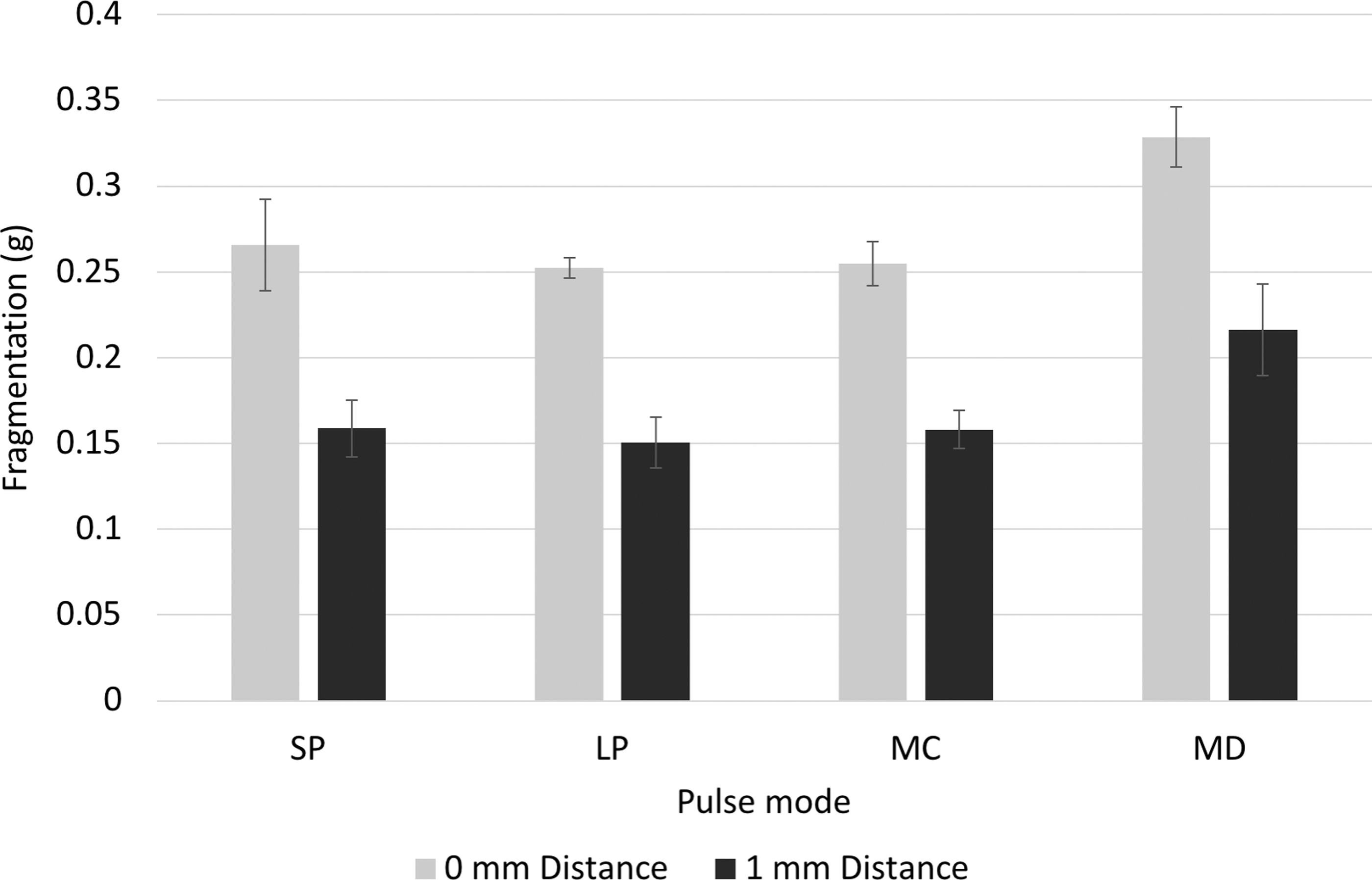
Mean fragmentation (mass loss) for SP, LP, MC, and MD modes at different fiber to stone distances when using a laser setting of 1.0 J × 10 Hz.

Additional experiments
Each pulse mode produced different vapor bubble shapes. SP produced an asymmetric bubble that is close to being rounded, while LP produced an asymmetric bubble that is pear shaped. MC and MD modes produced initial bubbles that resembled the SP bubble but were smaller in size with a second asymmetric bubble projecting from the tip of the initial bubble when it reached its maximum expansion. Furthermore, the size of the initial bubble was smaller for MC mode compared with MD mode, while the second bubble of the MD mode appeared to reach a greater distance. Figure 4 demonstrates still images of the fully expanded vapor bubbles of all four modes, and video clips are provided as Supplementary Video S1.
Still images of the SP
Optical pulse profiles for the different pulse modes are illustrated in Figure 5. SP was delivered over a total duration of 265 μs and LP over 390 μs. However, 90% of the pulse on SP and LP modes was delivered over 160 μs and 260 μs, respectively. With regard to Moses modes, MC had an initial pulse that was 100 μs and a second pulse of 285 μs, that is, 385 μs in total. MD had an initial pulse that was 200 μs and a second pulse of 240 μs. Moreover, there was a 100 μs interval between the first and second pulse for MD mode, making the total duration 540 μs. Figure 5 demonstrates differences in the pulse profile for the initial and second pulse for MC and MD modes. Regarding the total pulse energy, all pulse modes produced equivalent energy of 1 J when tested with the power meter.

Optical pulse profile for SP, LP, MC, and MD modes measured at 1 J.
Discussion
We studied the relationship of the laser fiber to stone working distance on two endpoints of laser lithotripsy. First, we assessed how much stone volume was ablated after a single firing of the Ho:YAG laser at 1 J of energy. We found that the stone crater volume is significantly affected by the working distance. For all pulse modes tested, the greatest ablation volume is produced when the fiber is in contact with the stone. At 1 mm working distance, MD mode produced deeper stone craters with larger volume. Second, we studied fragmentation efficiency using different pulse modes with the fiber tip either in contact with the stone (0 mm) or 1 mm working distance. We found that for both 0 and 1 mm fiber to stone distances, MD mode resulted in a significantly higher fragmentation compared with all other pulse modes.
Our findings are consistent with previous studies that have demonstrated that holmium laser energy transmission in saline is inversely proportional to the distance from the target tissue. 2,3 van Leeuwen et al. showed that energy transmission following a pulse of 1 J was 80%, 75%, 45%, and 25% at distances of 0.5, 1, 2, and 3 mm, respectively. The percentage change in stone crater volume in our study is lower than the transmission data reported by van Leeuwen et al.; however, the difference could be attributed to the fact that only part of the energy reaching the stone gets absorbed. 10 The influence of working distance on laser ablation has also been qualitatively assessed when using the thulium fiber laser. 3 Less stone ablation was observed with high-speed imaging when the working distance was increased to 0.5 mm and 1 mm.
We found that MD mode results in superior fragmentation efficiency when used both in contact and at 1 mm working distance compared with LP, SP, and MC mode. Contrary to what we expected to find, there were no significant differences when comparing MC mode with SP or LP for fragmentation efficiency. Elhilali et al. were the first to report on differences in fragmentation when using SP compared with Moses modes. 7 They found that the Moses mode resulted in significantly higher ablation volume compared with SP at 1 mm distance. However, they did not assess ablation at contact with the stone and did not specify which Moses mode (distance or contact) was used in their study.
Our data demonstrate that MD leads to superior fragmentation, even when the stone is in contact, and is a significant new finding. Furthermore, our study did not find any significant difference between SP and LP modes. The majority of studies that have assessed this have found no difference in fragmentation between SP and LP modes, 11 –13 while two studies found SP to be superior to LP mode, 14,15 with a further study demonstrating LP mode to be superior to SP mode. 16 We found an increase in the crater area when positioning the laser fiber at 0.5 mm compared with 0 mm distance for LP, MD, and MC modes but not SP mode. This may be because the laser beam diverges when exiting the fiber causing the energy to reach a larger area when the fiber is separated from the stone, compared with when it is in contact with the stone surface. 17 Furthermore, MC mode produced the largest area at 0.5 mm distance but with lower crater width-to-depth ratio compared with LP and MD modes. This might be because MC mode has a smaller and more focused initial bubble with most of the energy delivered during the second pulse thus increasing the fluence at the stone surface.
The difference in findings between the ablation crater volume and fragmentation experiments in our study can also be explained. The amount of stone material that can be removed from a single stone location would increase when increasing the number of pulses, that is, frequency. However, after a limited number of pulses with the fiber at a fixed distance, no more ablation can be achieved at that location. 18 If a limit is reached, then this might explain why there were no differences between SP, LP, and MC modes when assessing fragmentation efficiency. Our analysis of the crater dimensions showed that MD mode produced deeper craters compared with other pulse modes at 1 mm distance. One hypothesis is that MD mode results in greater ablation because the lines created by a moving laser fiber that is continuously firing at constant distance from the upper stone surface will be flooded with water. Water will become more obstructive to laser energy transmission as the line deepens and reduces fragmentation efficiency for SP, LP, and MC modes more than MD mode.
This study has several limitations. Because experiments were conducted using fixed stone blocks with smooth horizontal surfaces, to do a consistent evaluation and obtain accurate results, retropulsion and how it may affect fragmentation could not be assessed. Despite the two different BegoStone compositions used, evaluations and comparisons were always done within samples of the same composition, although preliminary data were similar for the two.
The implications of our in vitro study are that for the best fragmentation efficiency, the most important concept when performing laser lithotripsy is to position the laser tip in contact with the stone surface. Just being 1 mm off the stone in SP mode resulted in a 40% reduction in fragmentation. The actual percentage of time a surgeon spends firing the laser in contact with the stone is not known and may vary considerably. For those with access to the Moses technology, based on our findings, we recommend utilizing the MD mode for contact laser lithotripsy as opposed to the MC or other pulse modes. This ensures that fragmentation is maximal, both in contact and when the distance between the laser fiber and stone changes. This may have clinical benefits during ureteroscopic laser lithotripsy, especially when using dusting techniques such as chipping and dancing that require constant back and forth movement on the stone surface. 9,19 Future work is needed to understand the role of pulse width/modulation and working distance on stone retropulsion.
In conclusion, holmium laser lithotripsy is significantly affected by the fiber tip to stone working distance, with the greatest ablation volume obtained with the fiber in contact with the stone. Use of MD mode in contact with the stone, and at 1 mm distance from the stone, led to the greatest fragmentation, suggesting this mode may have clinical advantages during
Footnotes
Acknowledgments
This work was performed, in part, at the University of Michigan Lurie Nanofabrication Facility. This study was supported by a scientific research grant from Boston Scientific.
Author Disclosure Statement
Khurshid R. Ghani is a consultant for Lumenis and Boston Scientific.
Abbreviations Used
References
Supplementary Material
Please find the following supplemental material available below.
For Open Access articles published under a Creative Commons License, all supplemental material carries the same license as the article it is associated with.
For non-Open Access articles published, all supplemental material carries a non-exclusive license, and permission requests for re-use of supplemental material or any part of supplemental material shall be sent directly to the copyright owner as specified in the copyright notice associated with the article.