
Abstract
Introduction:
Prestenting of the ureter is commonly performed to allow for passive dilation and better access to the urinary system during subsequent procedures. There is no level 1 evidence on the duration of prestenting and EAU guidelines suggest a 1–2 weeks duration.
Materials and Methods:
Our primary aim is to investigate the optimal duration required for prestenting in a porcine model. Our secondary aim is to compare the ureteral wall compliance between the stented and the unstented ureters. Methods: Three female pigs between 40 and 50 kg were used. We modified a human protocol for performing intravenous pyelograms in our study to obtain ureteral measurements on days 0, 5, 7, and 14. Unilateral stenting on days 0, 5, and 7 was performed. On day 14, bilateral nephroureterectomy was performed, and ureteral compliance was measured in the stent and unstented ureter.
Results:
There were significant ureteral dilation between days 0 and 5 for all three pigs (p 1 = 0.001, p 2 ≤ 0.001 and p 3 = 0.01). The rate of dilation appears to plateau after day 5 (p 1 = 0.416, p 2 = 0.344, and p 3 = 0.774). Ureteral compliance in the stented ureter is better than in a nonstent ureter (p 1 = 1.44 vs 0.13, p 2 = 0.8 vs 0.04, p 3 = 0.62 vs 0.2). An unexpected observation was the ureteral dilation and increased tortuosity in the unstented ureter in two of the three pigs (p 1 = 0.152, p 2 = 0.007).
Conclusion:
Our results suggest that optimal prestenting may be achieved in 5 days in a porcine model. It can potentially form the basis to start randomized human trials.
Introduction
Ureteral stents are an integrated part of any urologic practice. Prestenting of the ureter is commonly performed to allow for passive dilation of the ureters in case of difficult access to the ureters. 1 –3 A dilated ureter provides for better access to the proximal urinary system in subsequent procedures. In a large multicenter international study evaluating prestenting for ureterorenoscopy, prestenting is associated with the lower complications rate, and higher stone-free rate (SFR). 4 However, stent discomfort occurs in up to 80% of patients and is a significant source of stent-related morbidity for patients requiring ureteral stent placement; therefore, it is essential to minimize the duration of prestenting. 5
There is currently no consensus on the optimal duration required for prestenting in the adult population. Current international guidelines, institutional practice, and expert opinions suggest a period of 7–14 days or has remained silent entirely. 3,6 To our knowledge, there is currently no study looking at the time required to achieve sufficient ureteral dilation for subsequent procedures. Our study aims to look at the optimal duration needed for prestenting and investigate the effects ureteral prestenting has on the ureter in a porcine model.
We performed unilateral stenting in pigs and measured the ureteral diameter for a period of 14 days using intravenous pyelograms (IVP). After baseline IVP measurements, we randomly selected a ureter for stenting. On day 5, the stent was removed from the animal, IVP performed, and restenting performed. We repeated this process subsequently on days 7 and 14 before we euthanize the pig to harvest the ureters and kidneys for pressure–flow study.
Materials and Methods
Three female Yorkshire-Landrace pigs weighing between 40 and 50 kg were used in this Institutional Animal Care and Use Committee (IACUC)-approved project. Each pig underwent 1 week of mandatory acclimatization before undergoing general anesthetic by a fully qualified veterinary.
On day 0, we performed an IVP to obtained baseline ureteral measurements. As there are no established protocols for animal intravenous pyelography, we modified a human protocol for our purposes. After anesthesia was given, an elastic body binder was applied to compress the ureters at the lower flanks to improve contrast collection in the renal pelvis (Fig. 1). An iodine-based contrast was given as a bolus infusion (calculated at 2 mg/kg) through an intravenous cannula placed at the right ear. The body binder was released, and a series of IVPs were obtained 15 minutes after bolus infusion was given.
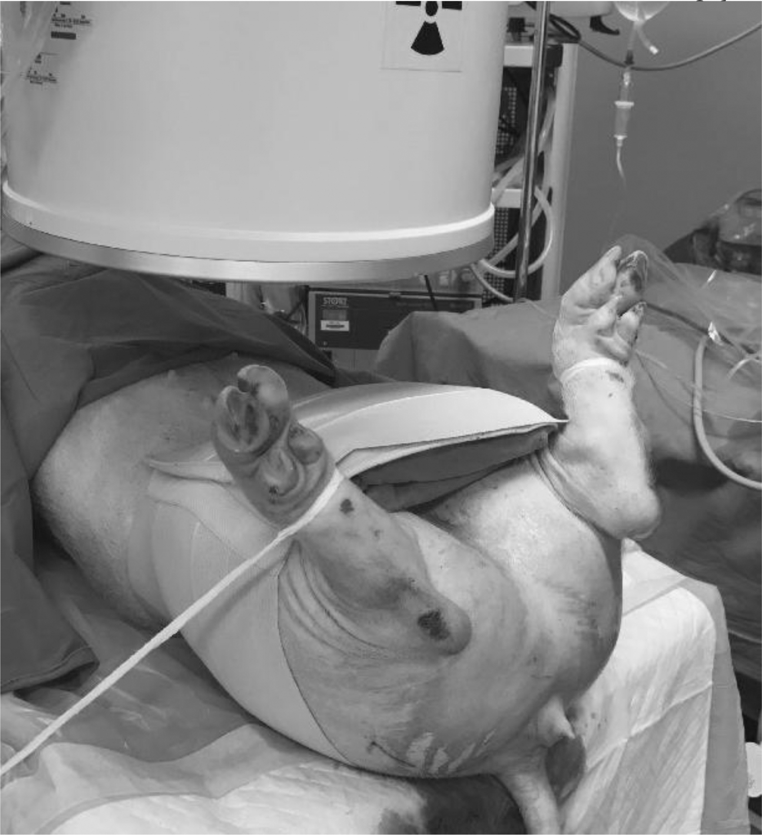
Abdominal binder.
A rigid cystoscopy was then performed using a cystoscope (Karl Storz, Germany, 22F, 22 cm 30°) and a randomly selected side was stented with a Polaris 4.8F × 26 cm stent (Boston Scientific) under imaging guidance. The pigs were monitored poststenting for behaviors suggesting of discomfort or pain after each IVP and stenting session. Water source was continuously available to prevent dehydration and acute renal damage.
On days 5, 7, and 14, the indwelling stent was removed, and IVP was repeated using the method previously described.
Analysis and statistics
The IVP image was then read by a blinded member of the team who is unaware of the side of stenting. Each ureter was measured at four consistent levels using the midline vertebral body as the reference (Figs. 2 and 3) on days 0, 5, 7, and 14. At least three readings were obtained, and the mean value was calculated for each level. These four measurements were again averaged to obtain the mean ureteral diameter of each side on days 0, 5, 7, and 14. The difference in each ureteral diameter across the 14 days was analyzed using mixed linear model. The software R was used for the analysis. 7

Intravenous pyelogram day 0.
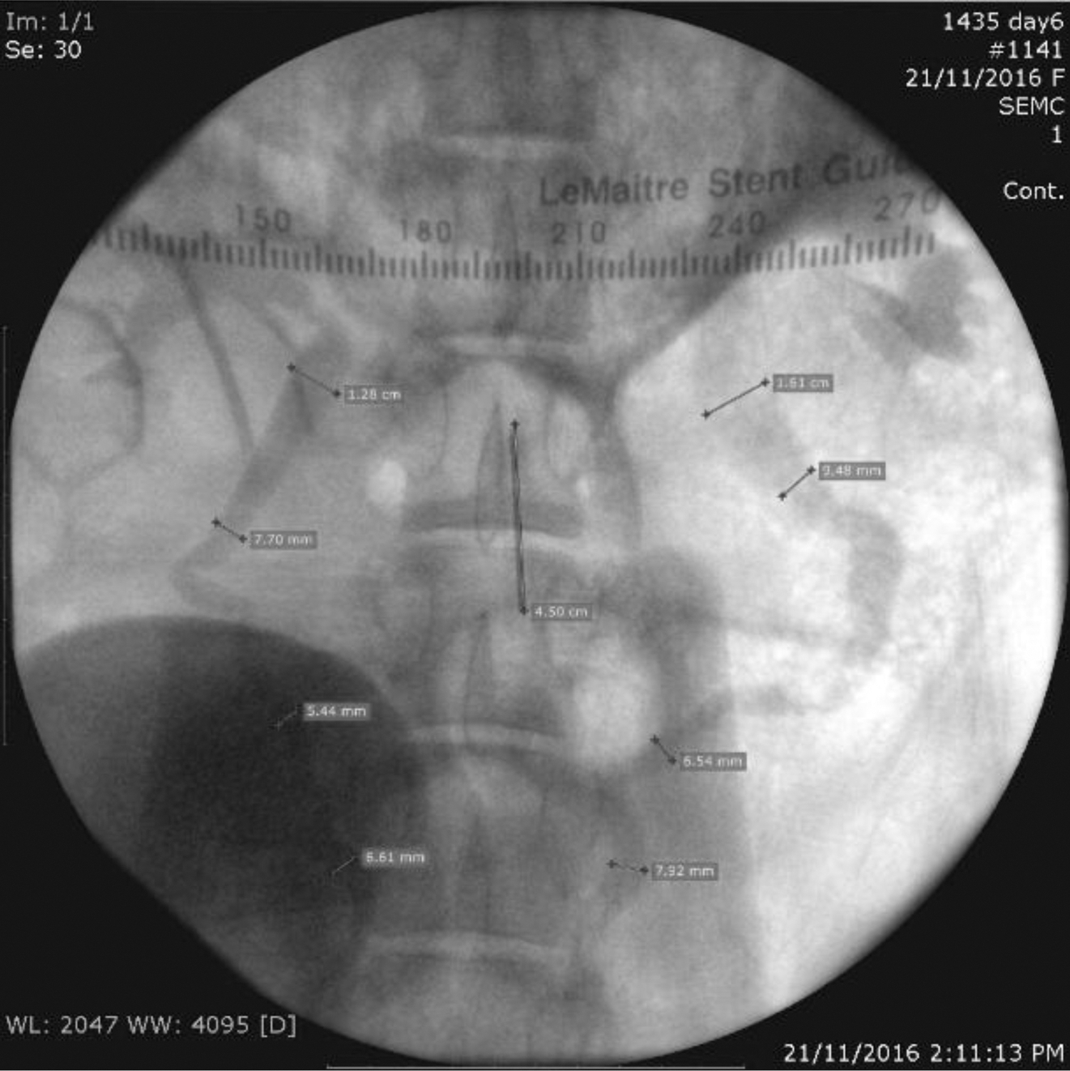
Intravenous pyelogram day 5.
On day 14, the animals were euthanized after IVP and bilateral laparoscopic nephroureterectomy was performed. A volume–pressure study was conducted on the freshly harvested kidney and ureter within 1 hour after harvest to maintain tissue integrity. A 19G epidural catheter, connected to a pressure conductor (Argon Medical Devices), and an infusion catheter were placed into the distal ureter and secured with a silk tie. Methylene blue-stained saline at room temperature was used as the infusion medium to ensure any leakage (which will result in a drop in pressure) can be detected immediately (Fig. 4). The infusion medium was slowly hand infused into the ureter and pressure reading was obtained until the pelvic caliceal ruptured. Ureteral compliance is defined as volume divided by pressure and was calculated using MMS software v9.1 (Medical Measurement Systems B.V., The Netherlands) for each kidney and ureter.

Ureteral compliance setup.
Results
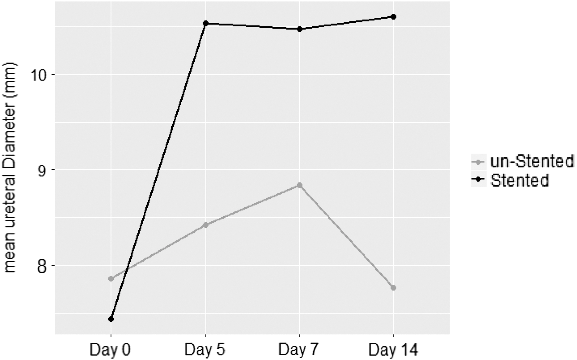
All three pigs effectively underwent 14 days of stenting and were euthanized uneventfully. Two pigs had their right ureters stented, and the remaining pig had the left ureter stented for 14 days. Table 1 gives the mean bilateral ureteral measurements across 14 days for each ureter of each pig, p-values were derived after comparison against day 0. Tables 2 –4 show the mean change in ureter diameter estimated from linear mixed model with different reference level. Figure 5 shows the trend of overall mean ureteral diameter for the 14 days.
Mean ureteral diameter (mm).
Mean Ureteral Diameters 0
Denotes stented side mean ureteral diameter.
Bold type indicates statistically significant values.
NS = not significant.
Compared Against Day 0
Bold type indicates statistically significant values.
Compared Against Day 5
Bold type indicates statistically significant value.
Compared Against Day 7
Bold type indicates statistically significant value.
At day 5, compared with baseline measurements, the diameter of stented ureter significantly increased ∼3 mm on average (Table 2) from a mean ureteral diameter of 7 mm (Fig. 5).
When compared with day 5, there were no significant increases in ureteral diameter after either 2 or 7 additional days of stenting. On day 14, there was a reduction (0.7 mm) in the diameter of the unstented ureter.
Similarly, compared with that on day 7, there is no significant variation in mean diameter of stented ureters. Again there was a small significant decrease observed in ureteral diameter in the unstented side.
Ureteral compliance
Ureteral compliance was measured immediately after the kidneys and ureters were harvested. Table 5 gives the results of the volume–pressure study. Compared with the unstented ureter, we found that in the stented side, a significantly larger volume of fluid can be accommodated in the kidney and ureter before pressure increases substantially. This translates into better elasticity of the ureteral tissue, which can accommodate a larger volume of fluid before any rise in pressure.
Ureteral Compliance
Denotes stented side.
Discussion
Prestenting is effective in improving stone removal rates. Jones et al. first reported that a preexisting stent was associated with improved stone clearance in 1990. 8 They found that by placing a ureteral stent after an uneffective stone removal attempt increases the likelihood of effective stone removal in a subsequent setting. This finding was collaborated in 2005 in the pediatric population by Hubert and Palmer. 9 In the second study, the prestenting duration was between 2 and 8 weeks with 3 weeks being the median duration. Rubenstein and coworkers also reported that prestenting increases SFR with minimal complications. 10 Several authors have since shown similar improvement in SFRs, although a recent systemic review did not find any improvement in operating time or complication rates. 11 –13 However, ureteral stenting comes with its morbidity and can affect patient comfort. Joshi and colleagues report up to 80% of patient-reported urinary symptoms and pain that interfered with daily activity and decreased quality of life. 5
There has been a significant amount of interest in using stent design, stent coating, drug-eluting stents, or medications to reduce stent discomfort. 14 –16 However, no modality has been shown to eliminate stent discomfort, and minimizing the stent duration remains the best and proven way to reduce stent discomfort.
There has been no human study on the optimal duration of prestenting caused by unnecessary exposure to radiation. International guidelines suggest a prestenting period of between 7 and 14 days as the duration required for optimal ureteral dilation. To the best of our knowledge, this recommendation is based on expert opinion, and institutional behavior and the optimal duration of prestenting are unknown.
We performed unilateral stenting on pigs to study the temporal effect of ureteral dilation for 2 weeks. Also, we intentionally stented a single side to account for animal variation, and at the same time, study the impact of prestenting (if any) on the unstented side, which is not routinely investigated in clinical practice.
Our results showed that the stented ureter undergoes significant dilation between days 0 and 5. Although there are varying degrees of ureteral dilation up to day 14, the magnitude plateaus after the initial 5 days. The increased dilation was statistically significant across all three pigs when compared against baseline reading. Although analyzed individually, Pig 1 continued to have significant dilation beyond 7 days of prestenting. However, combined analysis of all three pigs failed to show any significant dilation beyond day 5. Our result strongly suggests that a period of 5 days may be all that is necessary to reach optimal dilation and achieve higher SFR as reported in other studies. We are currently unaware of any evidence suggesting that an earlier prestenting duration of <5 days is sufficient to achieve optimal dilation. A period of 5 days was selected so as to reduce the amount of general anesthesia that a human patient will undergo within a short period of time in future studies. With our results, however, we aim to conduct future studies looking at stent dwell times of 5 days or less.
In addition to ureteral diameter measurement, we performed ureteral compliance to provide a quantitative measurement of the degree of ureteral dilation. Ramsay et al. have shown that prestenting in the ureter causes a reduction in ureteral peristalsis because of retroperistalsis, decrease in the frequency, and incomplete coaptation and can lead to pooling of urine in the ureters. 17 The observed ureteral dilation in our study may be caused by pooling of urine in the ureter rather than the actual increase in capacity of the ureter. As the results in Table 5 shows, there is a significant difference in the volume–pressure relationship between the stented and unstented side. Although ureteral compliance is only an improvisation of bladder compliance, it shows that stented ureters can tolerate a larger volume of fluid before pressure rises. The calculation reflects a breakdown in myogenic resistance to increased capacity rather than stasis alone. We know that ureteral peristalsis is myogenic driven and this brings to question the specific microscopic alteration that occurs in the muscularis layer that may explain for increased compliance and change in peristalsis.
Recent work by Janssen and colleagues found that expression of the Sonic Hedgehog (Shh) signaling pathway and downstream protein expression of Gli1 differ between the stented and unstented ureter. 18 An interesting observation that was noted in our study for the 2-week duration was the increase in ureteral diameter on the unstented side. Although not statistically significant, Table 1 shows that the unstented ureter in pigs 1 and 2 also underwent a small degree of dilation, which is evident on IVP as well. This is an unexpected finding, and we hypothesize that the presence of a foreign body (stent) triggers a systemic response and results in dilation bilaterally. This unknown pathway may identify biomarkers that can either be harnessed to detect sufficient ureteral dilation without the need to physically place a stent in situ or lead to the discovery of a bioactive agent directly responsible for ureteral dilation. Our understanding of ureteral physiology is incomplete, and we hope to bridge the knowledge gap through further study of histopathology on animal ureters.
There are various drawbacks in our study that need to be addressed. The choice of animal was affected by availability, cost, and logistics to achieve an as-best mimic of an adult human. The porcine model is a surrogacy of the human urinary system although there are similar macro- and microscopic architecture, distribution of neuroreceptors, intraureteral pressure, and electrophysiology. 19,20 Physiologic studies on the porcine model have been performed recently as a surrogate to improve our understanding of the human ureter. 18 The number of animals in our study is also small, but we argue that even at such sample size, a robust statistical difference is present.
In our study, ureteral compliance was measured ex vivo because of the lack of dedicated animal pressure instruments. As such, each kidney–ureter unit was transported separately in physiologic saline medium immediately to an offsite location for compliance measurements within 15 minutes of harvest. Although the optimal conditions would be to perform ureteral compliance measurements in vivo to maintain neural control and smooth muscle tone, we believe our results are still valid as both unstented and stent kidney–ureter measurements were conducted under the same laboratory conditions. Despite these limitations, we believe the study result reflects human physiology and provides a basis for which randomized studies can be performed on human subjects.
We acknowledge the presence of dynamic variables that can impact on the outcome of our study and we tried to account for variability where possible. Owing to the dynamic nature of peristalsis, multiple images were taken to best show ureteral diameter. We tried to select images that showed maximal dilation to obtain the ureteral measurement. Another dynamic variable is the level of which ureteral measurement was taken. There was increased tortuosity of the ureters for the 2-week periods, and to achieve consistency we used vertebrate as a landmark for measurement. This may be suboptimal as it may not be the similar point of previous measurements. The final dynamic variability is the pressure afforded by the abdominal binder during bolus infusion of contrast to improve contrast hold up.
Reduction of the stent duration remains the only proven way of reducing stent symptoms. Our data suggest that the duration of prestenting is ∼5 days. This is a significant reduction that can significantly improve the quality of life for patients requiring ureteral prestenting. The data from our study may form the basis at which human studies may be conducted alongside validated stent symptoms questionnaires.
Conclusion
We have shown that the optimal duration of prestenting is 5 days in our animal model. Large-scale prospective human studies are required to provide conclusive evidence on the duration of prestenting in humans, which have a potential to improve quality of life in patients requiring prestenting.
Footnotes
Acknowledgments
This study was made possible through a grant of SGD$25,000 (SHEF -28170930) from the Singhealth Foundation Grant, Singapore Health Services. We would like to thank Singhealth Experimental Medicine Centre and Mr Fong Ngan Keong, Department of Diagnostic Radiology, Singapore General Hospital, for their contribution to the project.
Authors' Contribution
K.S.L. was responsible for design, application of funding, conduct of laboratory experiments, drafting of article, and data analysis. Y.W.L., D.Z.P.Y, and Y. Hao conducted data analysis; L.G.N. was involved in urodynamic and design of the trial; H.S.S.H. supervised the study; and A.S.P.S. was involved in design of trial and supervision.
Author Disclosure Statement
No competing financial interests exist.