
Abstract
Objective:
To assess optical performance and diagnostic capability of the Endockscope system (ES) vs the standard endoscopic system (SES) using four rigid/semi-rigid endoscopes. The ES combines a smartphone, lens system, and a rechargeable light-emitting diode (LED) light source to provide a low-cost alternative ($45) to the standard camera and high-powered light source ($45,000) used in endoscopic procedures.
Materials and Methods:
Video clips (<20 seconds) of standard rigid nephroscopy, semi-rigid ureteroscopy, rigid cystoscopy, and laparoscopy in two adult male cadavers were recorded using the ES combined with either the Apple iPhone X or Samsung Galaxy S9+ and also with the high-definition SES (Karl Storz). Sixteen urologists blinded to the camera modality assessed the image resolution, brightness, color, sharpness, and overall quality using a Likert-type scale; acceptability for diagnostic purposes was judged on a binary scale (yes/no).
Results:
For rigid cystoscopy, there was no statistical difference between both ES systems and the SES. For semi-rigid ureteroscopy the two ES systems performed equal to or better than the SES. For rigid nephroscopy, the ES plus Galaxy was comparable to the SES, except in brightness (p < 0.05), whereas the ES plus iPhone was inferior in various parameters. For laparoscopy, the ES plus Galaxy was inferior to the SES in brightness and overall quality (p < 0.05); the ES plus iPhone was inferior for all laparoscopic image parameters compared with the SES. For diagnostic purposes, the ES plus Galaxy was equivalent to the SES for all endoscopes; the ES plus iPhone was equivalent to the SES for cystoscopy, ureteroscopy, and nephroscopy.
Conclusion:
The ES plus the Apple iPhone X or Samsung Galaxy S9+ offers comparable imaging and provides diagnostic information equivalent to the standard system for rigid endoscopy of the kidney, ureter, and bladder; the Galaxy S9+ provides comparable imaging and diagnostic capabilities for evaluation of the abdomen.
Introduction
Globally, many urologists do not have access to state-of-the-art endoscopes secondary to financial limitations. 1 This creates an obstacle for urologists in impoverished countries and directly contributes to the vast inequities in health care. 2 For example, flexible ureteroscopes are the urological standard for upper tract endourological procedures; however, given the immense cost of these fragile endoscopes and the need for frequent repair, this technology is often unavailable in impoverished nations. 3 On average, a flexible ureteroscope costs upward of $29,000, whereas rigid ureteroscopes, which are far more durable but typically are relegated to the distal/mid ureter, cost ∼$6500. 4 Aside from the expense of the endoscope, the standard camera and high-powered light source add $45,000 to the purchase price.
Accordingly, our research team sought to develop an affordable camera/light source alternative that would increase the mobility, availability, and dissemination of endoscopic equipment worldwide. Given the global prevalence of smartphones, we developed a three-dimensional (3D) printed attachment that connects a smartphone to the eyepiece of an endoscope combined with a 1000 lumen light-emitting diode (LED) cordless light source, all for a cost of $45. 5,6 Initial data from users of this system (i.e., Endockscope) in eight financially challenged countries corroborated its utility. 7
The Endockscope system (ES) was originally developed to work in conjunction with a flexible fiberoptic cystoscope. Earlier generation Apple and Samsung models (iPhone 6S, 7, and Galaxy S8) in conjunction with the ES were found to provide comparable imaging and diagnostic capabilities to the standard camera system and high-powered light source. 6 To facilitate proper sterile handling, the ES, when connected to the eyepiece of an endoscope, was contained using a transparent, sterile isolation bag. This bag allows for precise visualization of the smartphone, and is composed of a fluid strike-through resistant film, also possessing a draw string that allows for secure yet efficient opening and closing. With respect to the ES components, the 3D printed attachment may be created to be single use and disposable or reusable as there are several biocompatible and sterilizable materials available on the market. Several studies have explored the feasibility of the manufacture of plastic surgical instruments for use in the OR using 3D printing technology. 8
Currently, the Endockscope is not compatible with existing digital endoscopes, since these endoscopes serve as self-contained units with no physical eyepiece onto which the ES may be affixed. While the Endockscope presently cannot compete with the sophisticated camera technology of digital endoscopes and the associated high-powered light source, we believe it is a crucial first step in expanding the availability of endoscopy, where it currently does not exist or is limited due to resource constraints. Herein, we sought to test use of the ES with the more commonly available rigid and semi-rigid endoscopes used in urology: cystoscope, ureteroscope, nephroscope, and laparoscope.
Materials and Methods
Design of ES
A computer-aided design (CAD) file was created using SolidWorks software (Dassault Systemes Solidworks Corporation, Waltham, MA) to manufacture a custom-fit attachment to securely assemble an off-the-shelf smartphone case with a detachable 8 × zoom lens to the standard 32 mm eyepiece of an endoscope. The 3D attachment was printed with acrylonitrile–butadiene–styrene plastic using an u-Print SE Plus 3D printer (Stratasys Ltd., Rehovot, Israel). Additionally, a second CAD file was created and 3D printed to allow for attachment to the endoscope's light post of a 1000 lumen LED cordless light source, with a high and low setting (Fig. 1). 5,6 On the highest setting, the flashlight has a battery life just over 24 hours. 6
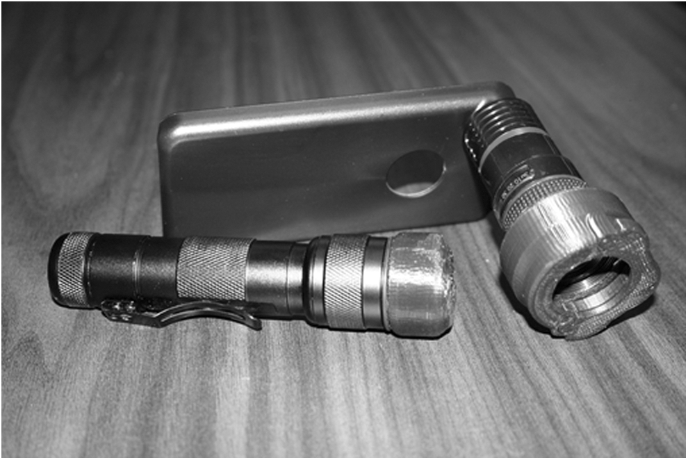
Endockscope with LED battery-powered light. LED = light-emitting diode.
Video endoscopy procedure
Video endoscopy was performed in two adult male cadavers using saline irrigation for cystoscopy, ureteroscopy, and nephroscopy; for laparoscopy, carbon dioxide was used to create a pneumoperitoneum. All endoscopes were handled by an experienced endourologist (Z.O.). Using the ES system, a Karl Storz Hopkins (Karl Storz Endoskope, Tuttlingen, Germany) rigid cystoscope (17F), rigid nephroscope (19.5F), semi-rigid ureteroscope (7F), and a 30° laparoscope (10 mm) were combined with either an Apple iPhone X (Apple, Inc., Cupertino, CA) or Samsung Galaxy S9+ (Samsung Electronics Co Ltd., Seoul, South Korea) (Fig. 2). Videos obtained with a standard Karl Storz imaging system used the conventional high-powered light source and camera system. A standardized evaluation of the bladder (rigid cystoscope), kidney (rigid nephroscope), ureter (semi-rigid ureteroscope), and intraperitoneal abdominal cavity (laparoscope) was performed with each of the 3 video arrangements, producing 12 unedited video segments, each ≤20 seconds.
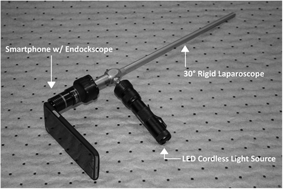
Assembled Endockscope with 30° rigid laparoscope.
Image evaluation
Twelve urology faculty members and four urology residents, blinded to the endoscopic system, evaluated the videos in a random order for each of the four endoscopic procedures. The 12 faculty urologists varied in age (35–71 years old) and subspecialty training (endourology, minimally invasive surgery, andrology, pediatric urology, urologic oncology, and female urology and pelvic reconstructive surgery). The four residents varied in length of residency training (1–5 years). Five image parameters were evaluated for each video (image resolution, brightness, color, sharpness, and overall image quality) using a 1 to 5 Likert scale (1 = poor, 5 = excellent). The videos were also assessed for acceptability for diagnostic use utilizing a yes/no binary scale.
Statistical analysis
A paired, two-tailed, t-test analysis was completed to analyze and compare the smartphone ES combinations and the standard Karl Storz HD camera system. Significant, statistical differences were defined as a p-value <0.05. Acceptability for the binary rating of diagnostic acceptability was quantified through the comparison of the percentages of the evaluators' responses. Further subanalysis of senior faculty, junior faculty, and residents was performed to evaluate statistical differences within each imaging parameter. Senior faculty (>20 years in practice) and junior faculty (<20 years in practice) were compared in a subanalysis. One-way analysis of variance was performed using responses from each imaging metric (seven senior faculty vs five junior faculty vs four residents) with statistical significance defined as a p-value <0.05.
Results
Evaluation of rigid cystoscopy
For rigid cystoscopy, there was no statistical difference (p > 0.05) for all imaging metrics when comparing the ES paired with the Galaxy S9+ or with the iPhone X to the standard imaging system. Among the reviewers, 67% found the ES paired with the Galaxy S9+ to be acceptable for diagnostic use, whereas 80% of reviewers found both the iPhone X and the standard imaging system to be acceptable for diagnostic purposes (Table 1).
Reviewer's Evaluation with Rigid/Semi-Rigid Endoscopes
p-values were obtained from comparison of smartphone vs standard Karl Storz endoscopes. Significance is indicated through bolded text.
Evaluation of rigid nephroscopy
The ES paired with the iPhone X was not significantly different from the Karl Storz HD camera in image resolution, brightness, and color; however, it was inferior in sharpness and overall image quality (p < 0.05). The ES with the Galaxy S9+ underperformed in brightness (p = 0.02), but was similar in all other imaging parameters when compared with the standard system. In addition, 100% of the evaluators found the standard system to be acceptable for diagnostic use, whereas 94% and 88% found the iPhone X and the Galaxy S9+ acceptable, respectively (Table 1).
Evaluation of semi-rigid ureteroscopy
There were no statistically significant differences between the iPhone X and the standard system (p > 0.05). Surprisingly, the ES paired with the Galaxy S9+ outperformed the standard system in image resolution and sharpness. Furthermore, 81% and 100% of evaluators determined the iPhone X and Galaxy S9+ images, respectively, to be acceptable for diagnostic purposes, whereas 88% of evaluators found the standard system to be acceptable for diagnostic use (Table 1).
Evaluation of laparoscopy with a 30° lens endoscope
The ES paired with the iPhone X significantly underperformed the standard camera system in all metrics (p < 0.05), with only 23% of evaluators deeming it acceptable for diagnostic purposes. In contrast, the ES paired with the Galaxy S9+ was comparable for image resolution, color quality, and sharpness, with 92% of evaluators considering it to be appropriate for diagnostic use (Table 1). The standard system was deemed appropriate for diagnostic uses by all of the evaluators (100%).
Subanalysis between senior faculty, junior faculty, and residents
There were no statistically significant differences between the responses of senior faculty, junior faculty, and residents under any parameter and camera system in rigid cystoscopy, nephroscopy, and semi-rigid ureteroscopy. There were also no statistical differences between each cohort under any imaging parameter with the standard system and iPhone X in laparoscopy. However, there were statistical differences among image brightness responses while viewing with the ES plus Galaxy S9+ under laparoscopic evaluation.
Discussion
As surgical technology advances in quality and capability, the cost of instrumentation has further widened the gap between global health care and the availability of these highly sophisticated resources. Currently, over 4 billion people worldwide lack access to adequate surgical health care, both diagnostic and therapeutic. 9 Due to cost, endoscopic equipment is unavailable, outdated, or poorly maintained. Despite the economic challenges, in these same countries, smartphone technology is abundant. With this in mind, we developed the Endockscope, initially to reduce the cost and increase the availability of flexible cystoscopy. 6 Having witnessed the sanguine results with this far less-expensive arrangement for flexible cystoscopy, we then sought to determine if the ES would be of value when applied to rigid endoscopes, which are more common worldwide. 6 Our study supports the use of the iPhone X and Galaxy S9+, in conjunction with the ES and LED light source, as both set-ups performed similarly to the standard camera and high-powered light source system for diagnostic purposes. 6 In this study, we utilized the smartphone camera without any additional filters or supplemental enhancements. No significant artifacts, such as the Moiré effect, were observed by the reviewers (Fig. 3).

Images of cadaver model using the ES and Karl Storz imaging system. ES = Endockscope system.
The Apple iPhone X and Samsung Galaxy S9+ were analyzed against the standard imaging system given that these two smartphones are the latest iterations of the most popular brands worldwide at the time of this study. However, it is notable that the two models tested may not exist as the top-selling smartphones in developing countries that would benefit from the ES. Considering the pace of technological advancement, the stock camera app available on less costly devices may indeed perform similarly to the tested models; however, this was beyond the scope of our study. Although a digital divide persists between developing and developed countries, smartphone ownership has increased exponentially in impoverished areas over the past 5 years and is expected to continue to rise during the next decade. 10 In addition, the image quality of older smartphone models coupled with the Endockscope were shown to be acceptable for diagnosis in an earlier study. 6
The ES paired with the iPhone X attached to a rigid nephroscope was inferior to the standard system in sharpness and overall image quality; nonetheless, 94% of the evaluators found it still acceptable for diagnostic use. Surprisingly, the overall image quality did not correlate with the percentage of evaluators who determined the images to be acceptable for diagnostic use. A similar, although inverse, discrepancy was present when comparing the three camera systems during semi-rigid ureteroscopy. In this study, the ES paired with the iPhone X performed similarly to the standard system under all imaging parameters, and yet only 81% of evaluators found the device acceptable for diagnostic purposes.
Further analysis was performed to evaluate statistical differences among our respondents, particularly between senior faculty (>20 years of experience), junior faculty (<20 years of experience), and residents. Among the 60 analyses performed, there was only one significant difference: image brightness with the 30° laparoscope.
Most impressive was the performance of the ES when paired with the Samsung Galaxy S9+. It outperformed the standard system for semi-rigid ureteroscopy both in imaging parameters and acceptability for diagnostic purposes. This could be due to the smaller viewing field provided during ureteroscopy. The Galaxy S9+ also performed well against the standard system during laparoscopy, with two exceptions: brightness and overall image quality. For cystoscopy, while the imaging parameters were equivalent to the standard, paradoxically, the diagnostic acceptability was low at 67%. Conversely for nephroscopy, image brightness was inferior to the standard system, and yet the diagnostic acceptability was 88%.
For the iPhone X, the outcome was less favorable. It performed particularly poorly for laparoscopy. Indeed, less than a quarter of evaluators found it to be acceptable for diagnostic use. However, for cystoscopy and nephroscopy, this smartphone performed well. The ratings for ureteroscopy were mixed in that, while the imaging parameters were rated similar to the standard, the diagnostic acceptability was only 81%.
In a side-by-side comparison of camera quality between the iPhone X and Galaxy S9+ under conditions outside the medical field, differences are very subtle. 11 For example, although the Galaxy S9+ performs well under low-light environments, the iPhone X produces finer lines and colors more true to life. 11 Notably, almost all camera specifications, such as image resolution, stabilization, zoom, and 4k and 1080p video fps, are the same in each phone, with the only difference being in the variable aperture. 11 Of note, it is important to highlight the contrasting performances of each smartphone in distinct fields of observation. Interestingly, the ES plus Galaxy S9+ performed more effectively in a larger/open field of the abdomen (laparoscopy). However, the ES plus iPhone X performed poorly within the abdomen; both cameras performed adequately within a more confined space such as the ureter. These differences are likely due to the configuration of the respective image stabilization software of the device, along with the processor and varied physical camera attributes. 12 Of note, other devices such as the Google Pixel, LG G7 ThinQ, and Huawei provide comparable functionalities with regard to their camera systems. 13,14 As such, although only the iPhone X and Galaxy S9+ were utilized in this study, our findings may apply to these other camera phones.
Although both smartphones performed nearly similar under nonclinical imaging conditions, the Galaxy S9+ provided the most consistent results among the four different rigid endoscopes within our fresh tissue cadaver models. This paves the way for physicians globally to have a readily available, durable, and inexpensive camera and light source at their disposal. Of note, similar technology such as the Endoscope-i, Clearwater Clearscope, and Karl Storz Smartscope adaptors, in conjunction with an Apple iPhone smartphone, have been tested clinically in the setting of otolaryngology and neurosurgery. In concert with our report, these studies indicate that this technology is a portable, easy-to-use, and low-cost alternative to the office-based camera system. 15,16 When compared with the average cost devoted to the purchase and maintenance of both rigid and flexible endoscopes, the ES with its LED attachment costs $45.00 (not including smartphone) and facilitates the simple construction of a device that can deliver comparable diagnostic imaging capabilities while drastically reducing expenditures. 17,18
As newer and more advanced smartphones emerge each year with enhanced features, it is possible that the ES in conjunction with a next-generation smartphone may significantly alter the current standard of care, challenging previous visualization norms, not only in countries with less access to advanced technology but also in western clinics. The cost effectiveness of the ES alternative may provide several financial advantages both in the hospital and office setting.
The future of the ES, we believe, has only been scratched. The potential is present for the development of an app that would instantly connect an urologist anywhere in the world with an expert urologist in another country. The two individuals could view an endoscopic procedure in real time or review a recorded endoscopy together. In this manner, the diagnostic playing field could be leveled such that individuals across the globe would have access to similar diagnostic expertise. This foray into the world of telemedicine might well reduce medical costs while improving diagnostic quality. 19,20
Our study is limited in several ways. One could argue that a larger sample size of evaluators would have provided clearer data especially with regard to the conflict between diagnostic acceptability and image parameters for the Galaxy S9+ cystoscopy and the iPhone X ureteroscopy results. Also, we did not test for any intraobserver differences as each individual reviewed the videos once in the random order in which they were presented. Also, the 16 participating reviewers at our institution typically only use the standard equipment and thus may not be representative of the urologists who may want to utilize the Endockscope. Finally, the study was cadaver based; findings may be different in a clinical study in the setting of bleeding and tissue color variation (e.g., due to disease or blood flow).
Conclusions
The Endockscope with its rechargeable LED light source ($45) coupled to the Samsung Galaxy S9+ ($800) provides reasonable imaging parameters and comparable diagnostic capabilities to the standard camera/light system (total cost: $45,000) for multiple rigid/semi-rigid endoscopic urologic procedures: nephroscopy, cystoscopy, ureteroscopy, and laparoscopy. The iPhone X ($900) provides reasonable imaging parameters and comparable diagnostic capabilities for rigid nephroscopy, rigid cystoscopy, and semi-rigid ureteroscopy but not for laparoscopy.
Footnotes
Author Disclosure Statement
No competing financial interests exist.
Funding Information
No funding was received for this article.