
Abstract
Objective:
To assess the effectiveness of laser lithotripsy in different holmium:yttrium aluminum garnet (Ho-YAG) laser settings with a wide range of energies, frequencies, and power.
Materials and Methods:
Two types of phantom stones were utilized, including soft stone, which mimics uric acid stone, and hard stone, which mimics calcium oxalate monohydrate stone. The stones were made into a round shape measuring 10 mm in diameter. The lithotripsy settings were 1 J × 20 Hz, 2 J × 10 Hz, 1.5 J × 20 Hz, 3 J × 10 Hz, and 2 J × 20 Hz. The lithotripsy was conducted in a caliceal model with a 2-mm filter. All stone vanishing from the artificial calix was an end point of the experiment. All fragments that passed through the filter of each setting were dried and weighed to calculate the vaporizing effect as well as to compare among the different settings. Laser fiber degradation was compared by using these settings.
Results:
Disintegration efficiency was determined by time consumption and the amount of vaporized stone. The best time consumption was 8 min 51 sec for 2 J × 20 Hz for hard stone and 5 min 13 sec for this setting for soft stone. The most vaporizing effect for hard stone was 92.19% for 2 J × 20 Hz and 87.30% for this setting for soft stone. The most fiber tip degradation was 28 mm for 3 J × 10 Hz for hard stone and 4 mm for 1.5 J × 20 Hz for soft stone.
Conclusion:
The study revealed that the best setting for hard stone was 2 J × 20 Hz, which was the fastest for achieving maximum vaporization, whereas fiber degeneration was comparable to others. For soft stone, there was no difference among the settings.
Introduction
Nowadays, the holmium:yttrium aluminum garnet (Ho-YAG, holmium) laser is widely used for the surgical management of kidney stones. Owing to the laser's flexibility and ability to disintegrate any type of stone composition, the Ho-YAG laser has become the standard lithotripter for endoscopic stone surgery. 1,2 The effectiveness of stone disintegration depends on the settings of the laser, the size of the laser fiber, and the composition of the stone. 1,2 Although there have been many previous studies focusing on the efficacy of Ho-YAG lasers, 3 –6 the ideal recommended laser settings have not been studied in depth.
American Urological Association (AUA) guidelines for surgical management in urinary calculi extended the size of the stone for ureteroscopy to 20 mm, but the usual laser setting established for low power is compromised when dealing with larger stones. To achieve stone-free status, a larger stone usually requires more operative time than a smaller one. However, prolonged operative time has been considered a factor in post-operative infection or sepsis. 7 Conducting laser lithotripsy within a specified time is considered to decrease the risk of complications. Thus, a high power setting was utilized to achieve stone-free status within the limited time.
In this in vitro experimental study, we used two different phantom stones. The stones comprised hard stone represented as calcium oxalate monohydrate (COM) and soft stone that represented uric acid. Together with various Ho-YAG laser settings along a wide range of pulse energy and frequency for high power settings of 20, 30, and 40 W, the outcomes of this study comprised the percentage of stone vaporization, time consumption, and laser fiber degradation after laser lithotripsy.
Materials and Methods
Phantom stone preparation
Two different types of stone composition were made, namely soft stone and hard stone. (1) Soft stone was made from type 1 gypsum plaster or plaster of Paris (calcium sulfate dehydrate). The acoustic and mechanical properties closely resemble kidney stones that are composed of uric acid and magnesium ammonium phosphate hydrogen stone. 8 The preparation ratio used the appropriate type 1 gypsum plaster powder to water ratio of 1.5:1 by weight. (2) Hard stone was made from Panamix stone (Panamix, Thailand) (type 4 gypsum plaster), which has acoustic and mechanical properties similar to kidney stones composed of COM and brushite stone. Panamix stone powder was mixed to a water ratio of 100:28 by weight. 8
Both types of stone mix were poured into round-shaped molds that were 10 mm in diameter. All samples were allowed to cure for 24 hours at room temperature. After demolding, the samples were dried for at least 24 hours before testing. Stones with visible cracks or bubbles were excluded. Before the experiment, the artificial stones were submerged overnight in 0.9% saline and then soaked in a uniform manner.
Laser fibers
The experiment was performed by 200-μm core laser fibers (Luminis Slimline SIS200). The tip was newly cleaved by using ceramic scissors after each experiment to avoid any possible fiber tip degradation bias. The laser tip degradation was observed after the experiment finished.
Lithotripsy settings
The laser energy used five different settings, including 1 J × 20 Hz, 2 J × 10 Hz, 1.5 J × 20 Hz, 3 J × 10 Hz, and 2 J × 20 Hz. The laser machine was a Luminis Versapulse PowerSuite Holmium 100 W (Luminis).
Experiments
All phantom stones were irradiated with various energy settings of the Ho-YAG laser by using a 200-μm fiber. The fiber was hand held, and laser emission was carried out while the laser fiber was straight forward to the stone by an experienced urologist using the popcorn technique (non-contact mode). The experiment was conducted three times in each setting.
In a water basin, stone was left in an artificial calix that had multiple holes measuring 2 mm in size, as shown in Figure 1. Fragments smaller than 2 mm were able to pass through the hole in the artificial calix, which was the end point of stone degradation. Time to stone vanishing from the calix was recorded in each setting. Disappearance of the stone from the calix was assessed by the naked eye. All visible particles in the basin after passing through the artificial calix were considered as residual fragments, which were collected and compared with pre-irradiated stone by weight to calculate the percentage of vaporizing stone. The percentage of vaporizing stone was calculated by phantom stone weight subtracted with the remaining particles that passed through a caliceal model and divided by phantom stone weight.

Caliceal model in a water basin that has multiple holes that are 2 mm in diameter. The fragmentation that passes through the calix is called residual fragmented stone.
Statistical analysis
Statistical analysis was performed by using Predictive Analytics Software statistics, SPSS ver18.0 (SPSS, Inc., Chicago, IL). One-way analysis of variance (ANOVA) was used to compare the differentiation of time, fiber degradation, and residual fragments between each setting. Independent t-test was used to compare the differentiation of time, fiber degradation, and residual fragmentation between soft and hard stone. Pearson's correlation test was used to evaluate the relationships between time and remaining stone. A probability p-value <0.05 was considered statistically significant.
Results
There were three parameters to consider in this study, including the percentage of vaporizing stone, time consumption, and fiber tip degradation.
Percentage of vaporizing stone
The percentage of vaporizing stone is shown in Figure 2. For hard stone, the setting of 1 J × 20 Hz had the greatest amount of residual fragments. The percentage of vaporizing stone for this setting was the least at 80.97% compared with others, which was 91.27% for 2 J × 10 Hz, 88.35% for 1.5 J × 20 Hz, 90.90% for 3 J × 10 Hz, and 92.19% for 2 J × 20 Hz.
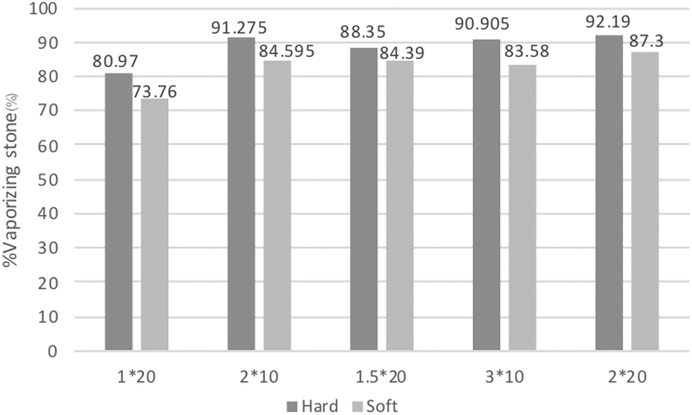
Column of %vaporizing stone calculated by collecting all fragmentation that filters through the caliceal model subtracted from the phantom stone's weight before irradiation.
For soft stone, there was no statistical significance among these settings (p-value = 0.163). Comparing hard and soft stone, hard stone had a higher percentage of vaporizing stone than soft stone 6.01% (p-value = 0.02). There was no correlation between the percentage of vaporizing stone and time.
Time consumption
Time consumption for stone degradation in each setting is shown in Figure 3. For hard stone, the setting of laser lithotripsy at 3 J × 10 Hz and 2 J × 20 Hz had the least time to vanish stone, whereas there was no statistical difference between them (p-value = 0.912). The setting for 3 J × 10 Hz had less time consumption than the setting for 1 J × 20 Hz for 8 min 1 sec (p-value <0.001), 2 J × 10 Hz for 5 min 12 sec (p-value = 0.013), and 1.5 J × 20 Hz for 4 min 12 sec (p-value = 0.046).
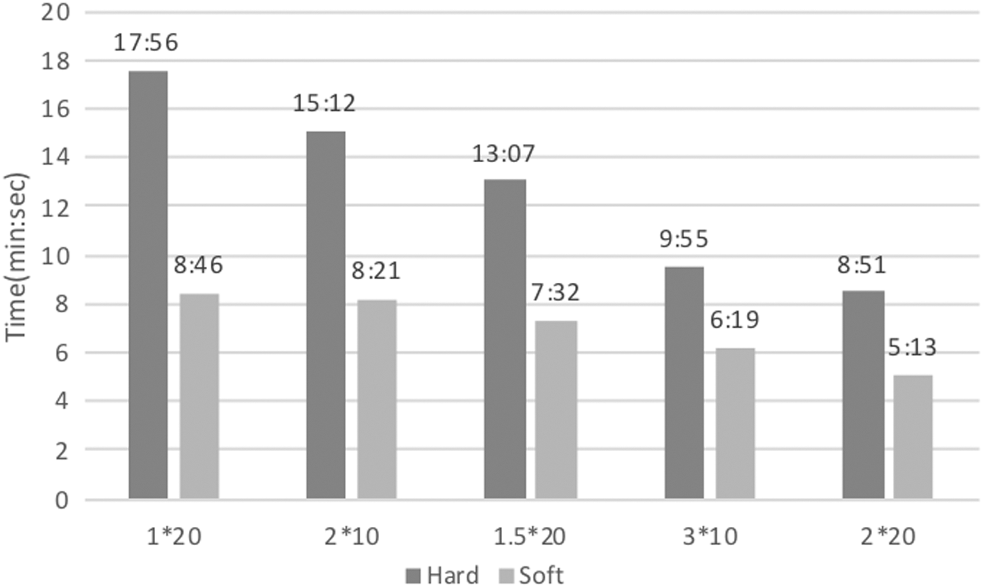
Column of time consumption (min:sec) in each setting to vanish 10-mm phantom stone.
The setting of 2 J × 20 Hz had less time consumption than the setting of 1 J × 20 Hz for 9 min 4 sec (p-value <0.001), 2 J × 10 Hz for 6 min 16 sec (p-value = 0.003), and 1.5 J × 20 Hz for 5 min 15 sec (p-value = 0.013).
For soft stone, there was no statistical significance among these settings (p-value = 0.27). When comparing hard and soft stone, hard stone had a longer mean time for consumption than soft stone at 5 min 49 sec (p-value <0.001).
Fiber tip degradation
For hard stone, fiber tip degradation after stone vanishing was 20 mm for 2 J × 10 Hz, 16.33 mm for both 1 J × 20 Hz and 2 J × 20 Hz, 17 mm for 1.5 J × 20 Hz, and 28 mm for 3 J × 10 Hz. The most fiber tip degradation was in the setting of 3 J × 10 Hz, which was statistically different from the setting of 1 J × 20 Hz, 1.5 J × 20 Hz, and 2 J × 20 Hz (p-value = 0.02). There was no difference in fiber degradation between 3 J × 10 Hz and 2 J × 10 Hz (p-value = 0.131) Figure 4.

Column of fiber tip degeneration (mm) in each setting to vanish 10-mm phantom stone.
For soft stone, there was no difference in fiber degradation among these settings (p-value = 0.23). Comparing hard and soft stone, hard stone had more fiber tip degeneration than soft stone at 16.43 mm (p-value <0.001). Time consumption was not correlated with fiber tip degradation.
Discussion
Newly updated guidelines for kidney stone removal tend to recommend increasing the stone size in the endoscopic procedure. From the previous study, the usual laser setting was established in low power to compromise larger stones. The aim of this study was to identify the best settings for laser lithotripsy in high power to deal with large and hard stones simulated from kidney stones in the retrograde intrarenal surgery procedure.
There were two techniques to remove stones after Ho-YAG laser lithotripsy, dusting or fragmenting with basket. Each technique has its own benefits. Dusting makes the stone fragments smaller than 2 mm, allowing them to easily pass out of the collecting system. The advantages include not having to do multiple access ureteroscopy to remove stones and decreased risk of ureteric avulsion, but with increased operative time. 1,3,9 –11 Popcorn or pop dusting, which is a non-contact laser lithotripsy approach, has become a useful technique to break up stones smaller than 2 mm, which is especially useful for large and hard stones.
In the experiment, a Ho-YAG laser was used to fire the diameter 1-cm phantom stone with the popcorn technique until vanishing in a caliceal model that had a 2-mm hole. Fragments <2 mm that passed through the hole were assumed to be dusting particles. The setting of 1 J × 20 Hz had maximum residual fragments. Therefore, it had the least % of vaporization among five settings (p-value = 0.036). The settings of 2 J × 10 Hz, 1.5 J × 20 Hz, 3 J × 10 Hz, and 2 J × 20 Hz had more %vaporizing stone, but no statistical difference among them.
Even though there was similar total power of 20 W in the setting of 1 J × 20 Hz and 2 J × 10 Hz, higher pulse energy (2 J) made more vaporization than the lower (1 J). However, there were no differences between pulse energy of 1.5 or 3.0 J at a similar power of 30 W. Thus, 1.5 J of pulse energy was the least pulse energy that made a high vaporizing effect similar to 2 and 3 J with high power settings.
The results showed the settings that made the fastest stone vanishing were 3 J × 10 Hz and 2 J × 20 Hz in hard stone. Compared with the same power at 30 W, 3 J × 10 Hz was faster than 1.5 J × 20 Hz (p-value = 0.046) and in the setting of 20 W, 2 J × 10 Hz tended to be faster than 1 J × 20 Hz, but was not statistically significant. This result was compatible with a previous study that encouraged HiE, LoF 3 –5 (high energy, low frequency theory). In soft stone, the result is not statistically significant with these settings. This study revealed that high pulse energy was not necessary to utilize for laser lithotripsy of soft stone.
Fiber tip degradation was determined among different settings for both soft and hard stone. For soft stone, there was no difference in fiber degradation for all settings (p-value = 0.23). For hard stone, the highest pulse energy (3 J) was the most fiber tip degradation. This finding was compatible with the burn-back effect, 12 which is more damaging to the fiber tip while using with high energy, short pulse duration, and hard stone. 13 This study used only short pulse duration for all settings to have maximum effect of fragmentation. Therefore, fiber tip degeneration was high.
Conclusion
According to the study, the best setting for the popcorn technique was 2 J × 20 Hz for hard stone. It was the fastest to achieve maximal stone vaporization, whereas fiber degradation was comparable with a lower setting. There was no difference among settings when carrying out the popcorn technique for soft stone.
Footnotes
Acknowledgments
This work was supported in part by the Department of Dentistry, Siriraj Hospital for the making of phantom stone.
Authors Disclosure Statement
No competing financial interests exist.
Funding Information
No funding was received for this article.