
Abstract
Introduction:
The use of fluoroscopy during percutaneous nephrolithotomy (PCNL) may lead to an overestimation of stone-free rates. The objective of this study is to demonstrate the feasibility of intraoperative CT-guided PCNL compared with standard of care (SoC) PCNL.
Patients and Methods:
A prospective feasibility study (20 patients undergoing PCNL with an intraoperative CT scan between June 2017 and February 2020) and a retrospective study of a historical cohort (20 consecutive patients undergoing SoC PCNL between September 2015 and September 2016) were conducted. All procedures were performed by an expert endourologist in a tertiary referral hospital. Follow-up was performed at 6 weeks postoperatively. The primary goal is to investigate the practicality and potential benefits and harms of intraoperative CT scanning during PCNL. Secondary outcomes are a stone-free rate after the 6-week follow-up, perioperative radiation exposure, the need for postoperative imaging, and peri- and postoperative complications. Statistical significance was considered at p < 0.05.
Results:
The initial stone-free rate in the CT scan group was 65% (n = 13). In 25% (n = 5) of patients, residual stone fragments were removed after the perioperative CT scan. In the SoC group, 85% (n = 17) of patients were thought to be stone free perioperatively. At the 6-week follow-up, 80% (n = 16) in the CT scan group vs 50% in the SoC group (n = 10) were found to be stone free. Radiation exposure, perioperatively, was higher in the CT scan group. Complications were comparable between groups. Limitations of the study are the nonrandomized design of the study and nonstandardized follow-up imaging.
Conclusions:
Intraoperative CT scanning during PCNL is feasible and gives a better estimate of any remaining stone fragments compared with fluoroscopy only.
Introduction
Percutaneous nephrolithotomy (PCNL) is effective for surgical removal of large renal calculi with reported stone-free rates ranging from 39% to 84% depending on the complexity of the case. 1 However, a surgeon's perioperative assessment of fluoroscopy during PCNL has been shown to be inaccurate. The study by Nevo et al. showed that 20% of patients who are considered to be stone free perioperatively still have significant residual stone fragments at postoperative imaging and are therefore still at risk for stone-related events. 2 Over the past decade, low-dose CT has become the most common imaging modality for renal colic due to its diagnostic accuracy for kidney stones with a sensitivity of 95% and specificity of 97%. 3 In addition, stone site and size can easily be measured.
During PCNL procedures, there is a risk of dislocation of calculi in the kidney to a place invisible or inaccessible to the surgeon. In addition, in case of multiple stones, it can be difficult to navigate through the kidney to visualize all stones. Using fluoroscopic guidance, large radiopaque stones will be identified. However, radiolucent stones, small stones, or stones overlying bony structures or the access sheath can easily be missed. This often leads to an overestimation of stone-free rates after PCNL. Whether residual stones are clinically relevant depends on the stone size and their composition. However, auxiliary treatments due to residual stones post-PCNL are not infrequent. 4 The aim of this study is to investigate the feasibility and potential added value of more accurate detection of residual stones during PCNL surgery.
Materials (Patients) and Methods
Study design and patient eligibility
A study protocol was developed and approved by the local ethics committee before initiation of the study. A prospective feasibility study was designed (patients undergoing an intraoperative CT scan during the PCNL), supplemented with a retrospective study of a historical cohort for comparison (patients who underwent a standard of care [SoC] fluoroscopy-guided PCNL). All patients in the prospective arm provided written informed consent. For the retrospective arm, the ethics committee waived the need for informed consent.
The prospective data were collected at the Radboud University Medical Center, Nijmegen, The Netherlands, between June 2017 and February 2020. Twenty patients with unilateral symptomatic nephrolithiasis with an indication for PCNL according to the EAU guidelines were included. 5 Exclusion criteria were (1) active urinary tract infection at the time of surgery, (2) anatomical abnormality as a barrier for PCNL access or prone positioning of the patient, (3) absolute indication for continuation of anticoagulant medication, (4) anesthesiology objections against prone positioning during the operation, and (5) pregnancy. An abdominopelvic noncontrast-enhanced CT scan was performed in all cases preoperatively to confirm the diagnosis and measure the stone load. The retrospective arm contained consecutive patients with identical inclusion and exclusion criteria and recruited between September 2015 and September 2016.
For all patients, preoperative abdominopelvic CT scans were evaluated for stone diameter (diameter of a single stone or the sum of diameters of multiple stones on axial CT images) and to calculate the S.T.O.N.E. score. 6 Preoperative (body mass index, age, sex, and use of anticoagulant drugs) and perioperative (type of dilation, access, and fragmentation) characteristics were extracted from the patients' medical files. The operative time was calculated as the time of cystoscopy to completion of the whole procedure. Effective fluoroscopy doses were calculated based on conversion factors calculated using PCXMC (Monte Carlo simulation program) based on a standard adult human. Peri/postoperative complications were reported according to the Clavien–Dindo classification system. 7 Perioperative stone-free rates were based on the perioperative findings of the CT scan (prospective arm) or based on fluoroscopy and direct inspection of the pyelocaliceal system (retrospective arm).
At the 6-week follow-up, stone-free rates were based on postoperative imaging (in the prospective arm, patients without residual stones on the perioperative CT scan did not undergo postoperative imaging and were also considered to be stone free at the 6-week follow-up). Necessary follow-up for inclusion was 6 weeks postoperatively.
Study device
PCNLs in the prospective cohort were performed in a hybrid operating room equipped with an Artis Zeego cone-beam CT scan (Siemens). This CT scan is equipped with robot-assisted positioning capability, offering flexibility in positioning of the patient while allowing visualization of the whole abdomen and pelvis. Due to the system's isocenter, the surgeon can work ergonomically even during long and complex procedures. The CT scan makes one rotation around a carbon surgical table, after which 2D reconstructions are available within minutes and 3D reconstructions can be made postoperatively if desirable.
Surgical procedure
A standard PCNL procedure was performed in all patients by an experienced endourologist with >10 years of experience and an average procedural volume of 25 PCNLs per year. All patients received prophylactic antibiotics before surgery. In brief, patients were under general anesthesia and in the lithotomy position, a 6F ureteral catheter was inserted at the affected side, and patients received a transurethral catheter. Under ultrasound and contrast-enhanced fluoroscopic guidance, a renal sheath was correctly positioned. The decision of the type of access (mini-PCNL or standard PCNL) and type of fragmentation/extraction (pneumatic, ultrasonic waves, laser, basket, and forceps) was at the discretion of the surgeon. In the prospective cohort, an intraoperative CT scan (with Amplatz in situ) was performed to confirm whether the patient was stone free. CT scan images were interpreted by the surgeon. If residual fragments were visible, an attempt was made to remove these. The need for a nephrostomy tube and/or a Double-J stent at the end of the procedure was at the discretion of the surgeon. Clinical follow-up was performed at 6 weeks postoperatively and the need for further imaging was evaluated individually.
Study objectives
This is a feasibility study with a primary goal to investigate the practicality and potential benefits (whether intraoperative CT scanning gives a more accurate estimate of residual stone fragments) and harmful effects (complications) of intraoperative CT scanning during PCNL. The secondary outcomes of the study are stone-free rates 6 weeks postoperatively after one procedure, comparing SoC PCNL procedures with PCNL procedures with intraoperative CT scanning. Stone free was defined as no visible residual stone on peri- and postoperative imaging. Further secondary outcomes are perioperative radiation exposure, the need for postoperative imaging, and peri- and postoperative complications.
Statistical design
Since this is a feasibility study, a formal sample size calculation was not performed. Analyzing 20 patients should suffice to investigate the proof of principle. In our institution, we perform 25 PCNL procedures yearly and planned to finalize the inclusion of patients within a year. Statistical analysis was performed using SPSS, version 27 (IBM, Armonk, NY). Continuous and binary variables are compared using the Mann–Whitney U test and chi-square test, respectively. Data are expressed as medians and interquartile rates (IQRs). Statistical significance was considered at p < 0.05.
Results
The surgical setup is represented in Figure 1. As an example, in Figure 2, a 3D reconstruction of an intraoperative CT scan is presented, showing residual stones next to the PCNL access sheath.
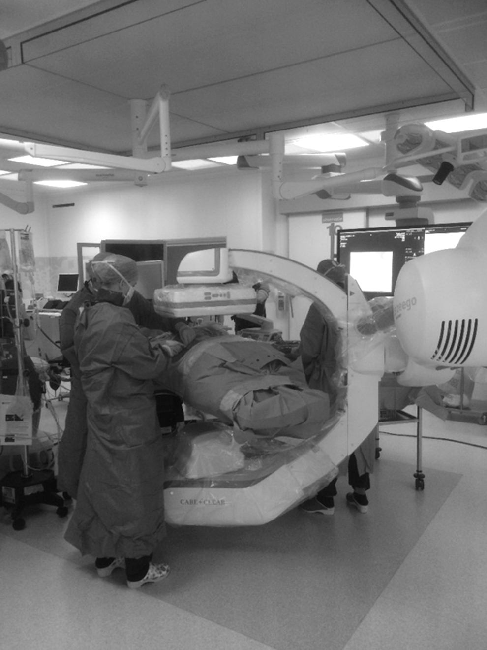
Image of the operating room with an intraoperative CT scan. The patient is positioned in a prone position completely covered by sterile surgical drapes. At the time of the CT rotation, the staff distance themselves from the patient behind a lead screen.
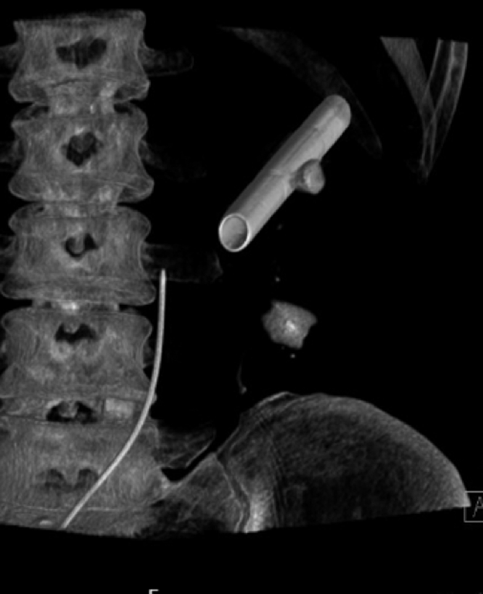
Three-dimensional reconstruction of perioperative CT scanning, showing a ureteral catheter and a 30F Amplatz not only with a large residual stone in the lower pole but also a stone adjacent to the Amplatz, which is often difficult to visualize on fluoroscopy. Taking this into account, the surgeon then knows he needs to carefully retract the Amplatz at the end of the procedure, allowing him to visualize and treat the stone.
Both treatment groups are comparable in their baseline characteristics with similar S.T.O.N.E. scores (Table 1). When comparing access to the kidney between both groups, there is a clear difference, with 75% (vs 20%) of patients in the CT scan vs SoC group undergoing mini-PCNL (p < 0.01). In addition, ultrasonic waves were used significantly less in this group compared with the SoC PCNL group (10% vs 45%) (p = 0.01) (Table 2). This difference can be explained by an evolution of preferred treatment strategies in our center. In one patient, it was impossible to gain access to the kidney.
Descriptive Characteristics of Preoperative Patients and Stone-Related Characteristics
Number of stones based on preoperative CT scan.
Stone size measured as maximal length in one dimension; if more than one stone was present, the sum of maximal stone lengths was calculated.
BMI = body mass index; IQR = interquartile range.
Description of Surgical Aspects of Both Cohorts
mSv = milliSievert; PCNL = percutaneous nephrolithotomy; SoC = standard of care.
As expected, the median operation time when performing a perioperative CT scan was longer compared with SoC PCNL, with a median of 159 minutes vs 117 minutes (Table 2). The fluoroscopy dose was significantly lower in the patient group undergoing perioperative CT scanning (0.45 mSv [IQR 0.28, 0.6] vs 0.75 mSv [IQR 0.58, 0.85], p = 0.01). The median radiation dose of perioperative CT scanning is 6.7 mSv (IQR 5.6, 7.4), as can be expected from an abdominopelvic CT scan without contrast (Table 2).
Surgical complications in the CT scan vs SoC group according to the Clavien–Dindo classification were as follows: grade I, 1 (5%) vs 1 (5%); grade II, 2 (10%) (one pulmonary embolism after discharge and one skin infection requiring medical treatment) vs 5 (25%) (three urinary tract infections and one skin infection); and grade III, 2 (10%) vs 1 (5%). There were no higher grade complications. Stone type distribution was similar between both groups (Table 3).
Peri- and Postoperative Stone-Free Rates and Need for Reintervention at 6 Months
For the perioperative CT cohort, stone-free rates are based on a perioperative CT scan, and for the historical cohort, stone-free rates are based on fluoroscopy.
URS = ureterorenoscopy.
The perioperative stone-free rate in the CT scan group was 65% (n = 13): in 39% (n = 5) of these cases, a patient was rendered stone free after removal of residual stones identified on the perioperative CT scan. In the remaining patients who were not stone free perioperatively, in 86% (n = 6), residual stones were identified, but were not removed because the remaining fragments were inaccessible. Three of these patients were treated conservatively; one was planned for a follow-up ureterorenoscopy and two for a follow-up PCNL. In one patient, we were unable to access the kidney. In the SoC group, more patients were considered to be stone free perioperatively (85%, n = 17).
At the 6-week follow-up, 50% (n = 10) of patients in the SoC group showed residual stones during follow-up imaging (45% [n = 9] underwent a CT scan, 45% [n = 9] a kidney, ureter, and bladder radiograph [KUB], and 10% [n = 2] did not undergo postoperative imaging). In contrast, in the six patients who had residual stones on perioperative CT scanning, three had no residual stones at follow-up imaging (10% [n = 2] underwent a CT scan, 20% [n = 4] a KUB, and 70% [n = 14] did not undergo postoperative imaging). Therefore, at the 6-week follow-up, 80% (n = 16) of patients in the CT scan group vs 50% (n = 10) of patients in the SoC group were confirmed to be stone free by peri- and/or postoperative imaging. Although these rates cannot be compared statistically, these data suggest clinical significance (Table 3).
Discussion
Large, symptomatic renal calculi often need active treatment and PCNL has been accepted as an effective treatment. Although minimally invasive, the operation is associated with a reasonable number of complications and does not always render the patient stone free. The latter largely depends on the size and complexity of the renal calculi, which can be described by stone scoring systems such as the S.T.O.N.E. scoring. 6 A multicenter external validation of different stone scoring systems showed a stone-free percentage of 80.3% to 84.2% for low-risk, 71.8% to 73.8% for intermediate-risk, 58.6% to 64.4% for high-risk, and 39.2% to 52.3% for extremely high-risk patients after PCNL. 1 However, reported stone-free rates are highly dependent on the accuracy of the detection method that is used as well. Sensitivity/specificity rates (%) for stone detection are 57/76 for a KUB, 84/53 for ultrasonography, and 95/97 for (low-dose) CT scanning. 3
Therefore, we hypothesized that by performing an intraoperative CT scan, a better estimation of the residual stone load after PCNL could be made and residual stones could be removed based on this imaging. The efficacy of perioperative CT scanning in detecting stone fragments has been published before in an ex vivo setting. 8 In addition, Vicenti and colleagues were the first to publish a case report of a patient in whom they were able to remove residual stones based on a perioperative CT scan, which were not visualized on high-resolution fluoroscopy, rendering the patient stone free in one procedure. 9
To the best of our knowledge, we are the first to report a prospective study on the feasibility and stone-free rate of this technique. First, our data confirm that performing a CT scan perioperatively gives a better estimation of residual renal calculi. While 85% of patients were judged to be stone free in the SoC group based on fluoroscopy, at the 6-week follow-up, only 50% of patients were confirmed to be stone free based on postoperative imaging (KUB or CT scan). In contrast, while 65% of patients in the group undergoing a CT scan perioperatively were confirmed to be stone free, at the 6-week follow-up, 80% were stone free, which is probably due to spontaneous passage of any small residual fragments.
Second, in 25% of cases, the perioperative CT scan proved to be useful in rendering patients stone free, as residual fragments were removed based on the findings of the CT scan. Although this is clinically relevant, we need to emphasize that this strategy is not always effective. In 25% of cases, we were unable to reach the remaining fragments identified on the CT scan. Although this information would allow the surgeon to approach the stone through another tract, this was not done as it would further increase the duration of the surgery and the risk of complications. With the current strategy, complication rates were comparable with other reports, with only one Clavien–Dindo Grade III complication in both groups and no Grade IV to V complications. 1 Still, we are convinced that an increase in the stone-free rate of 25% is clinically relevant. Furthermore, for 25% of patients in whom the residual stones were not removed in the same session, we were able to immediately plan a follow-up strategy.
Patients who receive a perioperative CT scan obviously receive a higher radiation dose perioperatively compared with the SoC group, which might be a concern for the patient. Literature shows that all patients with nephrolithiasis are at risk for increased radiation exposure, with CT scan being the most significant contributor to patient radiation exposure in this setting (current low-dose CT scans produce an effective dose radiation (ERD) of 1–5 mSv). No guidelines exist for maximal patient radiation exposure per year. However, there is an accepted recommended limit per adult for occupational exposure, which is not >20 mSv per year, on average over a 5-year period, and not >50 mSv in any 1 year, as established by the International Commission on Radiological Protection. 10 In the study by Ferrandino and colleagues, radiation exposure associated with an acute stone episode and 1-year follow-up at two large medical centers was 29.7 mSv. Of 108 patients, 22 even received >50 mSv. 11
Although intraoperative radiation exposure is higher in the CT scan group in our study, postoperatively only 30% of patients needed to undergo additional imaging (20% KUB and 10% CT scan), while 90% of patients in the SoC group needed to undergo additional imaging (45% KUB and 45% CT scan). None of the patients in this study were exposed to >20 mSv during their treatment follow-up. Furthermore, several studies have shown that residual stone fragments often have a significant impact not only on the need for auxiliary treatments but also on the need for future imaging. 12 –14 As our study was not designed to investigate the long-term impact on radiation exposure, no clear conclusions can be drawn. When perioperative CT imaging has completely proven its usefulness during PCNL procedures, future research can focus on reducing the perioperative radiation exposure with (ultra-) low-dose CT scanning, which has already proven its efficacy during follow-up of urolithiasis. 15 Similarly, although operating times will be longer when performing a perioperative CT scan and hence the costs associated with the prolonged procedure will increase, the reduced need for future imaging and reinterventions should outbalance this. One might say that the benefits of rendering someone stone free outweigh the impact of acceptable, additional, perioperative radiation exposure and increased operating time. Future cost–benefit analyses should be able to answer these questions.
This study has some strengths and limitations. This is the first prospective study on the efficacy of perioperative CT scanning during PCNL. Although no randomized controlled trail (RCT) was performed, we believe that this is not necessary for feasibility study purposes. Furthermore, the nonrandomized design of the study has some advantages in this setting. First of all, an RCT could lead to a high risk of performance bias: the surgeon would not be blinded to which patients received the intraoperative CT scan and could therefore influence outcomes as desired. Using a retrospective cohort as a comparator prevents this potential bias. Furthermore, procedures were performed by an experienced endourologist in a tertiary referral center in both treatment arms, warranting comparability of both groups.
A limitation of the study is the nonstandardized follow-up imaging, which does not allow us to accurately compare stone-free rates at the 6-week follow-up. However, in the intraoperative CT scan group, the stone-free rate was mainly CT based, in contrast to patients in the SoC group who often underwent less sensitive imaging. Hence, the impact of a perioperative CT scan in this study is more likely to be under- than overestimated. Therefore, the message of this study is not expected to change significantly if all patients would have undergone a postoperative CT scan. In addition, recruitment of patients took longer than expected due to scheduling issues because of limited capacity in the hybrid operating room.
Conclusions
Intraoperative CT during PCNL is feasible and gives a better estimate of any remaining stone fragments compared with fluoroscopy only. In case of residual stones, extraction during the same surgery is possible, but not always achievable through the same tract. Nevertheless, a plan regarding follow-up, additional imaging, or auxiliary procedure can immediately be made. We believe that intraoperative CT scans during PCNL should not be considered SoC treatment, but can be considered in selected cases. This study sets the stage for a future multi-institutional study on the indication and effectiveness of intraoperative CT scanning during PCNL procedures.
Footnotes
Authors' Contributions
T.V.d.B. was involved in investigation, formal analysis, writing—original draft, and visualization. X.Z. was involved in conceptualization, methodology, investigation, formal analysis, writing—review and editing, project administration, and supervision. A.K. was involved in investigation and writing—review and editing. J.F. was involved in conceptualization, methodology, and writing—review and editing. J.L. was involved in conceptualization, methodology, investigation, writing—review and editing, resources, and supervision. F.D. was involved in conceptualization, methodology, investigation, project administration, writing—review and editing, resources, and supervision.
Author Disclosure Statement
No competing financial interests exist.
Funding Information
No funding was received.