
Abstract
Objective:
Recently, retrograde intrarenal surgery (RIRS) using laser lithotripsy has become popular. However, the optimal laser energy setting for pop-dusting has not been established. In this study, we report our experiences of RIRS using the high-power (up to 100 W) pop-dusting (HPPD) technique.
Methods:
This study retrospectively assessed 82 cases with RIRS using HPPD. Patients who underwent abdominal CT or mercaptoacetyltriglycine (MAG3) diuretic renal scan at 3 months postoperatively were included in this study. Patient and stone characteristics and perioperative and postoperative outcomes were evaluated.
Results:
The average number of renal stones was 3.67 ± 4.11, and the average length of the largest stones was 13.30 ± 6.41 mm. The mean Hounsfield units was 959.99 ± 384.73. The operation time was 58.10 ± 26.67 minutes. The mean HPPD time was 11.93 ± 9.48 minutes, with settings of 1.97 ± 0.25 J and 48.78 ± 3.29 Hz. The stone-free rate was 89%. The mean hospital stay was 1.68 ± 1.29 days. Pelvicaliceal and ureter injuries were observed in 9.8% and 32.9% of the study population, respectively. However, there was no transfusion, subcapsular hematoma, persistent urinary leakage, ureteral or infundibular stricture, or renal functional deterioration. There was transient postoperative fever in 12.2% of the study population.
Conclusions:
HPPD could be performed safely during RIRS for renal stones without significant complications such as collecting system injury or bleeding. High-power laser mode (up to 100 W) can be a safe and effective choice for pop-dusting during RIRS, especially for large and hard stones.
Introduction
Over the past 20 years, treatment of renal stones has made many advances since the introduction of retrograde intrarenal surgery (RIRS). 1 RIRS is now considered a first-line treatment of renal stones sized smaller than 2 cm because of high success rates and low morbidities. 2 Furthermore, RIRS is rapidly expanding its indications, replacing percutaneous nephrolithotomy (PCNL) with increasing surgical outcomes. 3 The advancement of RIRS is mainly due to progress in stone breaking and retrieval techniques. Since it was first applied to the treatment of urinary stones in 1995, the Holmium: yttrium–aluminum–garnet (Ho:YAG) laser has been used as a standard method for treatment of renal stones. 4,5 In early stages of Ho:YAG laser lithotripsy, the low-power mode (less than 12 W) was generally used and was accepted to be sufficient for breaking urinary stones. 6 –8 However, indications of RIRS have been expanded, and the need for a method of more effective stone disintegration has increased. One decade ago, the use of high-power lasers for renal stones was introduced. Some studies have reported that it can effectively and safely remove stones in high-power mode of 60 W or more during PCNL. 9 –11
There have been many advances in the stone-breaking strategy of the Holmium laser. The currently used strategies can be largely divided into fragmentation with basketing, dusting, and pop-dusting (also known as the popcorn technique). Among those methods, the pop-dusting technique has been widely used because of its excellent advantage in reducing operation time, especially for large burden calculi. During pop-dusting, a laser fiber does not target the stone surface directly. The Holmium laser breaks floating stone fragments that make contact with the laser fiber incidentally due to the turbulent flow of irrigation fluid. 12 Generally, high pulse-power and high-frequency laser energy is used for the pop-dusting technique to produce a strong turbulent flow, known as the whirlpool effect. Theoretically, higher laser frequency can enhance the chance of contact between laser fiber and floating stone fragments, and higher laser power can break stone fragments into tinier particles. However, the optimal laser energy setting for pop-dusting in terms of pulse power and frequency has not been established.
The biggest concern about the high-power energy setting in RIRS is the thermal response to the tissue. This is because the temperature of the irrigation fluid inevitably rises during lasering and may cause thermal injury to the pelvicaliceal system. 13 Recently, Hein and colleagues reported an ex vivo study finding that if sufficient irrigation was secured, there were no major problems with thermal issues even if the laser energy was high. 14
High-power pop-dusting (HPPD) during RIRS is expected to shorten the operation time due to effective disintegration of renal stones and to reduce the need for active basket removal due to the smaller stone pieces. 15 In this study, we report the initial experience of RIRS using 100 W HPPD performed in a well-established surgical environment.
Materials and Methods
Study design
This study introduces 82 cases of RIRS using 100 W HPPD conducted by a single operator as a case series of retrospective design. Among RIRS cases between December 2018 and November 2019, all cases with mercaptoacetyltriglycine (MAG3) diuretic renal scan or abdomen CT at 3 months after surgery were enrolled.
Operation setting and operative technique
In all cases, RIRS was performed on a uro-table (Uroskop Omnia Max®; Siemens, Erlangen, Germany), and a fluid irrigation pump system (Lagis® Endosurgical, Taichung, Taiwan) was built and used to maintain the pressure of the saline bag. When performing HPPD, the pressure applied to the saline bag was maintained at 150 to 200 mm Hg. The saline bag was connected to the endoscope using 180 cm long TUR-Y Set™ (inner diameter 5.5 mm) (Sewoon Medical, Chungcheongnam-do, South Korea) (Fig. 1). The Ho:YAG laser and 200 μm laser fiber were used (Lumenis, Yokneam, Israel), and either the Flex-Xc (Karl Storz, Tuttlingen, Germany) or the LithoVue™ (Boston Scientific, MA, USA) were used.
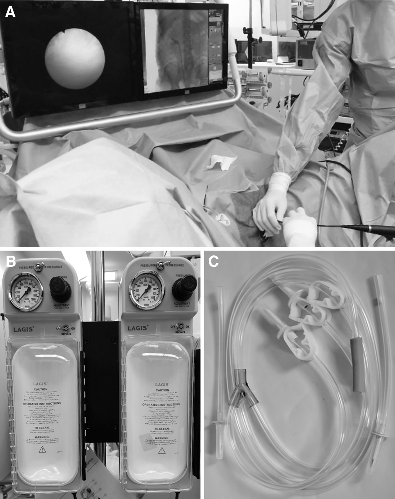
Surgical environment during retrograde intrarenal surgery.
In all patients, surgery was performed under general anesthesia. After inserting the guidewire under the rigid ureteroscope, retrograde pyelogram images were confirmed through a 5F open-end ureteral catheter. After inserting the Amplatz Super Stiff guidewire (Boston Scientific), a ureteral access sheath (UAS) (Navigator; Boston Scientific) was placed. The size of the UAS was 11/13F to 14/16F according to the width of the ureter in the retrograde pyelogram. The tip of the UAS was placed in the ureteropelvic junction, and a flexible ureteroscope was inserted. Before lithotripsy using laser, the structure of the pelvicaliceal system was observed, and the location, shape, and size of the stones were identified. During this navigation process, the calix suitability for HPPD was determined, and the stones were moved to the target calix using a basket. If the stone was too large or the infundibulum was too narrow to move directly by the basket, the fragmentation technique was used to move the stone to the target calix. The location for HPPD was determined by location of the calix, length of the infundibulum, and size of the calix. When the renal stone was well fragmented, it was crushed into pieces smaller than 1 mm using a dusting technique (Supplementary Video S1). During lithotripsy, some fragmented stones were removed for stone analysis. After removing all stones, the degree of injury to the pelvicaliceal system and the ureter was evaluated. 16 The degree of damage was judged based on the highest grade of injury. In all cases, Double-J stents were placed.
Follow-up
Patients were routinely discharged the day after surgery; however, when complications such as fever occurred, they were discharged after resolving the medical problem. On the first day after surgery, laboratory tests of complete blood count, electrolyte, blood urea nitrogen (BUN), creatinine, and urinalysis were routinely performed, and 3 months after surgery, the same tests were performed, in addition to kidney, ureter, and bladder radiograph (KUB) and abdominal CT or MAG3 diuretic renal scan. The stone-free rate was evaluated 3 months after surgery using KUB and CT.
Operation
The operation was performed by a urologist with at least 15 years of dedicated experience in endourology (i.e., more than 1000 cases for flexible ureteroscopic surgery, more than 2600 cases for rigid ureteroscopic surgery, and more than 650 cases for PCNL). HPPD was performed with a laser setting of 1.5 to 2.0 J × 40 to 50 Hz (60–100 W). In addition, the fluid management system, fluid irrigation set, and surgery environment were kept constant throughout the study period.
Statistical analyses
A paired t-test was performed to compare the clinical test results before and after surgery. Statistical analyses were performed using SPSS version 18.0. A p-value <0.05 was considered statistically significant.
Ethics statement
This study was performed in agreement with applicable laws and regulations, good clinical practices, and ethical principles as described in the Declaration of Helsinki. The institutional review boards of the involved institutions approved the present study (approval no. 2020-01-032-002).
Results
Patients
The mean age of the 82 patients who underwent RIRS with high-energy lasering was 54.79 ± 13.08 years. The mean maximum diameter of the largest stone was 13.30 ± 6.41 mm, and the mean number of stones was 3.67 ± 4.11. The average Hounsfield units of the largest stone was 959.99 ± 384.73. The stone-free rate evaluated at 3 months after surgery was 89.02% (Table 1).
Baseline and Perioperative Characteristics
ASA = American Society of Anesthesiologists.
Operative outcomes
The mean operation time was 58.10 ± 26.67 minutes. The 11/13F UAS was used in 16 (19.51%) cases, whereas the 12/14F UAS was used in 63 (76.83%) cases. The mean stenting period was 19.17 ± 12.90 days. The mean hospital stay was 1.68 ± 1.29 days (Table 1).
HPPD was used in all 82 cases, and the mean HPPD lasering time was 11.93 ± 9.48 minutes, with settings of 1.97 ± 0.25 J and 48.78 ± 3.29 Hz. Simultaneously, low-energy dusting technique was applied in 6 cases, and fragmentation technique was applied in 66 cases (Table 2).
Stone Breaking and Retrieval Procedures
Preoperatively, the mean BUN was 15.95 ± 9.86 mg/dL, the mean creatinine was 0.98 ± 0.68 mg/dL, and the mean estimated glomerular filtration rate (eGFR) was 86.41 ± 23.06 mL/minute/1.73 m2. Compared with the preoperative test, on the day after surgery, the mean BUN was 13.78 ± 8.54 mg/dL (p < 0.001), the mean creatinine was 1.01 ± 0.66 mg/dL (p = 0.228), and the mean eGFR was 84.55 ± 22.83 mL/minute/1.73 m2 (p = 0.166). In addition, compared with the preoperative period, at 3 months after surgery, the mean BUN was 15.18 ± 8.19 mg/dL (p = 0.116), the mean creatinine was 0.99 ± 0.71 mg/dL (p = 0.659), and the mean eGFR was 85.94 ± 20.74 mL/minute/1.73 m2 (p = 0.679) (Table 3).
Laboratory Findings Before and After Surgery
Paired t-test.
eGFR = estimated glomerular filtration rate; RIRS = retrograde intrarenal surgery; WBC = white blood cells.
Complications
Eight (9.76%) cases experienced direct injury to the pelvicaliceal urothelium by lasering (grade I: 4 [4.88%], grade II: 3 [3.66%], and grade III: 1 [1.22%]). Ureteral injury was observed in 27 (32.93%) cases (grade I: 7 [8.54%], grade II: 16 [19.51%], and grade III: 4 [4.88%]). Fever occurred in 10 (12.20%) patients after surgery, and no other perioperative complication was observed (Table 4).
Ureter Injuries and Postoperative Complications After Retrograde Intrarenal Surgery with High-Power Pop-Dusting
Direct injury to the pelvicaliceal system by lasering.
Highest grade when there are multiple injuries.
eGFR change greater than 20%.
Discussion
The present study showed that HPPD at ∼100 W during RIRS was safe and feasible in a well-established surgical environment with an expert surgeon.
Because HPPD uses high energy, it has the advantage of being time-efficient, and it can easily fragment stones to a small size that minimizes the need for basketing. Although this was a study without a comparison group, operations were completed in a relatively short time, with an average of 15.60 ± 10.43 minutes lasering and 6.51 ± 7.81 minutes basketing with a mean maximum size of 13.30 mm.
In this series, during HPPD, the vortex formation in the calix was increased and the sedimentary stones were very effectively fragmented by the laser (Supplementary Video S1).
There have been no reports of changes in temperature inside the renal pelvis during RIRS in clinical practice. In addition, even though RIRS is currently widely implemented, there is no concrete guideline on laser energy and irrigation management in RIRS. 11 Several studies have been reported showing concerns about potential thermal damage when lasering is performed at high-power settings during RIRS. Kallidonis and colleagues reported that with 40 W laser internal temperature could increase up to 10.5°C from a baseline value in an in vivo study. 17 However, information on irrigation pressure was not provided in their study. Aldoukhi and colleagues reported in vivo study results using a porcine model. They evaluated the caliceal fluid temperature according to the irrigation flow rate under 40 W laser activation. They demonstrated that fluid temperature can be maintained under 50°C with irrigation rate of 40 mL/minute irrigation. However, with irrigation rate of 15 mL/minute, the temperature could be increased up to 70°C. 18 In both these studies, animal models were used, in which the renal pelvis volume is much smaller compared with that of the human body, and critical information about entire fluid delivery system was limited.
Protein denaturation of the urothelium occurs above 43°C. 19 Hein and colleagues recently tried to predict temperature change according to irrigation fluid rate and laser energy through an in vivo study. 14 They suggested a formula to estimate the temperature rise with ΔT = 15K × (power [W]/irrigation [mL/minute]). When using this formula at a power setting of 100 W, an irrigation rate of more than 65 mL/minute should be maintained to prevent a temperature rise of 23°C or more, based on a room temperature of 20°C. Although the irrigation rate could not be confirmed accurately during RIRS in this study, when the irrigation rate was measured under the same conditions performed in this study—same fluid management system with 200 mm Hg, TUR-Y set, and LithoVue, the mean velocity of the perfusate ejected from the endoscope was about 89.3 mL/minute (data not shown in results). A UAS was used in all cases during the surgery, and the operator and assistant ensured that the temperature of the irrigation fluid did not rise to 43° or higher by continuously monitoring the drainage irrigation fluid through the UAS.
In addition to thermal damage, another concern with using high-power lasering is the risk of direct injury to the urinary system. Since Ho:YAG laser energy is well absorbed by water, it can be used safely if the tip of the laser fiber is more than 2 mm from the urothelium of the urinary system. Furthermore, HPPD is a suitable lithotripsy method for mimicking the risk of damage to the urothelium because floating stones are crushed by incidental contact with the laser fiber due to the turbulent flow in HPPD. However, in the real surgical environment, the kidneys and upper ureter constantly move with the patient's breathing. In addition, due to stone dust created by lasering, the field of view became frequently poor and possibly results in damage to the urinary system by the laser. Therefore, the operator needs to be careful to prevent this complication. It is necessary to observe the movement of the urinary system in accordance with the patient's regular breathing during the surgery and to confirm that the tip of the laser fiber is in a safe position throughout the respiratory motion. All damages to the urinary system were recorded during the surgery, and direct damage to the urothelium by lasering occurred in about 9% of patients. Ureteral injury was evaluated through a flexible ureteroscope while removing the UAS and was found to occur in 33% of the cases. However, ureteral injury was considered an event independent of HPPD and was an acceptable complication that can occur during RIRS when compared with previous studies. 20
Despite the risk of potential damage, the results of this study show that HPPD is a safe stone-breaking strategy. There were no complications related to lasering, such as extravasation, urinary leakage, bleeding, hematoma, anemia, infundibular narrowing, or ureteral stricture.
The limitations of this study include the lack of histologic evaluation of pelvicaliceal system and the lack of temperature measurement of the irrigation fluid in the pelvis during the surgery. In addition, the irrigation rate was not accurately presented. Because this study is a case series, it is not possible to demonstrate the relative safety and superiority of HPPD, and evaluation of long-term safety over 6 months was not possible as well.
However, when evaluated with clinical indicators, HPPD was suggested to be an efficient and very safe method. Finally, it should be careful to apply HPPD to every RIRS routinely. The safety shown in this study cannot be reproduced depending on surgical environments because the surgical modalities and operation settings vary by operator. As several studies have reported that the risk of thermal damage is very low when effective perfusion is achieved, 14,18 the current study presented a reproducible surgical environment in which HPPD can be safely performed. This case series has a significance that this study is the first report to show the availability of HPPD (about 100 W) in RIRS by a relative large number of cases.
Conclusions
In the operative environment where sufficient irrigation can be maintained safely, 100 W HPPD can be performed during RIRS without serious complications. In addition, HPPD can facilitate the treatment of relatively large burden renal stones with RIRS.
Footnotes
Author Disclosure Statement
No competing financial interests exist.
Funding Information
This work was supported by the National Research Foundation of Korea (NRF), with a grant funded by the Korean government (MIST) (no. 2017R1E1A1A01077487).
Supplementary Material
Supplementary Video S1