
Abstract
Transurethral resection of bladder tumor (TURBT) is still the gold standard for the diagnosis, treatment, and staging of nonmuscle invasive bladder cancer. En bloc resection of bladder tumor (EBRT) has been recently introduced to overcome the limitations of conventional TURBT. EBRT potential advantages are (1) complete resection, (2) a more precise and controlled resection (potentially fewer complications), (3) better sample orientation for histopathology analysis, (4) presence of detrusor in the specimen, and (5) less tumor seeding on normal urothelium by tumor fragments. This article aimed to present a step-by-step technique of conventional TURBT and EBRT with thulium laser support. We also aimed to provide tips and tricks for a correct surgical procedure and postoperative patient care. Finally, clinical outcomes of TURBT versus EBRT were reviewed.
Featured Video
https://stream.cadmore.media/player/fb7752ae-050d-4d0f-b301-4c931b90ff4f

Indications
Transurethral resection of bladder tumor (TURBT) is one of the most performed urologic procedures worldwide. The first description of a transurethral procedure for treating bladder tumors dates back to 1853 when Desormeaux extracted a papilloma through the urethra using his urethroscope. 1 The introduction of the resectoscope by Sterne in 1923 and its improvement by McCarthy adding a cutting loop were the landmark of transurethral surgery because the loop was able to resect and not only fulgurate tumors. 2 Nesbit made a further modification, allowing the loop to extend 1 cm beyond the beak of the resectoscope and opened the door of the modern TURBT. 3 TURBT is still the cornerstone in the management of bladder cancer (BCa) and has not only a diagnostic purpose, providing samples for histopathology analysis, but also other important aims (therapeutic and prognostic). The main goals of TURBT are (1) to resect completely and accurately all visible tumors (visually complete resection, observation of muscle at the resection base), (2) to provide muscularis propria of all tumors for histopathology analysis (en bloc resection or separate specimens for dome and base of tumors), (3) to send proper tissues to pathologists, allowing them to stage and grade BCa (avoidance of electrocautery damage as much as possible), and (4) to establish important clinical prognostic factors, such as number, size, and configuration of tumors. 4,5 Despite performing an accurate and proper resection, nonmuscle-invasive BCa is a recurrent disease, with a rate of recurrence up to 61% and 78% at 1 and 5 years, respectively. 6 Tumor recurrence depends on various clinical and pathologic risk factors such as tumor size and grade, multifocality, incomplete resection, and the presence of carcinoma in situ. 5 Despite the absence of a unanimous consensus in the available guidelines, a repeat/restaging resection is indicated 1–6 weeks after TURBT to exclude residual disease in case of (1) incomplete initial TURBT, (2) large and/or multifocal tumors, (2) absence of muscolaris propria on initial resection, (3) pT1 tumor, (4) pTa high-grade tumor, and (5) presence of variant histology. 4 Indeed, residual tumor was found in 17%–67% of patients after pTa and in 20%–71% after pT1 cancer, and most residual tumors (36%–86%) were found at the original resection site. 7 Furthermore, upstaging occurred in up to 8% (pTa to ≥pT1) and 32% (pT1 to ≥pT2) of cases. 7
It is clear that, despite familiar to urologists, TURBT is far from being an “easy” surgery and may potentially harm patient outcomes. Indeed, several early recurrences are truly persistent cancers left behind during the first resection. 8
To obtain optimal results, TURBT needs to be standardized. This article aims to describe all necessary steps performed during the procedure to achieve a “good” TURB and to describe the modification of the classical technique (Supplementary Full Video).
Preoperative Preparation and Patient Positioning
Urethrocystoscopy is necessary for the diagnosis of lower urinary tract tumor. 5 Medical history should be carefully investigated, including exposition to risk factors for BCa (smoking and occupational exposure). 9
Informed consent should be obtained from all patients before surgery. The patient must be informed of the nature of the disease, common operation-related risks (fever, infection, hematuria, bladder perforation, incomplete tumor removal, TUR syndrome, and ureteral meatal stenosis or reflux), and possible alternatives. 10,11
All patients undergoing TURBT should perform a routine preoperative work-up: full blood count, serum creatinine and electrolytes, electrocardiogram, urinalysis, urine culture, and coagulation status. Patients taking antiplatelet or anticoagulant treatment should stop before the procedure, according to cardiology/neurology consultation.
We give all patients one single dose of intravenous antibiotic prophylaxis, 1 hour before starting the surgery. The use of antibiotic prophylaxis in TURBT remains controversial. 12
Spinal anesthesia is usually preferred over general anesthesia (also according to anesthesiologist's and patients' preferences). Spinal anesthesia was also demonstrated to reduce immediate postoperative pain and 5-year recurrence rate. 13,14
The patient is placed in a lithotomy position. In our operating room, the endoscopic tower is usually positioned on the right, such as the table with all the endoscopic equipment (see chapter: list of instruments). Gravity fluid irrigation system and power generator(s) are placed on the left side (Fig. 1 and Supplementary Video S1).

Operating room setup and patient positioning for transurethral resection of bladder tumor.
What Should Be on the Table? What Should Be Available and Ready to Use?
Table setup (Fig. 2. and Supplementary Video S2)
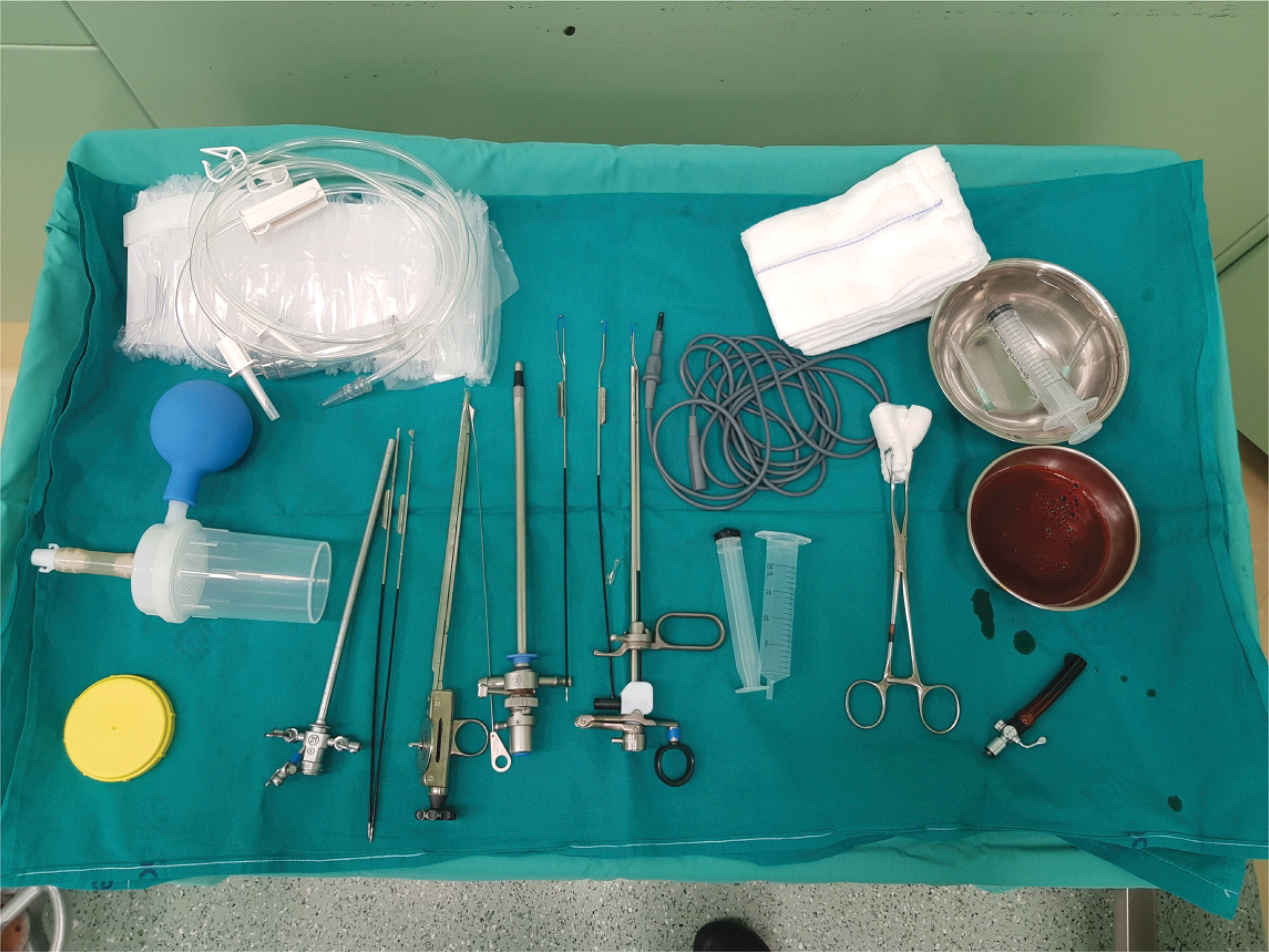
Instruments on the table.
OTIS urethrotome
SACHSE urethrotome
Ch. CYSTOSCOPE
RESECTOSCOPE Munchen or Iglesias with loop, Collin's knife, and rollerball
OPTICS: 0°, 12°, or 30° angle.
ELLICK evacuator
or 22 Ch THREE-WAY DUFOUR CATHETER
GUIDEWIRE: Polytetrafluoroethylene or hybrid (available)
DOUBLE J STENT (available)
Ch. OPEN-ENDED URETERAL CATHETER (available)
Surgical Steps
Access to the bladder and preliminary inspection
Introduction of the resectoscope should be performed under direct vision to observe the whole urethra, which could be affected by urothelial tumors. Inspection of the whole bladder mucosa should be performed to identify bladder tumors (Supplementary Video S3). Number, size, macroscopic appearance, and location should be described. 15
Resection of the tumor(s)
The area to resect should be divided into sectors, and each sector should be completed before moving on to the next sector. Resection should start from the upper part of the tumor and move down to the tumor base. Hemostasis should be achieved at the tumor base before moving to the next sector (Supplementary Video S4). When resecting on the lateral wall, surgeon should be aware of possible obturator nerve stimulation, which leads to the adductor's muscle spasm and bladder wall movement, causing accidental bladder perforation (Supplementary Video S5). Before starting the resection, coagulation of the mucosa should be performed to test obturator sensitivity. Resection on the lateral wall should be carried with the resection loop positioned parallel to the mucosa to use its edge and avoid deep perforation in case of spasm (Supplementary Video S6).
Resection on the bladder dome could be helped using the nondominant hand to compress the lower abdomen, obtaining lowering of the bladder dome and easier approach to it (Supplementary Video S7).
Biopsy of tumor base
After resection of the exophytic area is completed, additional deep and marginal specimens should be taken and separately sent for pathology analysis. 16 The presence of detrusor muscle (except in TaG1/LG tumors) is really important and is a surrogate marker of the quality of resection, also predicting recurrence at first endoscopic follow-up. 17
Hemostasis
Hemostasis should be achieved by targeted electrocauterization of visible bleeding vessels, paying attention not to fulgurate ureteral meatus (Supplementary Video S8).
Troubleshooting
Both cold knife urethrotomes (Otis for the anterior urethra, Sachse for the posterior urethra) and 0° optic should always be available to manage possible urethral strictures. Urethral strictures are frequent when previous endoscopic surgeries are performed. A guidewire and a ureteral Double-J stent should also be available, to be used in case of meatal resection to guarantee a correct wound healing and avoid stenosis.
Bladder perforation might accidentally occur during TURB because of obturator nerve jerk or deep resection. The risk of perforation is greater in patients with low body mass index and women who have thinner bladder walls. Reducing the diathermy current or using bipolar diathermy with a medium distended bladder, and the use of neuromuscular blockade or an obturator nerve block may reduce the risk of perforation. 18
When perforation is extraperitoneal, complete tumor resection and hemostasis should be performed quickly, keeping low intravesical pressure to reduce fluid extravasation and cell seeding (Supplementary Video S9). When perforation is on the bladder dome and intraperitoneal leakage occurs, hemostasis should be achieved and the procedure should be suspended. Early postoperative intravesical therapy should be omitted in case of perforation. Open or laparoscopic repair should be considered immediately or delayed if there are any signs of bowel perforation, peritonitis, or severe intraperitoneal urinary leakage. 19 In case of doubt, cystography can be helpful in diagnosis.
TURBT can be a challenging procedure in the presence of BCa in a diverticulum because of a narrow neck or an acute angle of entry. Moreover, the lack of detrusor muscle makes the diverticulum thinner and prone to perforation in case of deep resection. 20
Urologists often deal with men suffering from BCa who need bladder outlet relief at some point in their life or an unexpected bladder tumor during a planned transurethral resection of the prostate (TURP). One of the authors of this article published in 2018 a randomized trial regarding the oncologic safety of concomitant TURBT and TURP, showing that simultaneous surgeries had no difference in terms of recurrence, with a reduction in time of the first recurrence. 21 A recent systematic review confirmed that simultaneous TURB and TURP can be safely performed with no increased risk of bladder recurrence and progression. 22
Modification of Technique
To increase the performance and to improve the quality of traditional monopolar TURBT, new imaging technologies (photodynamic, narrow-band imaging), energy sources (bipolar energy, lasers), and techniques (en bloc resection) have recently been introduced in clinical practice, showing promising results. 23 En bloc resection of bladder tumor (EBRT) has recently gained increased interest (Supplementary Video S10). EBRT relies on the concept that removal of the tumor in its integrity rather than in pieces would achieve (1) complete resection, (2) a more precise and controlled resection with potentially fewer complications (bleeding, perforation), (3) better sample orientation for histopathology analysis, (4) presence of detrusor in the specimen, and (5) less tumor seeding on normal urothelium by tumor fragments compared with traditional TURBT. 24 The main goal of ERBT is complete resection and presence of detrusor in the specimen to make repeat/restaging TURBT useless (Fig. 3). EBRT can be performed with the support of either mono/bipolar energy (standard loop or Colling's knife) or laser fiber (mainly holmium or thulium). Despite those benefits, EBRT has also significant limitations. 24 First of all, there is no standardization in surgical steps. Second, EBRT can be inappropriate for multifocal tumors, flat lesions, and tumors >3 cm that may require additional equipment for specimen extraction. Finally, EBRT can be challenging in the presence of tumors located at the bladder dome.

En bloc specimen of 1.5 bladder tumor.
EBRT with laser support: physical propriety
Holmium and thulium are the most suitable lasers for EBRT because of their shallow penetration (0.4 and 0.2 mm, respectively), and excellent hemostatic propriety. 25 Thulium laser operates at 1940–2013 nm wavelength, in a continuous wave mode, which enables accurate cutting of the tissue. 25 Holmium laser works at 2100 nm wavelength, in a pulsed wave mode. Each pulse generates a steam bubble that detaches tissue layers by splitting them apart. 25
EBRT with thulium laser support: surgical technique
The procedure is performed in a standard lithotomy position. Laser energy is transmitted using an 800-nm front-firing fiber, introduced in a continuous-flow resectoscope with a dedicated working channel, and mounted with a 30° optic. Laser power is usually set at 20–30 W. Sodium chloride 0.9% solution is used as continuous irrigation. A standard urethrocystoscopy is the first step. After ureteral orifices identification, a superficial and circumferential incision of the mucosa, with a 5 mm safety margin of normal-appearing tissue, is carried out all around the lesion. Once the mucosa is incised, the resection is more superficial along the peripheral part of the tumor and proceeds deep to the detrusor resecting from the periphery to the tumor base, using the vaporesection effect of the thulium laser and the blunt dissection of the resectoscope tip. The irrigation fluid helps in uplifting the lesion. The detrusor layer should be reached at the beginning of the dissection, which should carry on along the muscular plane from the periphery to the center of the tumor base. During resection, bleeding vessels are punctually coagulated (Supplementary Video S11).
The resection should be performed filling the bladder to a medium capacity to avoid overstretching of tissue, which could make it difficult to identify the dissection plane.
The shallow penetration of the thulium laser virtually eliminates obturator nerve reflex in a tumor located in the lateral wall.
Once the tumor has been detached from the bladder wall, additional resection or biopsies of the tumor base can be performed and sent for histologic assessment separately if the surgeon has any doubt of tumor left in the site of resection.
The resected tumor can be retrieved using different approaches. Small tumors (<3 cm) can be evacuated through the resectoscope sheath or extracted using a standard Ellik evacuator or a grasper inserted into the working channel. Tumor retrieval can be challenging in the case of larger specimens (3–4 cm), which can be extracted through a nephroscope sheath using laparoscopic forceps or an endo bag used for vascular surgery. 24 Larger tumors can also be retrieved by the division of specimen into two to three pieces after resection. 26
EBRT with holmium laser follows the same principles of en bloc resection thulium laser. Laser energy is usually set at 1–2 J and frequency at 40–50 Hz using a 550-μm end-firing laser fiber.
Postoperative Care
A three-way silicone Dufour tip 20 Ch. or 22 Ch. catheter is applied after surgery with continuous saline irrigation. Postoperative single instillation is controversial. 27 Saline irrigation and catheter are removed on the second postoperative day and the patient is discharged after spontaneous micturition. If an experitoneal bladder perforation occurs, we suggest suspending irrigation and remove the catheter after 7 days.
Clinical Outcomes of Standard TURBT vs EBRT
Compared with standard TURBT, EBRT owns the fundamental principles in cancer surgery, which are resection of the tumor in one piece, complete resection with a safe margin, intact specimen for accurate histologic assessment, and avoiding fragmentation with lowering floating cells and tumor seeding. These advantages have led EBRT to gain global interest in the past 10 years.
A recent review of randomized clinical trials showed that EBRT had a longer operative time than standard TURBT 28 ; however, the advantages of EBRT stood out in the lower rate of bladder perforation and shorter irrigation time but no difference was found in catheterization time and length of hospital stay. 28 Surprisingly, the presence of detrusor muscle in specimens was similar between TURBT and EBRT. 28 Finally, the recurrence rate was also similar at 12, 24, and 36 months. 28
Current evidence showed that EBRT, especially with laser support, had a better safety profile than TURBT, significantly lower catheterization time, hospital stay, obturator nerve reflex, bladder perforation, and hospital stay. 28 –30 Moreover, EBRT demonstrated a higher rate of detrusor muscle than TURBT and a lower recurrence rate, particularly at 24 months. 28,29 Nevertheless, the mentioned results were considered to be at low certainty of evidence. 28
Despite lacking high-quality data for making a robust recommendation, several important messages can be suggested for resecting bladder tumors.
EBRT should be considered for treating nonmuscle invasive BCa; bipolar energy is widely available and en bloc resection can be achieved using a standard loop or Colling's knife. Large (>3 cm) and multiple tumors (>4) and tumors located at the dome/anterior wall are challenging to resect en bloc, particularly for novices. Conventional TURBT should be preferred in those cases. If a circumferential margin of 5 mm and a correct plane development can be ensured, no additional biopsies of the base and margins are routinely required, but a second look resection should be performed in case of doubt of tumor persistence. A single dose of immediate intravesical chemotherapy or Bacillus Calmette–Guérin (BCG) therapy is safe to administer after EBRT. 29 Follow-up surveillance should follow current guidelines regardless of the surgical approach.
Footnotes
Authors' Contributions
L.D. carried out conceptualization and supervision. M.A. and D.C. took care of methodology and article writing. A.C. and F.E. performed video editing. R.M.S. was in charge of supervision.
Patient Consent Statement
The author(s) have received and archived patient consent for video recording/publication in advance of video recording of procedure.
Recommended Videos from Videourology
1. Videourology 2015 Vol. 29, No. 3.
Laser En Bloc Resection of Bladder Tumors for Staging and Treatment of Primary Bladder Cancer.
Mathias Wolters, Mario W. Kramer, Stephan Jutzi, Florian Imkamp, Mahmoud Abbas, Udo Nagele, Axel Stuart Merseburger, Markus Antonius Kuczyk, and Thomas R.W. Herrmann.
Author Disclosure Statement
No competing financial interests exist.
Funding Information
No funding was received for this article.