
Abstract
Introduction:
Laser lithotripsy can cause excessive heating of fluid within the collecting system and lead to tissue damage. To better understand this effect, it is important to determine the percentage of applied laser energy that is converted to heat and the percentage used for stone ablation. Our objective was to calculate the percentage of laser energy used for stone ablation based on the difference in fluid temperature measured in an in vitro model when the laser was activated without and with stone ablation.
Methods:
Flat BegoStone disks (15:5) were submerged in 10 mL of deionized water at the bottom of a vacuum evacuated double-walled glass Dewar. A Moses 200 D/F/L laser fiber was positioned above the surface of the stone at a distance of 3.5 mm for control (no stone ablation) or 0.5 mm for experimental (ablation) trials. The laser was activated and scanned at 3 mm/second across the stone in a preprogrammed pattern for 30 seconds at 2.5 W (0.5 J × 5 Hz) for both short-pulse (SP) and Moses distance (MD) modes. Temperature of the fluid was recorded using two thermocouples once per second.
Results:
Control trials produced no stone ablation, while experimental trials produced a staccato groove in the stone surface, simulating efficient lithotripsy. The mean temperature increase for SP was 1.08°C ± 0.04°C for control trials and 0.98°C ± 0.03°C for experimental trials, yielding a mean temperature difference of 0.10°C ± 0.06°C (p = 0.0005). With MD, the mean temperature increase for control trials was 1.03°C ± 0.01°C and for experimental trials 0.99°C ± 0.06°C, yielding a smaller mean temperature difference of 0.04°C ± 0.06°C (p = 0.09).
Conclusions:
Even under conditions of energy-efficient stone ablation, the majority of applied laser energy (91%–96%) was converted to heat.
Introduction
Ureteroscopic laser lithotripsy has become the dominant modality for the treatment of urinary stones, and the introduction of high-power lasers (≥100 W) has widely expanded the laser settings that are available. However, use of higher power settings, especially higher pulse frequency, can result in excessive temperature elevation of the fluid within the collecting system and thermal injury to adjacent structures. 1 –9 In June 2021, Olympus issued a voluntary recall of its Soltive™ Super Pulsed Thulium Fiber Laser System after several patients suffered ureteral thermal injury from laser lithotripsy procedures. 10 To properly manage thermal risk, a better understanding of fluid heating during laser lithotripsy is needed.
While much of the applied laser energy during laser lithotripsy creates vapor bubbles and heats the fluid, it seems intuitive that some portion of the applied laser energy will be used for stone comminution and lessen the amount converted to heat. However, the percentage of laser energy used for comminution and the percentage converted to heat have not been clearly established. Furthermore, in two previous studies, laser stone ablation has been reported to more greatly increase the temperature of fluid in the collecting system compared with laser activation in the absence of stone. 3,11 This is surprising as stone breakage is driven by a thermally induced breakdown of the mineral that is highly endothermic. 12,13
To more rigorously explore the apportionment of energy to stone comminution and to fluid heating, a vacuum evacuated double-walled glass Dewar was created to provide precise, quantitative assessment of temperature change during laser activation. The objective of this study was to compare the difference in fluid temperature elevation when 75 J of laser energy was delivered 3.5 mm from the stone surface (no stone ablation) vs 0.5 mm from the stone surface (stone ablation).
Materials and Methods
Experiments were conducted within a custom vacuum evacuated double-walled glass Dewar (160 mm height, 32 mm diameter, and 10 mm wall thickness) (Fig. 1). Ten flat circular BegoStone disks (15:5 formulation; 32 mm diameter and 4.5 mm thickness) were cured and dried overnight. BegoStone disks were hydrated in deionized water at room temperature (22.02°C ± 0.20°C) before use and secured at the bottom of the Dewar. Two Type-T needle thermocouples (Physitemp) were affixed to the opposite sides of the inner wall of the glass cylinder with the tip of the needle 6 mm above the surface of the stone and 1.5 mm away from the wall. Thermocouple measurements were recorded once each second.
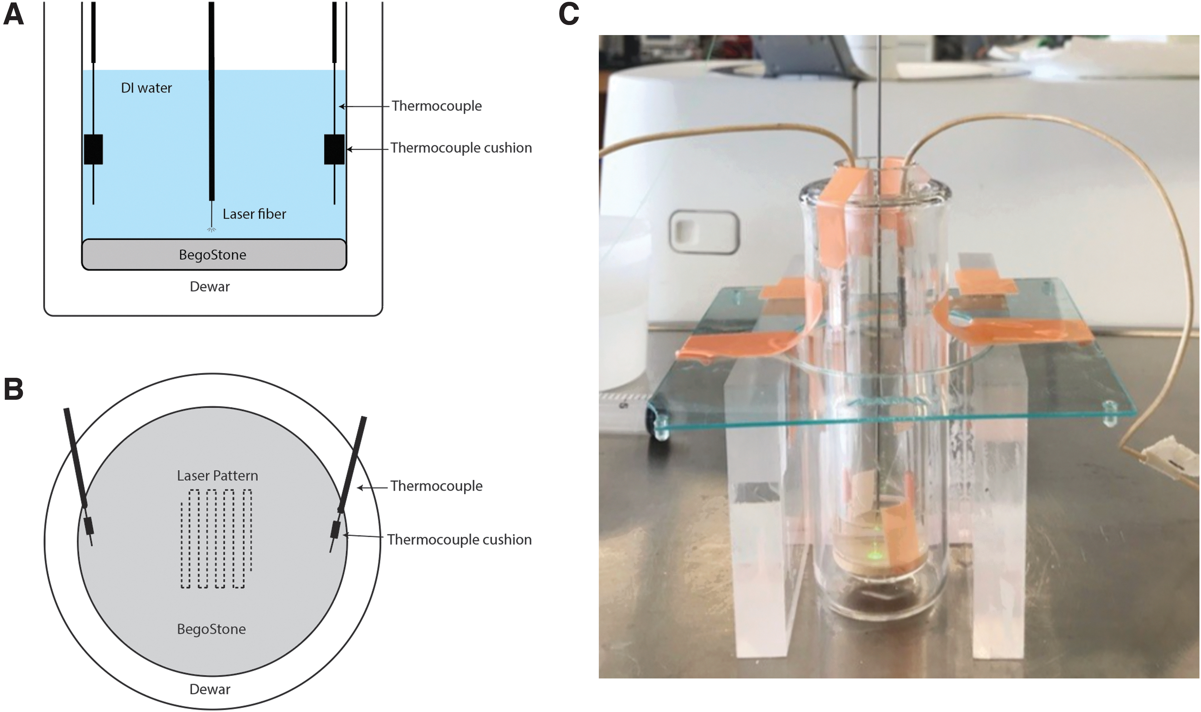
For each laser trial, the Dewar was filled with 10 mL of room temperature deionized water. A Moses 200 D/F/L laser fiber (230 μm core) was positioned at a standoff distance with the tip either 3.5 mm (control) or 0.5 mm (experimental) above the surface of the stone. Baseline temperature was recorded for 20 seconds, and then the laser (120 W holmium:YAG pulse 120; Lumenis) was activated at 2.5 W (0.5 J × 5 Hz, short pulse [SP]). The laser fiber was scanned across the stone surface at 3 mm/second for 30 seconds using a three-dimensional positioning system following a preprogrammed pattern (MATLAB; MathWorks). A total of 75 J of energy was delivered. After laser activation was complete, the laser fiber was removed and the fluid within the Dewar was manually mixed to determine fluid temperature.
After each trial, the glass Dewar was emptied, rinsed, dried, and refilled with 10 mL of room temperature deionized water. Laser pulse energy was measured with a power meter (Ophir) before and after each laser trial. The fiber was cleaved when the pulse energy had decreased by 0.10 J below the target output of 0.50 J.
To generate paired comparisons, two trials were performed for each stone: first at 3.5 mm laser standoff (control trial), and then later at 0.5 mm laser standoff (experimental trial). After the control trial, the stone was visually examined and placed in a room temperature water bath for temperature equilibration.
Fluid temperature was determined once per second by calculating the average value of the two thermocouples. Temperature rise was calculated as the difference between baseline temperature (20 seconds) and plateau temperature (170 seconds). A paired two-tailed t-test was conducted to compare the mean temperature rise from control and experimental trials. Statistical analysis was performed using Microsoft Excel (Redmond, WA). After completion of the SP laser trials, the study was repeated in ten additional BegoStone discs with Moses distance (MD) pulse mode at 0.5 J × 5 Hz.
Results
Control trials with the laser fiber tip positioned 3.5 mm above the surface of the stone produced no visual ablation on the stone surface. Experimental trials with the laser fiber tip positioned 0.5 mm above the stone produced a staccato groove consisting of a series of equally spaced pinpoint ablation craters (Fig. 2).
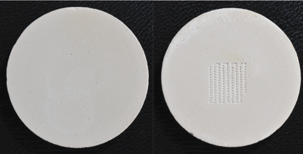
BegoStone disc after control trial (3.5 mm laser fiber-to-stone standoff distance; left) and after experimental trial (0.5 mm standoff distance; right). The pattern of laser ablation is apparent in the experimental trial. Color images are available online.
Fluid temperature in the Dewar increased upon activation of the laser in each trial. A small peak was noted in each temperature curve corresponding to the start of manual mixing, followed by a decline toward a plateau. Temperatures appeared to rise more steeply and to a higher peak temperature in control trials compared with experimental trials (Fig. 3A, B). Error bars between control and experimental temperature curves did not overlap as temperatures settled to plateau values in SP trials (Fig. 3A), while error bars did overlap with MD trials (Fig. 3B).

In SP mode, the mean temperature increase for control trials was 1.08°C ± 0.04°C and for experimental trials 0.98°C ± 0.03°C (Table 1). Assessing the differences in paired trials, this yielded a mean temperature difference of 0.10°C ± 0.06°C (p = 0.0005).
Temperature Change of Fluid Within the Glass Dewar for Each Trial (Short Pulse)
Temperature difference was calculated between the paired trials for each stone. (p = 0.0005).
In MD mode, the mean temperature increase for control trials was 1.03°C ± 0.01°C and for experimental trials 0.99°C ± 0.06°C (Table 2). The mean temperature difference was 0.04°C ± 0.06°C (p = 0.09). Experimental trials resulted in 91% and 96% of the temperature elevation of control trials for SP and MD modes, respectively. Thus, only 9% and 4% of the energy applied for SP and MD modes, respectively, during experimental trials did not contribute to temperature elevation.
Temperature Change of Fluid Within the Glass Dewar for Each Trial (Moses Distance)
Temperature difference was calculated between the paired trials for each stone. (p = 0.09).
Discussion
In this study, the increase in fluid temperature for laser activation without stone ablation was on average greater than the temperature increase with stone ablation, indicating that 91% to 96% of the applied laser energy was converted to heat. The experimental trials were designed to maximize efficiency of laser stone ablation, by ensuring each pulse impacted a fresh location on the stone surface. Irrespective of pulse mode, the vast amount of applied laser energy is converted to heat even during efficient laser lithotripsy.
There are theoretical reasons to believe there may be differences in fluid heating based on pulse mode. While Ho:YAG laser comminution is largely attributable to photothermal effects, there is suggestion that the photoacoustic effects may contribute particularly with shorter laser pulses. 14 –16 The formation and collapse of vapor bubbles with greater intensity, as seen with photoacoustic processes, might be expected to yield a greater temperature rise than a photothermal mechanism where more energy may be expended to elevate the stone to threshold temperature and then drive chemical breakdown of the stone. In addition, one might expect MD mode to deliver energy to the stone through a more efficient vapor channel, thus resulting in more energy used for stone comminution and less for fluid heating. This hypothesis is supported by previous work demonstrating that the single-pulse crater volume for MD was greater than SP at a 0.5 mm distance. 17
However, in the current study, the temperature difference between control (3.5 mm standoff distance) and experimental (0.5 mm standoff distance) with MD mode was smaller than seen with the SP mode and did not reach statistical significance. While these data suggest that the fluid temperature rise may be minimally greater with MD than SP, both sets of data support the conclusion that only an extremely small fraction of energy is used for stone comminution.
Interestingly, two other research groups found that fluid temperature increased by a greater amount during laser activation in the presence of a stone compared with laser activation without a stone. 3,11 In these studies, a gypsum model or calcium oxalate stone was placed in a fluid-filled tube where laser activation occurred. A subsequent comparison of no stone with calcium oxalate stone in an ex vivo model also demonstrated a marginal increase in temperature with stone present. 4 Although the cause of these findings is uncertain, the occurrence of a laser-induced exothermic reaction has been suggested. 4 However, calorimetric data on gypsum indicate that thermal breakdown is accompanied by dehydration, and this is an endothermic process that absorbs heat from the system. 12,13 This is likely to be true of most kidney stone materials as well because dehydration reactions are almost always endothermic.
Alternative explanations for the findings in these previous studies include volume displacement of some of the fluid by stone in the testing apparatus and interruption of normal fluid flow patterns. The presence of a gypsum stone displaces some water and would be expected to lower the heat capacity of the system by ∼1 J/mL°C of displaced water. With a lower heat capacity, delivery of the same energy would result in a greater temperature. Alternatively, the presence of stone may disrupt irrigation flow patterns and prevent complete mixing of fluid throughout the whole volume of the tube, and in this way artificially increase the measured average fluid temperature.
To avoid these same issues in the current study, a BegoStone disc was consistently in place during laser activation, while only the standoff distance between the laser fiber and stone changed. Therefore, there was no change in fluid volume displacement or flow patterns between the control and experimental trials. Manual mixing of the fluid after each laser trial ensured that fluid mixing was complete and homogenous.
The experimental trials in this study were designed to reflect conventional pulse energy settings and optimize stone ablation efficiency to produce the maximal difference from control trials. This was accomplished by using a small (0.5 mm) laser fiber to stone distance and low pulse frequency. The laser fiber was scanned across the stone surface at 3 mm/second to maximize stone exposure by ensuring that each laser pulse was delivered on a region of untreated stone, thus avoiding overlap of laser ablation craters. A standoff distance of 0.5 mm for experimental trials also ensured that the fiber would not drag against the stone during scanning while keeping in close contact with the stone.
Even with these conditions of maximal efficiency, the percentage of energy not converted to heat within the system and therefore available for stone ablation is quite small (9% for SP and 4% for MD). During clinical laser lithotripsy, with lower laser pulse strike rates, 18 the percentage of applied energy actually used for stone ablation is likely much lower than the 4% to 9% determined here under optimal conditions.
Several limitations should be noted. BegoStone is a commonly used stone model but may not fully replicate the thermal and chemical properties of real stones. If we use a stone with different chemistry, it is conceivable that different results could be obtained (e.g., uric acid may have exothermic decomposition), although our hypothesis is that calcium oxalates stones will also only undergo endothermic decomposition and thus track the results seen with BegoStone. As previously discussed, experimental trials were done under conditions to maximize stone ablation efficiency. The percentage of energy calculated in this study that is available for stone ablation provides an upper bound of what is scientifically possible but is likely several folds lower in the clinical environment. In addition, only a single-pulse energy was tested with two different pulse modes in this study.
It is conceivable that energy transmission to the stone may be enhanced with greater pulse energy resulting in greater temperature difference between the experimental and control groups. However, this is unlikely as King and colleagues demonstrated >80% transmission of 1 J laser pulses with and without modulation through 1 mm of water. 14
The knowledge that only a small percentage of applied laser energy is available for stone ablation, with the rest converted to heat, emphasizes the importance of properly managing laser settings and energy delivery during laser lithotripsy to avoid thermal injuries. Furthermore, the results from previous in vivo and in vitro studies assessing temperature elevation in a variety of model systems with laser activation, but without stone present, can be applied to the clinical environment with a high degree of confidence. 19 Furthermore, the method of assessment developed in this study could be used to generate an efficiency metric for comparing different laser wavelengths, laser parameters, or lithotripsy techniques.
Future work is planned to assess the energy required for ablation of each of the common urinary stone compositions. This thermally isolated Dewar and stone ablation assay will also be used to assess temperature change, and hence the efficiency of energy delivery of laser pulse modulation strategies at varying laser fiber-to-stone distances.
Conclusions
Laser activation in a 10 mL fluid volume for 30 seconds at 2.5 W without stone ablation results in a 0.10°C ± 0.06°C greater temperature elevation compared with laser activation with stone ablation using the SP mode, and 0.04°C ± 0.06°C for MD. This difference equates to only 9% and 4% of applied laser energy being available for stone ablation, respectively. Even under conditions of energy-efficient stone ablation, the vast majority of applied laser energy (91%–96%) appears to be converted to heat.
Footnotes
Authors' Contributions
J.J.D. performed the experiments and collected the data, with assistance from M.M.L., N.R.K., and W.W.R. A.J.M. provided the custom Dewar. J.J.D. and W.W.R. analyzed the data and wrote the article. T.L.H., K.R.G., and A.J.M. provided critical revisions. W.W.R. conceived and managed the study, with input from J.J.D., T.L.H., K.R.G., and A.J.M. All authors reviewed the final article.
Author Disclosure Statement
K.R.G. has consulting relationships with Boston Scientific, Olympus, Karl Storz, and Coloplast. W.W.R. has a consulting relationship with Boston Scientific. All other authors have no disclosures.
Funding Information
Funding for this study was provided, in part, by a Faculty Catalyst Award, Department of Urology, University of Michigan, and, in part, by a research grant from Boston Scientific.