Expert Panel
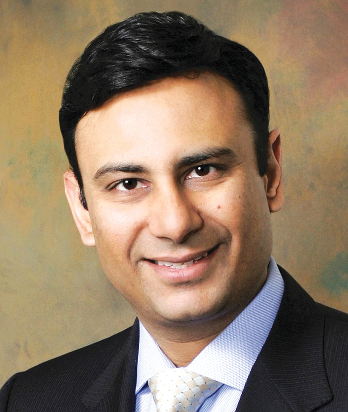
Dr. Mantu Gupta is professor of urology, Icahn School of Medicine at Mount Sinai, chair of urology at Mount Sinai West and Mount Sinai St. Luke's Hospitals, and director of endourology and stone disease for the Mount Sinai Health System. Dr. Gupta is recognized as a world leader in the research and treatment of urinary stone disease, ureteropelvic junction obstruction, urinary tract obstruction, and upper tract urothelial malignancies, having pioneered many of the techniques in current use. He is recognized as one of the leading endoscopic, percutaneous, and minimally invasive surgeons in the world, having performed over 10,000 major endourologic procedures. Dr. Gupta is unique in that he employs a nurturing, compassionate, and holistic approach to the management of stone disease, utilizing alternative medical, nutritional, and preventive strategies. Prior to joining Mount Sinai, Dr. Gupta served as associate professor, director of endourology and director of the Kidney Stone Center at Columbia University Medical Center for the past 18 years. The recipient of numerous honors and awards, Dr. Gupta was given a Presidential Gold Medal by President Ronald Reagan for his academic achievements and has served as a visiting professor both within the United States and throughout the world. He is a frequent lecturer and moderator at national and international meetings and has published extensively in major peer-reviewed journals and authored numerous book chapters. In addition, Dr. Gupta teaches various surgical skills courses throughout the world and has served as editor and guest editor for numerous publications including the Journal of Endourology, the Journal of Urology, the Indian Journal of Urology, and Urology. Dr. Gupta was a member of the prestigious Honors Program in Medical Education at Northwestern University where he earned a combined BS-MD degree with distinction and was elected to the Alpha Omega Alpha Honor Society. He subsequently did his urology residency at the top ranked University of California at San Francisco and completed a fellowship in endourology at Long Island Jewish Medical Center under the tutelage of the founding father of endourology.
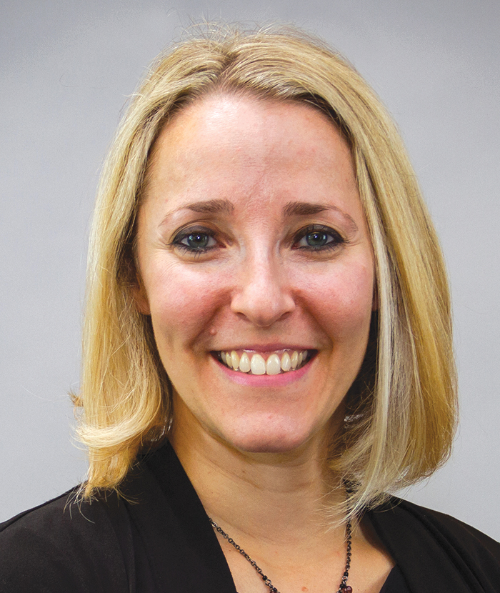
Dr. Michelle Jo Semins is a urologic surgeon and chief of the division of urology at West Virginia University Wheeling Hospital since 2022. She is also clinical professor at the West Virginia University School of Medicine. She received her undergraduate degree from the University of Michigan in 2001 and her medical degree from the University of Pittsburgh in 2005. She then completed her general surgery internship in 2006 and urology residency training in 2011 at Johns Hopkins Hospital with additional specialty training in the field of endourology. Dr. Semins has a clinical research focus in kidney stones, and she started the multidisciplinary kidney stone clinic at her hospital in 2012. She is a past awardee of the Young Investigator Research Grant by the Northeastern Section of the American Urological Association (AUA). Her special interests are reducing radiation exposure to both patient and provider, managing kidney stones during pregnancy, and minimizing infections in the management of kidney stones. She is a published author of 40+ articles, 50+ abstracts, and 9 book chapters, and serves as the associate editor for the journal Urology. Dr. Semins is active in the Northeastern Section (NS)AUA/AUA. She was selected to participate in the 2018–2019 AUA Leadership Program, sits on the NSAUA Board as the Pennsylvania Representative, and is past chair of the NSAUA Scholarship Research Committee. She currently serves on the AUA Quality Improvement and Patient Safety Committee, the AUA Urologic Video Education Committee, and the AUA Publications Committee. She is chair of the Taskforce on Diversity and Inclusivity for the Society of Endourology. She is also an active member of the Society of Women in Urology and Research on Calculus Kinetics Society. Dr. Semins is a recognized leader in the field of endourology and has been invited faculty at conferences and courses both nationally and internationally. She was named Young Urologist of the Year by the AUA in 2016.
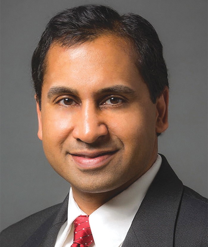
Dr. Ravi Munver is vice chair of urology, director of minimally invasive and robotic urologic surgery, chief of living donor kidney surgery, and the founding director of the robotic surgery and minimally invasive urologic oncology fellowship at Hackensack University Medical Center. He is a core professor of urology at Hackensack Meridian School of Medicine. Dr. Munver received his undergraduate education at Cornell University where he graduated magna cum laude and Phi Beta Kappa with a BA from the College Scholar Honors Program. He received his medical education at Cornell University Medical College and completed residency training at Duke University Medical Center. He received advanced fellowship training in endoscopic, laparoscopic, and robotic surgery at New York-Presbyterian Hospital/Weill Cornell Medical Center and is a Fellow of the American College of Surgeons. Dr. Munver is an internationally renowned leader in endoscopic, laparoscopic, and robotic surgery. He is a passionate academician and educator and has lectured in 26 countries spanning 5 continents. He teaches at national and international conferences and regularly serves on the faculty of courses sponsored by the American Urological Association and the Endourological Society. As an active educator, he has mentored more than 50 residents and 15 fellows. He has authored more than 450 scientific manuscripts, articles, book chapters, and abstracts. He has received awards from the National Institutes of Health (NIH), American Urological Association, Endourological Society, and Society of Laparoscopic and Robotic Surgeons. Dr. Munver's clinical interests include endourology, minimally invasive and robotic urologic surgery, prostate enlargement, and urologic oncology. In addition, Dr. Munver has a unique interest in optimizing clinical outcomes through innovation and technology. His extensive research has allowed him to develop a unique approach for the prevention and management of urologic complications. Dr. Munver's career has been spent at prestigious academic institutions where he has held numerous leadership positions. He is also very active in the American Urological Association, the New York Section of the American Urological Association, Endourological Society, and Society of Laparoscopic and Robotic Surgeons. Dr. Munver was awarded the Alpha Omega Alpha Volunteer Clinical Faculty Award by the AOA National Honor Medical Society and the Golden Apple Award for excellence in teaching. Dr. Munver has been recognized for his compassionate care of patients and received the Gold DOC Award for Humanism in Medicine. He is known to be humble, honest, hardworking, and the embodiment of a servant leader who is known for personally stepping up to support and help others in need. He remains devoted to advancing medicine with a personal touch.
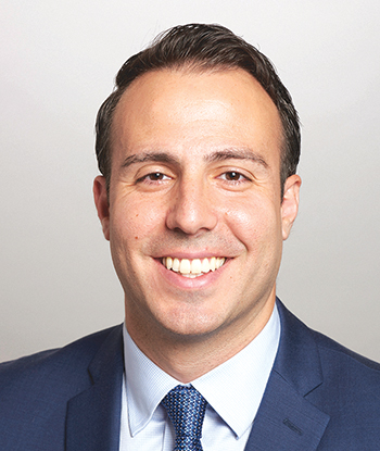
Dr. William Atallah is an assistant professor of urology at Kidney Stone Center of Mount Sinai, as well as the Chief of Endourology at Elmhurst Hospital. William Atallah, MD, MPH, specializes in endourology and urinary stone disease. He treats kidney stones using a variety of techniques such as extracorporeal shockwave lithotripsy, ureteroscopy, percutaneous nephrolithotomy, and mini-percutaneous nephrolithotomy. Besides treatment of the stone, Dr. Atallah focuses on kidney stone prevention using dietary changes. He also treats patients with flank pain, upper tract urothelial cancer, hydronephrosis, blood in the urine, difficulty urinating, slow urinary stream, urinary retention, hydroceles, and phimosis. He received both his MD and MPH degrees at St. George's University, located in Grenada, West Indies. Dr. Atallah subsequently did his urology and general surgery residency at SUNY Downstate University Hospital. A fellowship in endourology was completed at the Mount Sinai Health System under Dr. Mantu Gupta, director of endourology and stone disease at Mount Sinai. During his fellowship, he won 1st place in the clinical category of the annual essay contest of the Endourological Society and was asked to speak in Paris, France, for his work: “A prospective randomized controlled trial comparing the efficacy of low-oxalate diet versus Vitamin B6 and magnesium supplementation versus both in idiopathic hyperoxaluria.” Currently a member of the American Medical Association, the American Urological Association (AUA), and the Endourological Society, he has published his work and presented at multiple conferences. At the AUA general conference, he presented his work about the impact of the African American race on outcomes after prostatectomy in an equal access center. In Vancouver, Canada, at the World Congress of Endourology, he presented on how continuing aspirin does not increase blood loss from percutaneous nephrolithotomy.
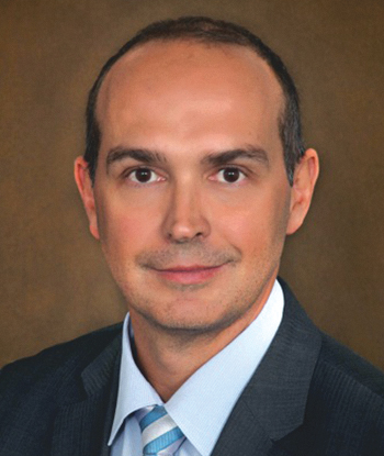
Dr. Wilson Molina is a professor of urologic surgery at the University of Kansas. Dr. Molina is a urologist dedicated to kidney stone disease. Dr. Molina was born and raised in Sao Paulo, Brazil, where he completed his medical school and general surgery and urology residency at the Federal University of Sao Paulo. Just after completing his residency, Dr. Molina moved to Cleveland, Ohio, and began a fellowship in urology minimally invasive surgery at Cleveland Clinic. He went back to Brazil for a few years, where he worked as a full-time urologist in an academic program in Sao Paulo, as well as at a kidney transplant service in the most prestigious hospital in the country. During this time, he realized his passion for treating patients with kidney stones. In 2008, Dr. Molina moved back to the United States, this time to Denver, Colorado, where he completed his second fellowship in urology at the University of Colorado and Denver Health Medical Center. After his fellowship, he was hired as a faculty member at the University of Colorado and was an attending urologist at Denver Health Medical Center. He became the chief of the endourology-stone program and was responsible for the treatment of complex kidney stones cases. In September 2018, Dr. Molina moved to the Kansas City area, when he joined the department with the academic ranking of professor. Academically, Dr. Molina has been productive in his career. He has published more than 50 peer-reviewed articles, and he is an active member of the South Central Section of the American Urological Association (AUA), the AUA and the Endourological Society. He also has a passion for educating medical students, residents, and fellows and was awarded the Faculty Teaching Award in 2011, while at the University of Colorado. Dr. Molina is dedicated to the treatment of kidney and ureteral stones. He performs all of the most advanced minimally invasive endoscopic procedures for stones, including ureteroscopy, external shock wave lithotripsy, and percutaneous nephrolithotomy. He also performs endoscopic procedures for ureteropelvic junction obstruction, upper tract urothelial tumors, and ureteral strictures. His research is concentrated in stone disease, including development of new tools to treat stones more efficiently and less invasively with a faster recovery period for patients. In his spare time, Dr. Molina likes sports activities such as running, golfing, and soccer.
Dr. Gupta: Welcome. My name is Dr. Mantu Gupta. I am the professor of urology, chair of urology, and director of endourology and stone disease at Mount Sinai. I will be your moderator for today's discussion. We will be talking with experts in the field of endourology about the importance of monitoring intrarenal pressure and their initial clinical experiences using the LithoVue™ Elite System, the first ureteroscope with intrarenal pressure monitoring.
Our esteemed panel consists of four endourology experts. Dr. William Atallah is an assistant professor of urology at Mount Sinai and chief of endourology at Elmhurst Hospital. Dr. Wilson Molina is a professor of urologic surgery at the University of Kansas. Dr. Michelle Jo Semins is chief of the division of urology at West Virginia University Wheeling Hospital and a clinical professor at West Virginia University School of Medicine. Dr. Ravi Munver is vice chair and chief of minimally invasive robotic urologic surgery and a professor of urology at Hackensack University Medical Center.
To start things off, let us talk about why having the ability to monitor intrarenal pressure is important to you in your practice.
Dr. Semins: I think this is potentially going to become the standard of care at some point. For me, it is important to know the intrarenal pressures because a lot of evidence has shown that high intrarenal pressures are associated with complications. The biggest concern to me is with regard to sepsis, because I think infection is the most common post-ureteroscopy complication out there.
But it also has to do with barotrauma. It could potentially increase the risk of renal hemorrhage. Those are less common, and I think we know a little bit less about barotrauma. For me, clinically, I am concerned about infections. If I know what the pressures are, I could potentially decrease those complications, specifically infection, afterward. Research needs to be done to prove that, but reducing complications is my ultimate goal.
Dr. Molina: I think flexible ureteroscopy has been evolving for the past 25 years, with better ureteroscopes and better lasers, etc. Now comes a new era of being able to measure real-time intrarenal pressure. It is like driving a car for the past 25 years without a speedometer. We feel that we are safe at some point, but we do not have the measurements to really predict whether you are doing things correctly. Now, we have that. We will need to figure out what is the real intrarenal pressure that is safe or not, and then all the related parameters. As Dr. Semins already mentioned, we understand that postoperative pain and postoperative infection and bleeding are associated with intrarenal pressure. But the exact number and if it is the same number for every patient, we do not even know that, so that is why I am really excited for this new era of measuring intrarenal pressure in real time.
Dr. Munver: My perspective is a little different. I have been in practice now for two decades. My initial experiences with ureteroscopy complications were subcapsular hematomas and perinephric hemorrhage. When patients have these complications, it can lead to pain, needing a blood transfusion, maybe embolization, and then just waiting for these hematomas to resolve. Maybe having a Page kidney afterward. I did some research and looked at the background literature for these types of complications. Of course, we all talk about sepsis. And, yes, that is something that we are aware about. But the perinephric hemorrhage and the subcapsular hematomas are what I have looked at in the literature. We came up with an algorithm. We found there were certain criteria that made patients more prone to have bleeding complications. For example, renal parenchymal thickness under 1 cm. Maybe one specific calyx that was dilated because the stone is obstructing a calyx, not necessarily the entire renal pelvis and calyces, the duration of the operation, and then finally the type of irrigation. Whether you are using a single action pump or whether you are using manual irrigation or a pressure bag. And the only thing that was really controllable is the duration of the operation. So how quickly did the operation end at the degree of irrigation? When I started my practice, we went from where I was training in residency and fellowship to where I am currently as an attending physician. We were using single action pumps. We are using manual irrigation. Looking at my residents, fellows, other attending physicians, there is such a variety and diversity in terms of how these single action pumps are used in terms of how someone actually irrigates—whether they irrigate continuously, not at all, or just as needed. That led me to believe that intrarenal pressures were a substantial contributor to these types of complications. Now, I agree with everybody else about sepsis as well. When you have an infected stone and you have bacteria within the calyces or you have infected urine behind the stone, even though you treat someone and you get a negative urine culture, same concept. The intrarenal pressures are what can be controlled, and to this point, we have never had a modality where we are able to monitor intrarenal pressures. We know about the Whitaker test, we know about obstruction, we know that intrarenal pressures are significant. Now we have the first-generation ureteroscope that can monitor intrarenal pressure. I think there are many applications in terms of our understanding and doing studies to figure out what the optimal irrigation pressures are, including types of irrigation and scenarios that we can utilize to help improve the outcomes for our patients.
Dr. Atallah: I agree with everything my esteemed colleagues have mentioned. Being an endourologist, we all love our toys. And being an endourologist now in 2023 in this new era of all this new technology, having a ureteroscope like the LithoVue Elite is going to be a game changer. We are going to be able to answer all of these questions regarding postoperative pain, postoperative sepsis, and trauma to the kidney. We do not even know what an accurate pressure should be. I think this ureteroscope is going to allow us to have all those answers. And within the same kidney, within the same system, the pressure might be different in a dilated calyx versus a stenotic calyx like Dr. Munver was suggesting, or in the renal pelvis. Then we can discuss access sheaths. Are access sheaths going to decrease our intrarenal pressures? Some of us use access sheaths, some of us do not. Some of us use gravity irrigation, some of us use pressure. Some of us hand pump. This technology will allow us to all have our questions answered.
Dr. Gupta: I agree with all of you. I started my training in urology 33 years ago. I do not remember ever thinking about intrarenal pressure when we were doing ureteroscopy. We did whatever we wanted to. We used hand irrigation, manual irrigation, pumps, all sorts of different things, and never thought about it. Only in the past 10 years has this become an important topic. But, until now, it was just a conversation. It was a theoretical problem. No one had any data whatsoever to back up the claims that high internal pressure can lead to complications. Now we have a tool that can do that. All the fears that we have had over the years, are they founded or unfounded? We may find that it is not founded and the intrarenal pressure does not correlate with any of these things we were fearing. But we will know—if it does or does not, we will know at what level it does. Having that hard data is invaluable to all of us, I think, in our practices. I think that's why the LithoVue Elite is such a game changer and such an important innovation in the field of endourology. I am going to ask you to talk about your experiences with the LithoVue Elite System because all of us have had a chance to try it.
Dr. Atallah: The first time we used the LithoVue Elite here, we used an access sheath. We routinely do not use access sheaths. But, in this case, the stone was about just over a centimeter. So, we figured it would be more beneficial to use an access sheath. I was trained to leave the access sheaths in the proximal ureter. Then when our ureteroscope was in the kidney, and we were lasering the stone, the intrarenal pressure was elevating significantly. That allowed me to make an adjustment and put my access sheath into the renal pelvis. The intrarenal pressure decreased significantly once that was done. If I did not have a ureteroscope like the LithoVue Elite, I would have never noticed that and I would have just continued. The patient did fine postoperatively. The patient could have been fine if I left the sheath in the proximal ureter as well. The fact that we now know and make these adjustments is an invaluable tool. We also started using it for percutaneous nephrolithotomy (PCNL) because the resolution is definitely much improved from the first LithoVue. We have been doing more endoscopic-guided access for PCNL and that allows us to do completely fluoroscopic-free PCNLs. The LithoVue Elite can be used in many ways.
Dr. Molina: I was fortunate to do a handful of cases lately. And really, as I said, it has completely changed my perspective about what I have been doing. With some patients where I used the LithoVue Elite, I did bilateral ureteroscopy. The intrarenal pressure on one side was completely different compared with the other side on the same patient, and they did not have extra hydronephrosis or something like that. It was bilateral stones in different calyces, but completely different intrarenal pressures using the same irrigation system, et cetera. Since I am at an academic institution and residents are working with me, I am typically the one who hands the ureteroscope to them and then I will control the irrigation with single action pump until they experience some difficulties and then I will take over. Typically, working with good residents, they can complete the case. When I look at the intrarenal pressure graphic and using a single action pump, I changed how much irrigation I used. I think this is something that is important; you have the feedback in real time whether you are overdoing irrigation or not.
Dr. Semins: I am fortunate that I have gotten to use the LithoVue Elite like all of you. I have had good initial experiences, starting just with visualization. Compared with the first-generation LithoVue, there is definitely a big difference for me, specifically, when there is some blood in the system. The LithoVue first generation is still excellent visualization. But I really notice a difference when there is a little bit of blood in the system. It is more crisp and clear. So, I have been impressed with that. In terms of the pressure and accuracy and things that I have done to change my practice, I have been able to learn a lot about little maneuvers that help me to decrease the pressure during the case. There have been a couple patients where even if they are pre-stented, I can only get a 10/12 access sheath in. And those patients have had high pressures while I am working. Typically, even if they are low risk for sepsis, I think those are some patients who end up getting sepsis if you go a little bit too long or you have those persistent high pressures. Now with those patients, I am definitely more aware of the fact that their system is narrow and their ureter is narrow and my outflow is not quite as good. I may move the stone to a different calyx. If it is a smaller calyx with not a lot of outflow specifically to that calyx, I will actually move it to a larger upper pole calyx and work there, even if it was an interpolar calyx and it was perfectly fine where it was. I will find sometimes I have lower pressures. If I am trading things in and out and I do not need excellent visualization, I will completely turn my flow off. I have always been a big fan of the automated irrigation machines mostly because I think they are great for staff. I do not need to rely on somebody else. I adjust the actual flow with my stopcock. So, I will turn it completely off when I can and that brings the pressure down. I have also started using aspiration more throughout the case. I used to just aspirate if visualization was poor from contrast when I go into the system. Now I find myself aspirating if the pressure goes a little higher than I want, I will back up to the renal pelvis and suck out some fluid. I find that I have more sustained lower pressures for a period of time. So, I will do that periodically through the case. It has definitely affected how I practice. There have been some patients that I have been nervous about risk of sepsis because of struvite stone or history of sepsis with presentation and just frail patients. With those patients, I have been reassured when I am in there with a 12/14 sheath and their pressures remain low, like in the teens, the entire case. I have not had a patient so far where I have had infection after using LithoVue Elite, and have done some frail higher risk patients. So, overall, the experience has been great. I am looking forward to actually collecting data to see whether it translates into reduced complications by live adjustments that we make.
Dr. Molina: Dr. Semins mentioned “high pressure” and “low pressure.” What is high pressure? What is low pressure?
Dr. Semins: That is a great question. I think that needs to be defined.
Dr. Molina: Exactly. Everything is 40, it seems like, in urology. When it's above 40, we think it is high. But is it really high? Now, we have a new era; we can set up the parameters better than before.
Dr. Semins: I agree. I think there has been evidence over decades that the single action pump can be associated with a high peak, even when people think that they are gently irrigating.
1
–3
When they actually measure pressure, there are high peaks. One thing we don't know is whether a short high peak is more important, or if sustained moderate or high pressures are more important? And what is high pressure? Does the pyelovenous backflow occur at the same pressure for every individual, or does everybody have their own threshold? There was a recent study looking at pigs, and they found that pressures over 90 led to pyelovenous backflow.
4
So, I think it does need to be defined. That is what is so great about this device. It is going to answer many questions that have been unanswered for the past century.
Dr. Gupta: I do not use any pressure irrigation, I only use gravity. I usually hang the bag around 60 to 70 cm of water above the patient. I noted that LithoVue Elite intrarenal pressure monitoring is accurate. If you raise the bag a little bit, it goes up. You lower the bag, it goes down. It is almost an immediate change. I found that the magic number for me seemed to be around 60 cm of water pressure. With gravity, that is what it seemed to go to and then level off. Like inflating a tire, initially you could put a lot of air in and the compliance is good and not much changes. When you get to the higher level, like getting to the limit of the tire, all of a sudden, the pressures sharply rise. I think the compliance and the rate of rise are important. Once you get to a certain level, it rises faster and faster the more you put fluid in. Conversely, when you're aspirating, it goes down quickly. If you can get it down to 10 or 15 and then turn the irrigation back on, again, it takes a long time for it to reach that level where it is full. And then once it is level, it starts to go steeply. So those are interesting things that we would never know if we didn't have these curves that we could look at.
Dr. Munver: I agree with what Dr. Semins said about the visualization, especially during bleeding situations. I have talked to many urologists over the past several years who have used LithoVue and who do high volume ureteroscopic surgery for upper tract urothelial carcinoma. Depending on the situation, surgeons can encounter a significant amount of bleeding. When comparing reusable ureteroscopes with the first-generation LithoVue disposable ureteroscope, some urologists report that the LithoVue visualization is much poorer, and that it is not ideal for dealing with larger or more vascular upper tract urothelial carcinoma. I think with the LithoVue Elite, the enhanced visualization is a true advantage to allow us to use it for not just stones, but also upper tract urothelial carcinoma. I want to share a fun day that I had recently when I did three cases back to back to back. One case I had was a patient with a 1-cm stone in the renal pelvis. I did a ureteroscopy with no access sheath. No matter how gently I was irrigating—and I was in control of the irrigation. I had my resident and fellow doing the ureteroscopy. I was making sure that I was irrigating slowly and gently. As you were alluding to, Mantu, no matter how slow you irrigate, the intrarenal pressures can shoot up. I could not keep the pressures low, even when I was not irrigating. The pressures would still stay high above 60, 70, or 80 mmHg, and I would need to wait about 10-15 seconds for the pressures to decline. The second case I did was a patient with two renal stones—a 7-mm stone and a 2-mm stone. I used an 11F/13F ureteral access sheath with the tip of the sheath in the proximal ureter, not the renal pelvis. I noticed that with the same technique for irrigation, I was able to keep the intrarenal pressures around 40 to 50 mmHg. If I had to irrigate a little bit more I noted little pressure spikes up to 70 or 80mmHg, but I was essentially able to sustain lower pressures with more irrigation. The third case was a patient with a 2-cm renal pelvis stone. I used a 13F/15F ureteral access sheath with the tip positioned within the proximal ureter. No matter how I irrigated—whether I irrigated slowly, moderately, or literally, forceful and rapid irrigation, I could not get the pressures to increase beyond 20 mmHg. These observations made me believe that the access sheath truly helps minimize intrarenal pressures, even if the tip of the sheath was not within the renal pelvis. I think that this was a phenomenal discovery that helped reassure my concept of slow and steady manual irrigation. Basically, depressing the pump syringe till the chamber was empty, and then squeezing—again, ever so slowly. And, yes, sometimes, I may need to increase the rate or force of irrigation as necessary. However, this allowed me to confirm that having a larger access sheath or having an access sheath compared to not having an access sheath, would result in lower intrarenal pressures. No matter how fast I irrigated with the 13F/15F ureteral access sheath, I still could not get those pressures over about 20 to 25 mmHg, which I thought was really groundbreaking and an eye opening to me, my resident, my fellow, and everyone else in the operating room that day.
Dr. Molina: Dr. Munver, this last patient that you mentioned, was he a pre-stented patient?
Dr. Munver: No, he was not a pre-stented patient. I always pass the obturator up first, which was, in this case, 13F, which would have been the same as the outer diameter of an 11F/13F access sheath. Then we tried to make sure that the 13F went up. The reason I did it was because I did not know whether I was going to be pulling out larger stone fragments; I did not see the Hounsfield units so, I did not know how hard the stone was going to be. That is not what I typically do. Normally I use a 13F/15F access sheath in pre-stented patients.
Dr. Molina: The reason I am asking is that we know that the ureteropelvic junction (UPJ) can be completely different from one patient to the other. If the UPJ is narrow and does not drain well, we can see patients that we do an access sheath on the proximal ureter and there is no outflow. Then with patients, especially the pre-stented ones, where the UPJ is wide open, then you can see the flow coming out. I think more than where is the sheath sitting is more how is the outflow coming or not?
Dr. Munver: Agreed. I think all three of these patients had capacious UPJs. So, I think from that standpoint, we were comparing apples and apples. I did not know that ahead of time. But none of the patients had a narrow UPJ and none had a particularly wide UPJ. All of the UPJs were just right. I got lucky in that sense, as I was comparing similar diameter UPJs and placing the access sheath for second and third cases in the same location and not advancing the tips past the UPJ and into the renal pelvis. I think your point is extraordinarily valid. The majority of the time, if you use a 13F/15F ureteral access sheath, it is going to be in a pre-stented patient.
Dr. Gupta: I think there are at least three factors there that determine that pressure. I think one is the difference between the size of your ureteroscope and the sheath you are using. But, as Wilson pointed out, it is any obstruction that is above your sheath. So, if the UPJ is tight around your ureteroscope, it does not matter whether the sheath is below UPJ. That is the fulcrum, the point where the pressure is going to generate. But, also, it is the compliance of the patient's collecting system. The anatomy. If someone has a large to extra large renal pelvis or if someone who has had a prior UPJ, their system is accommodative. They can hold a lot of fluid without the pressure rising. There are other patients who have a tight intrarenal collecting system. And even a small amount of irrigation will increase their pressure. I think in the future, measuring the compliance of a system in some way, maybe using LithoVue Elite, would be something feasible. Almost like a urodynamics of the renal collecting system. And we all see collecting systems where 5 cc is enough. You can fill the entire collecting system just by putting 5 cc of fluid in. And other collecting systems where you could put 20, 30, 40, 50 cc, and they are still not completely full. So I think those are all important points.
Dr. Molina: What is fascinating is that in the past 5, 10 years, with the development of the new laser platforms, our tendency is to say, “OK. Now, I can dust this stone nicely. I can even try to not use a stent near an access sheath.” Correct? Now we are backing ourselves out to like, “OK. Maybe access sheath is something that we're never going to get rid of.” It is really fascinating how the field of endourology goes from, “Let's get rid of the access sheaths now,” to, “Hold on. Wait a minute. Maybe we should keep using access sheath and then stents post-op, et cetera.” It is really fascinating how we shift our field.
Dr. Gupta: I do not use access sheaths at all. Hardly ever. Extremely rare that I will use one. And what using LithoVue Elite taught me is that I can get away with not using an access sheath now, where I could not before, because now I can actually measure that pressure and know whether I am doing something bad. Before, you were always wondering. But using the gravity is important. But it teaches you a lesson. When you get up to that 60 level or the level you feel comfortable with – 40, 60, whatever it is. At that point, you know to stop and aspirate and start again. And I did not have that information before. I just did it on a feeling. Oh, maybe I should aspirate now. My visualization is not as good. I don't see things moving. The flow is extremely poor. Time to aspirate. Now I actually have a number that I can look at and know what to do. I was shocked at how well it went up the ureter. How easily it went up. I went up a few ureters without a guidewire. Just going right in. Not pre-stented. I was really surprised at that because I usually use a smaller ureteroscope and I am able to do that. But, I did not think I could do it with the LithoVue Elite. And we could. Not only that, but the brightness of the image, the clarity, and the resolution were much better than I was expecting and that I had seen before with the prior version of the LithoVue.
Dr. Molina: More importantly, now when you are going to look back in your patients, Mantu, and the patients that you kept the pressure below 60 and see whether they did better than the other patients.
Dr. Gupta: It is going to take a lot of data.
Dr. Molina: We will see when we correlate the data with the outcomes. That is really what is going to make a huge difference.
Dr. Munver: Yes. I completely agree with this because I feel that most of us who are interested in this technology are academic urologists. We are those who are really thinking about the potential benefits. Where can this take us? And how are we going to be able to transmit these data to the urologic community? I think it is fascinating for us to be discussing this, because we all have excitement and interest. We do not have all the answers. Some of the data that we are receiving is justifying some of the hypotheses we had. I sincerely believe that some of the data that we will be getting will allow us to formulate new conclusions.
Dr. Semins: So, Dr. Gupta, if you do not use access sheaths but you see higher pressures consistently throughout your case and you aspirate and it still comes right back in delicate narrow systems, would you consider placing an access sheath? Do you see yourself placing potentially more access sheaths with this information?
Dr. Gupta: Dr. Atallah and I did our first cases together. Both of us do not use access sheaths much. But for this case, what we did was we went up without an access sheath, and we were surprised how well it went up and went to the collecting system. Then we started to irrigate, and we noticed the pressures were going up to 60. We had set our limit at 30 and then we had changed it to 40. And we said, “Oh, no. It's going above our limit. Something's not right. How are we getting pressures of 60? 40 is our limit. We better put an access sheath in.” So, then we put the access sheath in, and when we put it up right up to the UPJ, it actually did not help much. This patient had a relatively tight UPJ. And we said, “Well, this is not helping at all. Now what do we do? It's still going up 60 or more.” And then we started using pressure irrigation, because we figured, “Oh, we're using an access sheath. I never use pressure, but let me use pressurized irrigation.” So, we used pressurized irrigation. Was going up to 90, 100. And we said, oh, now what do we do? So then what I finally did was put the access sheath a little further. And think it was a 12/14 to 11/13?
Dr. Atallah: 11/13.
Dr. Gupta: Then we put it past the UPJ and put the sheath up in the renal pelvis. And then the pressures were low, even with pressurized irrigation. So that was pretty eye-opening for us. But I mean, the way I do it with the bag, it is never going to go above that pressure, because it cannot. The fluid just stops going in. And then I need to aspirate. And the tight collecting system, it is a pain. We must keep aspirating every few minutes to get the pressure down or just keep going with the case, and hopefully the visualization is fine. I think having this information will change how I practice. I probably will use more access sheaths when I see those pressures going up, or we have poor irrigation. How about everyone else? What is the routine practice pattern you guys have in terms of access sheaths, and what size access sheaths and how you put it up. I know that Dr. Clayman has talked a lot about trying to use larger access sheaths to do a better job with our cases. And actually measuring the force at which you put the access sheaths up in terms of Newtons of force. I think he has come up with a magic number of 6. If it takes more than 6 Newtons of force to put an access sheath up, you should go with the smaller access sheath.
Dr. Molina: Dr. Clayman's group at Irvine, they are really pushing the envelope for bigger sheaths. For the past 4 or 5 years, I tried to use less and less because I'm using more and better lasers to dust. But now I am waiting to see what the new intrarenal pressure data will indicate and see whether I need to come back to where I used to have 80% of my cases using access sheath 5 or 10 years ago. Now I'm using just 20% or 30% maximum, especially those patients who cannot undergo PCNL for multiple reasons. But for small stones, I am not using them at all.
Dr. Semins: My philosophy is it is never all or none. I often use access sheaths if I am treating renal stones. That is because I am an extractor. Although I do use dusting sometimes. Sometimes I cannot get an access sheath up. I will still do the case, but I will switch my technique to dusting technique. The times when I do not use an access sheath is if I have a ureteral stone burden that I am treating and then they have a few small stones up in the kidney that I am just going to quickly grab and run. And it is easy to get in and out of the ureters. For those I might not do an access sheath. But, otherwise, I tend to use an access sheath to make extraction a little bit easier. My go-to is 12/14 access sheath because I think most studies out there have shown that's the optimal size to minimize injury and maximize outflow to where levels are, quote unquote, safe even though we do not really know what that safe number is. Sometimes, I cannot get 12/14 access sheath up, and then I will either use 11/13 or 10/12 access sheath. And before I open all the access sheaths, I usually will calibrate with a semirigid ureteroscope to see what I think I can probably access. I use 10/12 access sheath mostly as convenience for in and out, because that ureteroscope to sheath diameter ratio is important in terms of outflow. I am not sure that it actually helps me with lowering intrarenal pressure, but it is more convenient for just making extraction a bit faster for an extractor like myself. I think sheaths are here to stay.
Dr. Gupta: I think we'll conclude here by just saying, this is extremely important technology to have in our hands. And we're fortunate to have companies like Boston Scientific working on these technological advances. Just when we thought we saw it all in terms of stones. We're just beginning. It's amazing. It's a great field to be in. And it's such a pleasure to talk to all of you experts in the field and get your perspective on this.
IMPORTANT INFORMATION: These materials are intended to describe common clinical considerations and procedural steps but may not be appropriate for every patient or case. Decisions surrounding patient care depend on the physician's professional judgement in consideration of all available information for the individual case. Boston Scientific (BSC) does not promote or encourage the use of its devices outside their approved labeling.
This information does not constitute medical or legal advice, and Boston Scientific makes no representation regarding the medical benefits included in this information. Boston Scientific strongly recommends that you consult with your physician on all matters pertaining to your health.
Case studies are not necessarily representative of clinical outcomes in all cases as individual results may vary.
This educational roundtable was produced in cooperation with Drs. William Atallah, Mantu Gupta, Wilson Molina, Ravi Munver, and Michelle Jo Semins. The roundtable was sponsored by Boston Scientific. For information purposes only. The content of this roundtable is under the sole responsibility of Drs. Atallah, Gupta, Molina, Munver, and Semins and does not represent the opinion of Boston Scientific.
CAUTION: U.S. Federal law restricts this device to sale by or on the order of a physician.
All trademarks are the property of their respective owners.
© 2023 Boston Scientific Corporation or its affiliates. All rights reserved. URO-1693408-AA SEP 2023.





