
Abstract
Objective:
To characterize the pulse characteristics and risk of fiber fracture (ROF) of the pulsed-Thulium:YAG (p-Tm:YAG) laser and to compare its ablation volumes (AVs) against Holmium:Yttrium-Aluminium-Garnet (Ho:YAG) laser and Thulium fiber laser (TFL).
Materials and Methods:
p-Tm:YAG (100 W-Thulio, Dornier-Medtech©, Germany) was characterized using single-use 272 μm core-diameter-fibers. p-Tm:YAG characterization included pulse shape, duration, and peak power (PP) studies. ROF was assessed after 5 minutes of continuous laser activation (CLA) at five decreasing fiber bend radii (1, 0.9, 0.75, 0.6, and 0.45 cm). p-Tm:YAG, Ho:YAG (120 W-Cyber-Ho, Quanta®, USA), and TFL (60 W-TFLDrive, Coloplast®, Denmark) AVs were compared using a 20-mm linear CLA at 2 mm/second velocity in contact with 20 mm3 hard stone phantoms (HSP) and soft stone phantoms (SSP) (15:3 and 15:5 water to powder ratio, respectively) fully submerged in saline at 0.5 J–20 Hz or 1 J–10 Hz. After CLA, phantoms underwent three-dimensional (3D) micro-scanning (CT) and subsequent 3D segmentation to estimate the AVs, using 3DSlicer©. Each experiment was performed in triplicate.
Results:
p-Tm:YAG presents a uniform pulse profile in all of the available preset modes. PP ranged from 564 to 2199 W depending on pulse mode. No laser fiber fracture occurred at any bend radius. p-Tm:YAG achieved similar mean AVs to TFL and Ho:YAG for HSP (8.96 ± 3.1 vs 9.78 ± 1.1 vs 8.8 ± 2.8 mm3, p = 0.67) but TFL was associated with higher AVs compared with p-Tm:YAG and Ho:YAG (12.86 ± 1.85 vs 10.12 ± 1.89 vs 7.56 ± 2.21 mm3, p = 0.002) against SSP. AVs for HSP increased with pulse energy for p-Tm:YAG and Ho:YAG and (11.56 ± 1.8 vs 6.36 ± 0.84 mm3 and 11.27 ± 1.98 vs 6.34 ± 0.55 mm3, p = 0.03 and p = 0.02), whereas AVs for SSP were similar across laser settings for all laser sources. AVs with TFL were similar across laser settings for both phantom types.
Conclusion:
p-Tm:YAG combines intermediate PP between Ho:YAG and TFL, a uniform pulse profile, no ROF with increasing deflection and effective ablation rates. Further clinical studies are needed to confirm these in vitro results.
Introduction
Kidney stone disease is a common benign urological disease, with an increasing prevalence (almost 12%) due to global warming and modification in dietary habits. 1 A quarter of these patients will require interventional management of their urinary stones, hence the drive for technological and surgical advancements to limit the clinical and financial burden of urolithiasis on health care systems. 2 New laser technology has helped flexible ureteroscopy (FURS) gain popularity for treating larger stones as much as 2 cm of maximum diameter. 2
The Holmium:Yttrium-Aluminum-Garnet (Ho:YAG) laser has been the gold standard for endoscopic laser lithotripsy during FURS for the last 20 years, but has recently been challenged by the Thulium fiber laser (TFL). 3 A recent randomized clinical trial reported similar outcomes (stone-free rate [SFR] and zero-fragment rate [ZFR]) with both Ho:YAG and TFL during FURS. 4 Although Haas et al. used high frequency (80 Hz) settings for dusting lithotripsy, Ulvik et al. demonstrated higher SFR and ZFR with TFL, compared with Ho:YAG (92% vs 67%, p = 0.001 and 47% vs 34%, p = 0.006, respectively), using low frequencies (≤20 Hz). 5 A recent meta-analysis demonstrated TFL is associated with higher ZFR than Ho:YAG for renal (OR: 3.14, 95% CI: 1.69–5.86; p < 0.001) stones with lower intraoperative complication rate (Odds Ratio (OR): 0.34, 95% CI: 0.19–0.63; p < 0.001). 6 TFL’s uniform pulse shape and low peak power (PP) could explain its better efficiency during laser lithotripsy compared with Ho:YAG, emphasizing these physical properties could represent key determinants for an efficient laser-to-stone interaction. 7,8 In addition, TFL’s technology does not present a risk of fiber fracture (ROF) while deflecting the laser fiber (LF) for lower pole access, whereas LF breaks more frequently using short pulse duration and >1 J pulse energy with Ho:YAG because of a thinner laser beam (70 μm for TFL, 300 μm for Ho:YAG) and a low PP (500 W) for TFL. 9,10
Recently, a new pulsed-Thulium:YAG (p-Tm:YAG) laser has been proposed for endourological applications. 11,12 Several in vitro p-Tm:YAG studies have shown promising results including efficiency (dusting and fragmentation abilities) and safety (temperature profiles and retropulsion forces) aspects. However, p-Tm:YAG, Ho:YAG and TFL have never been compared in the same experimental conditions. 13 –17 Moreover, laser characterization (pulse shape, pulse duration, PP, and ROF) has never been explicitly investigated for p-Tm:YAG.
Accordingly, we aimed to characterize the new p-Tm:YAG laser (pulse shape, duration, and PP), determine its ROF, and compare p-Tm:YAG, Ho:YAG, and TFL efficiency, using ablation volumes (AVs).
Materials and Methods
Laser generators and fibers
A 100 W p-Tm:YAG (Thulio, Dornier-Medtech, Germany) with a 2013 nm wavelength was compared with TFL (60 W-TFL, Quanta-Systems, Italy) and Ho:YAG (120 W-Cyber-Ho, Quanta-Systems, Italy) generators with 1940 and 2120 nm wavelengths, respectively. Single-use 270 μm LFs were used for all laser sources.
Stone phantoms
Eight cubic centimeter hard stone phantoms (HSP) and soft stone phantoms (SSP) were made, according to a valid method, using a “powder to water” ratio of 15:3 (HSP) and 15:5 (SSP), respectively. 18 Phantoms were completely immersed in saline at ambient temperature 30 minutes before experiments. After laser emission, phantoms were dried at ambient temperature for 48 hours.
Experimental setup
Pulse shape
TFL and Ho:YAG pulse shapes have already been analyzed and published.
19
We assessed the pulse shape of p-Tm:YAG in air using 270 μm single-use LF, inserted into an integrating sphere with a photodiode sensor (Thorlabs©, USA) positioned at its side opening (Fig. 1A). The photodiode was connected to an oscilloscope (InfiniiVision-DSO5014A, Agilent-Technologies©, France) to obtain pulse shapes and measure pulse durations, with subsequent PP calculated as
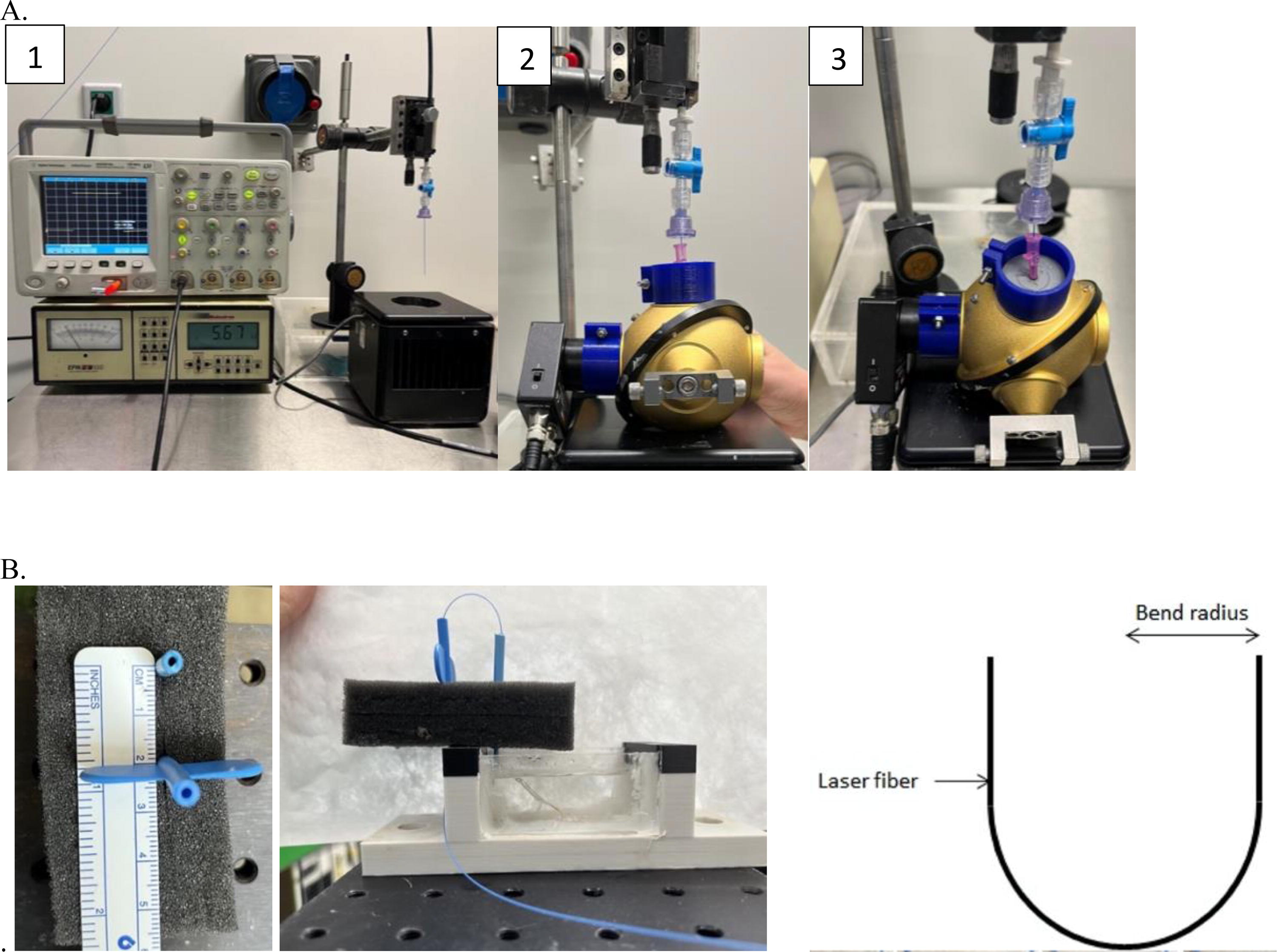
Experimental setup: pulse shape and peak power evaluation using an oscilloscope
Pulse Duration Evaluation and Peak Power Calculation Across Pulse Modes and Pulse Energy with the p-Tm:YAG
p-Tm:YAG, pulsed-Thulium:YAG.
ROF
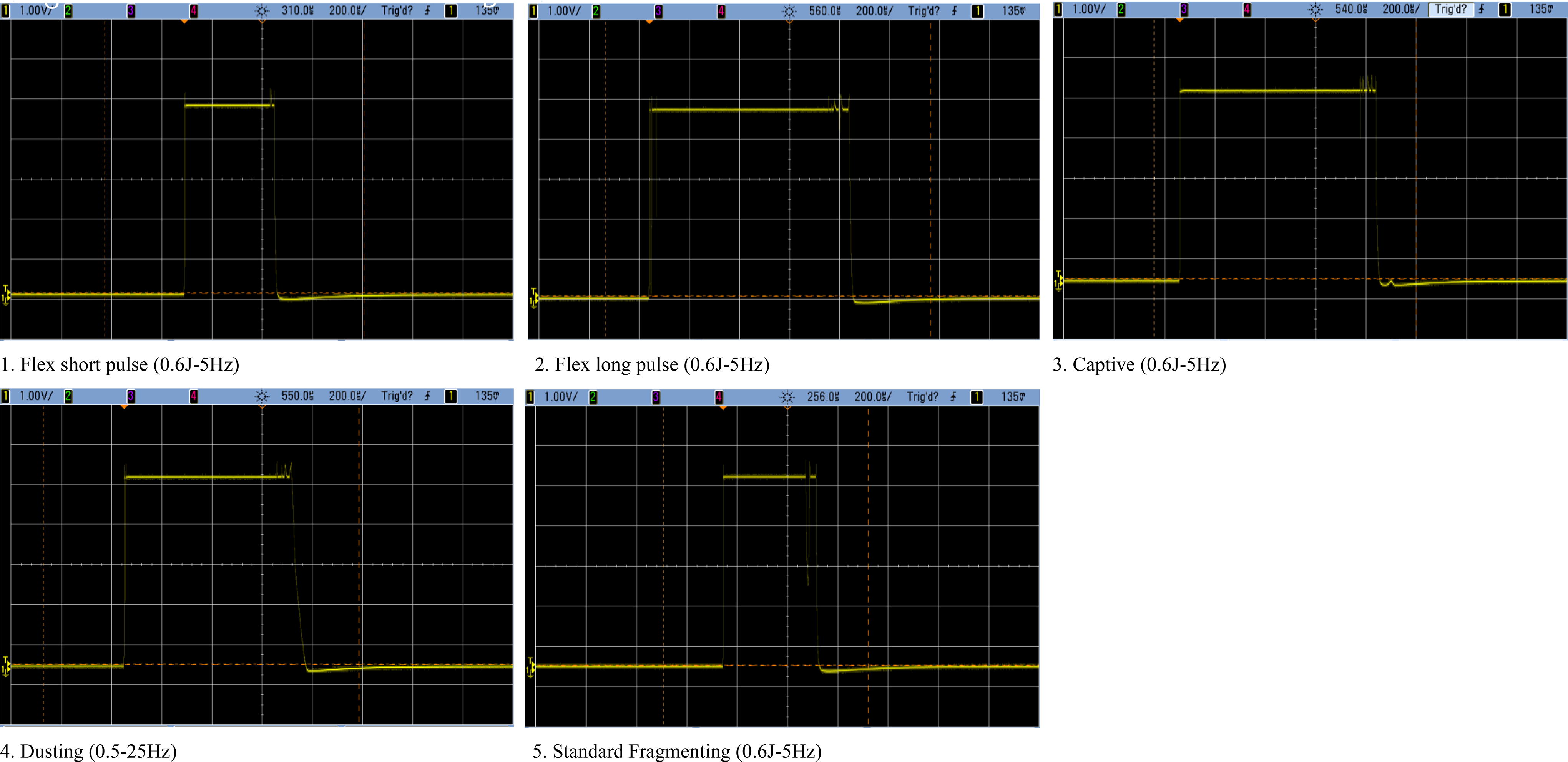
Single-use 270 μm LF were connected to the p-Tm:YAG generator. The fiber breakage threshold test was performed by bending the fibers at 180° with an initial radius of 1 cm, as previously described by Uzan et al. for Ho:YAG and TFL. 20 We used the same five different bend radii: an initial radius of 1 cm followed by tighter deflections at 0.9, 0.75, 0.6, and 0.45 cm. The LF tip was supported by soft silicon tubes, secured by plastics crews (to hold the LF without causing damage) and immersed into saline for 5 minutes of continuous laser activation (CLA), which was ceased only if the LF fractured (Fig. 1B). “Flex short pulse,” “flex long pulse,” “dusting,” “captive,” and “standard fragmenting” p-Tm:YAG pulse modulations were evaluated, with pulse energies and rates ranging from 0.15 to 1 J and 15 to 100 Hz, respectively (Table 2). Each test was performed five times.
Risk of Fiber Fracture According to p-Thulium:YAG Modes, Pulse Energy and Frequency, and Bend Radius
AVs
Wet HSP and SSP were fixed into a dedicated 3D-printed bench model, filled with saline at ambient temperature (Fig. 2). The LF was motionless, fixed vertically in contact with the stone phantom, whereas the stone phantom itself was displaced using a robotic translation table (8MTF102LS05, Standa©, Lithuania), respecting a 2 cm linear trajectory at 2 mm/second velocity (i.e., for 10 seconds of CLA). Laser and table were activated jointly. After drying, stone phantoms underwent three-dimensional (3D) micro-scanning (CT) (QuantumFX, Perkin-Elmer©, USA). AVs were calculated using a previously described technique (threshold segmentation of the air contained in the crater using 3DSlicer). 18 p-Tm:YAG (“captive”), Ho:YAG (“long pulse”), and TFL (“high PP”) were compared using two pulse energy-frequency combinations: 0.5 J–20 Hz (0.6 J for p-Tm:YAG due to preset restrictions for this pulse mode) and 1 J–15 Hz, representing a total delivered energy of 100 J (120 J for p-Tm:YAG) and 150 J, respectively. Each experiment was performed in triplicate.
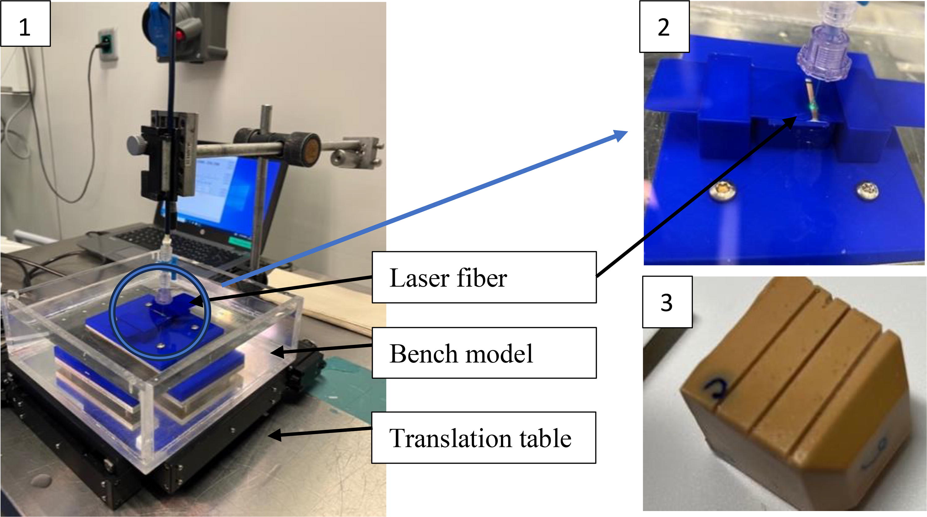
Ablation volume experiment using a translation table realizing a linear displacement of the stone while laser fiber was in a fixed position.
Statistical analysis
Continuous variables were reported as mean and standard deviation. Ablation experiments used Student’s t-tests and one way analysis of variance with multiple comparisons to compare two and more than two groups. p-Values < 0.05 were regarded as statistically significant.
Results
Pulse shape
The p-Tm:YAG pulse profile was uniform and stable over time and independent of the pulse mode and energy (Fig. 3, Supplementary Fig. S1). The calculated PP ranged from 564 to 2199 W, depending on pulse mode and energy (Table 1). Increasing pulse energy resulted in a higher PP/shorter pulse duration for the same pulse mode and rate. Considering 1 J pulse energy, both “captive” and “flex long pulse” modes presented PPs lower than 1000 W (864 and 888 W, respectively). Overall, “captive” and “flex long pulse” modes presented similar pulse shapes, durations, and PPs that could be equivalent to a p-Tm:YAG long pulse mode. On the opposite, “flex short pulse” and “standard fragmenting” presented similar pulse durations but equivalent to p-Tm:YAG short pulse modes. The lowest recorded PP was in “dusting” mode (0.6 J–25 Hz: 564 W) without the possibility to set the pulse rate lower than 25 Hz due to the fixed settings on the graphical user interface. The highest recorded PP was in “flex short pulse” mode (2 J–5 Hz: 2199 W).
Pulse shape analyses for pulsed-Thulium:YAG across pulse modes.
ROF
No fiber fracture occurred at any bend radius or laser settings (pulse energy, rate, or mode) (Table 2).
AVs
p-Tm:YAG, Ho:YAG, and TFL achieved similar overall AVs against HSP (8.96 ± 3.1 vs 8.8 ± 2.8 vs 9.78 ± 1.1 mm3, p = 0.67, respectively) (Table 3). Although p-Tm:YAG and Ho:YAG achieved similar AVs against SSP, TFL was associated with higher AVs than both p-Tm:YAG and Ho:YAG against SSP (12.86 ± 1.85 vs 10.12 ± 1.89 vs 7.56 ± 2.21 mm3, p = 0.002, respectively).
Holmium:YAG, Thulium Fiber Laser, and Pulsed-Thulium:YAG Ablation Volumes (mm3) After Linear Laser Emission on Hard and Soft Stone Phantoms Using 270 µm Laser Fibers
*Student’s t-test comparing laser settings across a laser source. **Student’s t-test comparing stone phantoms across a laser source. ***One way ANOVA comparing laser source across a same laser setting. ****One way ANOVA comparing Ho:YAG, TFL, and normalized p-Tm:YAG.
Normalized p-Tm:YAG: ablation volumes for 100 J delivered energy.
Bold data = statistically significant data.
ANOVA, analysis of variance; Ho:YAG, Holmium:Yttrium-Aluminum-Garnet; TFL, Thulium fiber laser.
AVs with p-Tm:YAG and Ho:YAG were not influenced by the stone phantom hardness (p = 0.56 and 0.54, respectively) whereas TFL achieved higher AVs against SSP than with HSP (p = 0.004).
At 0.5 J–20 Hz, TFL was associated with higher AVs compared with p-Tm:YAG and Ho:YAG for both HSP and SSP (p = 0.02 and 0.01). At 1 J–15 Hz, all three laser sources presented similar AVs against HSP (p = 0.3) and SSP (p = 0.1).
AVs significantly increased with pulse energy, for p-Tm:YAG (p = 0.03) and Ho:YAG (p = 0.02) against HSP.
Normalizing p-Tm:YAG AVs to 100 J delivered energy in dusting resulted in lower p-Tm:YAG AVs compared with Ho:YAG and TFL against HSP but remained intermediate against SSP.
Discussion
Laser technology
p-Tm:YAG differs from Ho:YAG and TFL lasers. It has a solid-state cavity laser architecture, similar to the Ho:YAG technology but uses laser diodes to produce light and a Thulium ions-doped crystal. Because of its favorable energy and power efficiency (i.e., a lower amount of light transformed into heat than a Ho:YAG laser), a single p-Tm:YAG cavity can emit as much as 100 W maximum power, whereas Ho:YAG is limited to 30 W for each laser cavity. Although the full 100 W power is not required for endoscopic lithotripsy during FURS, the p-Tm:YAG generator benefits from reduced noise from its cooling system (with fan water-cooling system compared with the vapor-compression refrigeration system for an equivalent high-power Ho:YAG). 12 p-Tm:YAG’s stimulated photons have a wavelength of 2013 nm, between Ho:YAG (2120 nm) and TFL (1940 nm) meaning the water absorption peak of p-Tm:YAG has been reported to be as much as 84% higher than Ho:YAG, but 50% lower than TFL. 16,17,21
Our study has demonstrated a uniform and stable p-Tm:YAG pulse profile despite pulse mode (similar to TFL) with its PP ranging from 564 and 2199 W (Fig. 3, Table 1). Its PP is therefore adjustable and lies between the PP achieved with Ho:YAG (1800–5000 W) and TFL (125–500 W), confirming the primordial role of PP in stone retropulsion. 19 As retropulsion correlates with PP, a wide range of PP would help to adapt the settings to the clinical situation. 19 Indeed, p-Tm:YAG was associated with lower retropulsion forces compared with Ho:YAG in a preclinical study especially using its long pulse duration modes, but a formal comparison against TFL was not performed. 14 Noteworthy, our experiments showed that increasing pulse energy resulted in higher PP levels regardless of the pulse mode, which is consistent with available data on Ho:YAG. 19 In distinction to this, TFL’s PP varies only slightly with pulse energy (550 W if pulse energy >1 J). This aspect, consistent with Ho:YAG, should be integrated into clinical practice to remain in safe conditions of laser activation. Finally, the “captive” modulation is supposed to limit stone retropulsion (<1000 W, even at 1 J), similar to the “flex long pulse” mode (864 and 888 W, respectively). More than a specific pulse modulation, “captive’s” PP and uniform profile could explain its low retropulsion level, described both in vitro (for p-Tm:YAG long pulse durations) and in vivo during FURS. 14,22
This present study has described the PP associated with commonly used pulse energies in the endoscopic management of urinary stones, trying to help Urologists better understand their future individual presetting, acknowledging that laser settings are not universal and could not be driven by companies. 23,24
AVs
Our experiments reported similar overall AVs for p-Tm:YAG, Ho:YAG, and TFL against HSP but that TFL had higher AVs compared with p-Tm:YAG and Ho:YAG against (Table 3). TFL’s low PP could not be high enough to fracture HSP as well as SSP, potentially explaining our findings. 7 Indeed, p-Tm:YAG and Ho:YAG were not influenced by the stone phantom hardness. Moreover, p-Tm:YAG demonstrated intermediate AVs at both 0.5 J–20 Hz (0.6 J–20 Hz for p-Tm:YAG) and 1 J–15 Hz between TFL and Ho:YAG.
Our findings confirm that p-Tm:YAG can fragment HSP stones with similar effectiveness as Ho:YAG and dust SSP with similar effectiveness to TFL, which is consistent with previous in vitro studies. 15,16 Petzold et al. first described the in vitro comparative dusting efficiency between p-Tm:YAG and Ho:YAG. 21 Using 400 μm LFs, the authors reported significantly higher ablation rates for Ho:YAG laser at slow displacement velocity (500 mm/minute) and short p-Tm:YAG pulse duration (p < 0.05). At high displacement velocity (1500 mm/minute) and p-Tm:YAG long pulse duration (>350 μs), p-Tm:YAG presented significantly higher mass loss than Ho:YAG. Comparing p-Tm:YAG and TFL with similar experimental conditions, Kraft et al. reported higher ablation rates (mass loss) for TFL using high pulse rates (≥50 Hz) but overall, the difference was not significant. 16 In another in vitro experiment, Kwok et al. showed p-Tm:YAG’s ability to dust any human stone type, producing a high percentage of particles <250 μm in the resulting dust and the remnants fragments, including cystine stones. 13 Regarding p-Tm:YAG’s ability to fragment stones, at all laser settings, Ho:YAG and p-Tm:YAG needed similar mean total energy (p = 0.97), which was lower than that for TFL. 15 In the present study, AVs increased with pulse energy, which is consistent with the previous Ho:YAG–TFL in vitro comparison. 18 Finally, p-Tm:YAG ablations rates (0.5–1.5 mm3/second) are also consistent with the only FURS evaluation, reporting a 0.75 mm3/second ablation speed. 22
From laboratory to clinical practice: strengths and limitations
This study first presents a laser characterization of the new p-Tm:YAG. Its technology translates into intermediate in vitro between Ho:YAG and TFL, for which future clinical comparative data between all laser sources will clearly be needed. However, based on the available data and these findings, p-Tm:YAG could represent a safe and effective alternative to TFL and Ho:YAG for endoscopic lithotripsy. 12,14 –17,21 Drawing from lessons from the worldwide TFL experience, more clinical trials are needed to define p-Tm:YAG’s place in the pipeline of laser technologies for endoscopic lithotripsy, as well as its safe and appropriate starting laser settings. Thus, our study protocol tried to adhere to low-frequency and low-power settings in each experiment, although we realized other laser settings could have been chosen. Indeed, we have compared a single pulse mode for each laser source in AVs’ experiments, supported by our PP results and previous publications. 19 “Captive” mode presented the best compromise for p-Tm:YAG, long pulse durations and high PP to be the best pulse modulation for Ho:YAG and TFL, respectively. 19 Regarding Ho:YAG, we emphasize not integrating Moses’ Technology, as it previously showed similar outcomes (retropulsion and ablation efficiency) to Ho:YAG in long pulse mode. 19 Furthermore, we compared two laser combinations in the ablation experiments (0.5 J–20 Hz [0.6 J–20 Hz for p-Tm:YAG] for dusting and 1 J–15 Hz for fragmentation settings) that may not fit to all clinical situations even considering “captive” was the preferred p-Tm:YAG mode in the single published clinical evaluation. 22 –24 Moreover, in the “captive” mode, the only pulse modulation that allows to set a low pulse energy and frequency, 0.5 J not being available and forcing us to set 0.6 J. We acknowledge this could limit the interpretation of our results, but we first compared Ho:YAG, TFL, and p-Tm:YAG technologies using AVs over ablation mass, which could represent the most accurate method for in vitro efficiency experiments. 18 Furthermore, we acknowledge that the applied energy rates in dusting and fragmentation were 100 J (120 J for p-Tm:YAG) and 150 J. This could limit our findings regarding the AVs and their increase with pulse energy, even if it was previously demonstrated for Ho:YAG. 25 Regarding the differences in total delivered energy among laser sources, normalizing p-Tm:YAG AVs demonstrated lower AVs for p-Tm:YAG in dusting compared with Ho:YAG against HSP, whereas stayed intermediate between Ho:YAG and TFL against SSP (Table 3).
As a limitation of the present experiment, we included only artificial stones and have not reported comparisons of fragments’ sizes generated from CLA. Thus, artificial and human stones may behave differently to CLA, according to various responses to acoustic or optical energy. 26,27 For example, King et al. reported higher AVs with human calcium oxalate monohydrate samples compared with 15:3 HSP (30 vs 19 μm3 and 24 vs 19 μm3 in Moses Distance and non-Moses Distance modes, respectively). 26,27 King et al. used optical coherence tomography and volume calculation as opposed to 3D segmentation in our study and repeated each experiment 10 times, whereas we performed ours in triplicate. 26 In general, artificial stone fragments are smaller than those with human samples. 18,28 Indeed, a 51.03 ± 121.7 μm overall mean fragments’ diameter was reported with TFL and 272 μm LF against stone phantoms, whereas Petzold et al. were not able to measure fragments produced from CLA with Ho:YAG or p-Tm:YAG (<125 μm). 18,21 On the contrary, fragments size studies on human stones have been conducted, trying to define stone dust, with Ho:YAG, p-Tm:YAG, and TFL, with size categories. 13 Furthermore, we acknowledge that different results could have been obtained using LF thinner than 270 μm, especially for TFL that allows 150 μm LF. 11 We intended to compare all laser sources using similar parameters, acknowledging that a direct comparison at their “best setting” could have been interesting.
Finally, we present the evaluation of a single p-Tm:YAG laser generator, that is, Thulio (Dornier-Medtech, Germany), emphasizing a second one (Revolix-HTL, Lisa-Laser©, Germany) could present different performances. Further in vitro studies could compare these two generators directly.
Conclusion
This study demonstrated that p-Tm:YAG laser represents a valuable alternative between Ho:YAG and TFL that translates to intermediate PP, a uniform pulse profile, and no risk of fiber fracture in vitro. p-Tm:YAG indiscriminately treated HSP and SSP, with intermediate AVs between Ho:YAG and TFL. Further preclinical and clinical studies will be needed to confirm these in vitro results and define p-Tm:YAG’s adequate role in the laser technology pipeline for advancing the practice of endoscopic lithotripsy.
Footnotes
Authors’ Contributions
M.C.: Protocol development, data collection and management, data analysis, and manuscript writing and editing. F.P.: Protocol development, data collection and management, data analysis, and manuscript writing and editing. O.T.: Protocol development, data analysis, and manuscript writing and editing. L.B.: Protocol development, data analysis, and manuscript writing and editing. C.S.: Manuscript writing and editing. S.D.: Protocol development and manuscript writing and editing. S.K.: Data collection and management. L.C.: Manuscript writing and editing. M.C.: Manuscript writing and editing. D.S.: Manuscript writing and revision and editing.
Research Involving Human Participants or Animals
The present study was fully conducted in vitro, without human samples or participants. Therefore, this study does not involve human participants or animals but adheres to the tenets of the Declaration of Helsinki.
Author Disclosure Statement
The authors declare that they have no conflict of interest. O.T. has declared as a consultant for Karl Storz, Coloplast, IPG photonics, Ambu, Quanta System, and Rocamed. S.D. has declared as a consultant for Boston Scientific Corporation and Coloplast. F.P. has declared as a consultant for Dornier-Medtech. D.S. has helped run courses supported by Karl Storz and Cook Medical and is vice chair of the Endourology Academy.
Funding Information
EUSP Scholarship of the
French Association of Urology Research Grant (FPT).
Supplementary Material
Supplementary Figure S1
Abbreviations Used
References
Supplementary Material
Please find the following supplemental material available below.
For Open Access articles published under a Creative Commons License, all supplemental material carries the same license as the article it is associated with.
For non-Open Access articles published, all supplemental material carries a non-exclusive license, and permission requests for re-use of supplemental material or any part of supplemental material shall be sent directly to the copyright owner as specified in the copyright notice associated with the article.