




Abstract
Introduction:
One advantage of supine percutaneous nephrolithotomy (sPCNL) is the ability to access anterior as well as posterior calyces, but the safety and efficacy of anterior calyceal entry has never been investigated to our knowledge. We prospectively evaluated patients scheduled for sPCNL comparing anterior and posterior calyceal access.
Materials and Methods:
After Institutional Review Board approval, we prospectively enrolled 100 consecutive patients undergoing sPCNL from February to September 2023. Primary outcomes included intraoperative complications, blood transfusions, 30-day complication rates, and emergency department (ED) visits or readmissions. Secondary outcomes included stone-free rates (SFR).
Results:
Seventy-six patients had anterior calyceal entry and 24 had posterior. No significant differences were found in terms of baseline demographics (age, body mass index), stone characteristics (location, density, complexity), or intraoperative features (operative time, location of access). Safety outcomes, including intraoperative complications (1% vs 4%), blood transfusions (3% vs 8%), 30-day complication rates (17% vs 21%), and ED visits (1% vs 0) or readmissions (11% vs 21%) were comparable between groups. Overall SFRs were equivalent (86% vs 90%).
Conclusion:
We found that anterior and posterior accesses in sPCNL offer similar safety and efficacy, with no significant differences in complications or SFRs. Surgeons can select either approach based on patient anatomy and surgical needs without concern for increased complications. Further research is necessary to confirm these findings and guide best practices for calyx selection in sPCNL.
Introduction
Traditionally, PCNL has been performed in the prone position for decades. However, the supine position has gained popularity due to its advantages in positioning, reduced anesthesia requirements, and simplified retrograde instrumentation. 1,2 During PCNL, entry into the appropriate calyx is imperative to achieve optimal outcomes. Both the cranio-caudad location (upper, middle, lower) and the anterior-posterior location of the selected calyx are critical to consider. With prone PCNL, puncture into an anterior calyx is generally avoided as it traverses more parenchyma and possibly more vessels, increasing the risk of bleeding, and usually does not allow access to other portions of the collecting system. 3 With supine percutaneous nephrolithotomy (sPCNL), however, one touted advantage is the ability to puncture anterior and posterior calyces without traversing through more parenchyma. In addition, there may be situations where anterior calyceal entry can allow easier and more complete access to the rest of the collecting system. Although these potential benefits to entering anterior calyces during sPCNL have been hypothesized, there is a lack of studies investigating its safety and efficacy. The aims of this study are to compare the safety and efficacy of anterior and posterior accesses in sPCNL.
Materials and Methods
Study design
After Institutional Review Board (IRB #14-00879) approval was obtained, consecutive patients scheduled for outpatient sPCNL were appropriately consented and prospectively enrolled in a nonrandomized fashion from February to September 2023. The decision to puncture anterior or posterior calyx was left to the discretion of the surgeon at the time of actual puncture in the operating room. All procedures were performed by two experienced fellowship-trained surgeons, (M.G.) and (W.A.), with the assistance of fellows and residents. Inclusion criteria included age >18 years and calculi located in the renal collecting system or proximal ureter. Exclusion criteria included specific anatomical variations such as horseshoe kidney, pelvic kidney, ptotic kidney, malrotated kidney, or urinary diversion, and the presence of a prior nephrostomy tube. Primary outcomes included intraoperative complications, blood transfusion, 30-day complication rate, and emergency department (ED) visits or readmissions. Secondary outcome was stone-free rate (SFR). Intraoperative complications were defined as any unexpected event occurring during the surgical procedure that required additional intervention or deviation from the planned surgical course, including injury to adjacent organs, excessive bleeding requiring intraoperative blood transfusion, or technical difficulties leading to a prolonged operative time beyond the expected duration for the procedure. The time interval considered for blood transfusions was 24 hours postoperatively, and ED visits and readmissions were tracked from the time of discharge up to 30 days postoperatively.
Surgical technique
Intravenous antibiotics were administered according to American Urological Association guidelines on antibiotic prophylaxis. Patients were positioned in a modified Barts flank-free position. Cystoscopy and occlusion balloon catheter placement over a guidewire were performed. Saline distention of the collecting system allowed visualization of all calyces. Ultrasound (US)-guided access was established into an anterior or posterior calyx during each procedure and confirmed by US. Calyces were labeled as anterior or posterior purely based on axial and longitudinal ultrasonic views of all calyces to visualize the entire collecting system. With ultrasonography, compared with fluoroscopy, it is much easier to discern anterior vs posterior calyces, especially in the supine position. The final determination of whether an anterior or posterior calyx puncture was accessed was assessed by rigid and flexible nephroscopy and intraoperative fluoroscopy, which make it quite clear. Figure 1 illustrates the ultrasonographic and the anatomical differences as viewed in the supine position. The choice of accessing an anterior or posterior calyx was at the surgeon’s discretion, considering anatomy, location of stones, and surrounding organs. Our main landmarks included the 12th rib, the iliac spine, and the posterior axillary line. With the use of US we were able to delineate visceral structures including lung, liver, spleen, and bowel, allowing us to avoid puncture into those structures. In cases of multiple accesses, we agreed to include only patients with either anterior or posterior calyceal accesses and not mixed. Dilation methods in standard PCNL utilized balloon dilation (Nephromax, 24F, Boston Scientific, Inc), and in nonsuction miniPCNL utilized metallic dilators and sheaths (Storz MIP-M, 17.5F, Storz Medical Inc). Nephroscopy and lithotripsy were conducted with gravity irrigation using 3-L normal saline bags at a standardized height of 80 cm above the patient. Lithotripsy was performed using a dual energy lithotripter (Swiss LithoClast Trilogy, Boston Scientific Inc). All procedures were performed tubeless (no nephrostomy tube). Stent placement was left to the surgeon’s discretion. Foley removal was performed 1 hour following the procedure. Same day discharge was planned in all patients. Stent removal was performed at 10 days in patients with stents, and US or kidney-urterer-bladder x-ray (KUB) were performed 1 month after the procedure, including metabolic counseling.

Demonstration of anterior or posterior calyxes:
Statistical analysis
Demographic and stone baseline characteristics, including age, sex, body mass index (BMI), diabetes, prior stone surgery, S.T.O.N.E. score, stone burden (summing the maximum diameter of each stone observed on imaging), and preoperative degree of hydronephrosis, were collected. Intraoperative findings, including access location, total access time, puncture operator (attending, fellow, resident), needlestick attempts, tract length, fluoroscopy time, and total operative time, were prospectively recorded. Descriptive statistics were calculated, with categorical variables reported as frequency and percentage and continuous variables reported as median and interquartile range (IQR). Categorical variables were compared using chi-square or Fisher’s exact tests, and continuous variables were analyzed via Mann–Whitney U tests. All statistical tests were two-sided, and p < 0.05 was considered statistically significant. SPSS software was used for all statistical analyses (IBM SPSS Statistics, Version 25, IBM Corp., Armonk, NY, USA, 2017).
Results
Study population
The study cohort consisted of 100 prospectively enrolled patients who underwent sPCNL (Fig. 2). Table 1 summarizes the baseline demographics and stone characteristics. Seventy-six patients had anterior calyceal access (anterior group) and 24 had posterior calyceal access (posterior group). The median age of the cohort was 62 years (IQR 48–71) and the median BMI was 27.06 kg/m2 (IQR 24.42–31.73). Overall, 15% of the participants were pre-stented, and Guy’s stone score was 3–4 in 23% of the patients. Full staghorn stones were observed in 11% of the patients, and the median stone burden was 25 mm (IQR 15–36). There were no significant differences between the anterior and posterior groups in terms of age, BMI, gender, diabetes, ASA scores, prior PCNL, pre-stenting rates, laterality, S.T.O.N.E. and Guy’s stone scores, stone burden, density and composition, and preoperative hydronephrosis (Table 1).
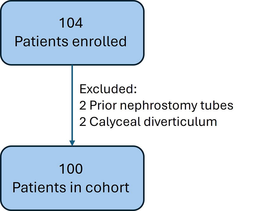
Enrollment flow diagram.
Baseline Demographic and Stone Characteristics of the Study Cohort, Stratified by Anterior and Posterior Accesses.
Bold indicates significant.
ASA, American Society of Anesthesiologists; BMI, body mass index; IQR, interquartile range; PCNL, percutaneous nephrolithotomy.
Intraoperative findings
The location of the access did not differ significantly between the groups, with most accesses being into the lower pole (76% anterior group vs 67% posterior group, p = 0.462) and subcostal (85% anterior group vs 75% posterior group, p = 0.231). Most puncture operators were either fellows or attending surgeons (59%), with no significant difference between the groups (p = 0.597). Most accesses required only one needlestick attempt (54%), and the number of attempts did not differ between the groups (p = 0.344). Other intraoperative findings, including multiple access rates, type of sheath used, tract length, fluoroscopy time and dose, and the operative times for all sPCNL steps, also showed no significant differences between the groups (Table 2).
Intraoperative Findings, Stratified by Anterior and Posterior Accesses.
Bold indicates significant.
IQR, interquartile range; US, ultrasound.
Study outcomes
The results of the study outcomes for intraoperative complications, blood transfusions, 30-day complications rates, ED visits, readmissions and SFRs are summarized in Table 3. Intraoperative complications were uncommon, occurring in 2% of the cohort, and blood transfusions were administered to four patients (4%). There were no differences between the groups for these outcomes (p = 0.424 and p = 0.243, respectively). The overall 30-day complications rate was 18% for the entire cohort, with no significant difference between the groups; 17% in the anterior group and 21% in the posterior group (p = 0.762). Major complications rates (Clavien-Dindo ≥3) were also not different between the groups (3% vs 12%, p = 0.088). No visceral or pleural injuries occurred in our cohort. Rates of ED visits and readmissions were 1% and 13%, respectively. In the anterior group, only one patient visited the ED (1%) and eight patients were readmitted (10%). In the posterior group, none visited the ED (4%), whereas five patients were readmitted (21%). There were no significant differences in ED visits or readmissions between the groups (p = 0.314).
Comparison of Study Outcomes Between Anterior and Posterior Groups.
Bold indicates significant.
Flank pain, hematuria, and stent-related urinary symptoms.
Due to fever/infection, hemoglobin drop requires transfusion and pseudoaneurysm.
ED, emergency department; SFR, stone-free rate.
Forty-five patients underwent both KUB and US after their sPCNL and the overall SFR was 87% (39/45 patients). The SFRs were 86% (30/35 patients) in the anterior group and 90% (9/10 patients) in the posterior group, with no statistical difference (p = 0.725) (Table 3).
Discussion
Historically, PCNL has primarily been performed in the prone position (pPCNL), which limited the selection of the target calyx due to positional constraints. Puncturing an anterior calyx was traditionally considered risky due to the greater distance the needle had to traverse, increasing the likelihood of bleeding complications. 3,4 Targeting the posterior calyces for puncture during prone PCNL is generally preferred as it aligns with the avascular plane defined by Brodel’s line, thereby reducing the risk of significant hemorrhage. However, it is important to note that Brodel’s line accurately corresponds to the location of the posterior calyces in only approximately two-thirds of cases, which may lead to variability during the procedure. 5 The advent of the supine position brought many advantages, including easier access to different parts of the renal collecting system, both anterior and posterior calyces. In certain cases, anterior access may be needed to achieve better surgical outcomes, such as in cases where posterior access is not feasible due to anatomical considerations, large stone burden in the anterior calyx, or when posterior access may result in an inadequate tract to address stone fragments effectively. As with the extension of less conventional PCNL surgical techniques, such as nonpapillary approach, without compromising the safety or efficacy of the procedure, anterior access in sPCNL may be advantageous, but needs further investigation due to limited literature. 6 Therefore, our study aimed to prospectively compare the safety and efficacy of anterior vs posterior accesses during US-guided sPCNL, using complication rates and SFRs.
Since kidneys exhibit significant mobility between prone and supine positions, and maximal stone clearance is essential, 7 accessing the appropriate calyx during the planning and performance of sPCNL is crucial for a successful surgical outcome and patient safety. Most of our patients had multiple stones located in multiple calyces, involving both anterior calyx, posterior calyx and renal pelvis. The location of the stones was part of the surgeons’ considerations about the choice of accessing, and it is not possible to get granular detailed information on each case and tabulate it in any meaningful way. In general, if the primary targeted stone is located in an anterior calyx, we prefer entering the anterior calyx, knowing full well from our experience that anterior calyces do allow access to the majority of the collecting system in the supine position, and is also excellent for entering the renal pelvis and traversing the ureteropelvic junction (UPJ) down the ureter, and that posterior calyceal entry in these situations make it very difficult to enter an anterior calyx, especially if it is parallel to that anterior calyx. If the primary stone was in a posterior calyx, either an anterior or posterior calyceal access we generally choose a posterior entry, especially if there are no other collecting system stones, but in certain situations, especially with multiple stones, an anterior approach is still preferred due to the ease in accessing the entire collecting system. This perhaps explains why in this series there were more anterior calyceal entries than posterior. Our study found that surgeons chose the anterior calyx more frequently in sPCNL (76% of cases) when the choice of access was at their discretion. We observed no differences between the anterior and posterior groups in baseline demographics, stone characteristics, or intraoperative findings. Specifically, BMI, stone location, and stone complexity were not associated with the decision between anterior or posterior access, suggesting that multiple factors influence this decision, with no single factor being the sole determinant. Furthermore, comparisons of total operative time (59 minutes vs 63 minutes), total access time (120 seconds vs 118 seconds), and total fluoroscopic time (29 seconds vs 33 seconds) demonstrated no significant differences between the anterior and posterior groups, highlighting the straightforward nature of the procedure with either calyx type.
The primary safety outcomes, including intraoperative complications, blood transfusion administration, 30-day complications rates, and ED visits or readmissions, did not differ between the anterior and posterior groups, suggesting that anterior calyceal entry is as safe as posterior access. Intraoperative complications were very uncommon, occurring in only one case in each group, and blood transfusions were administered to two patients in each group, reflecting known rates of approximately 5%. 8 The overall 30-day complication rates in the anterior and posterior groups were 17% and 21%, respectively, consistent with rates from previous studies. 9,10 No visceral or pleural injuries were reported, and major complication rates were low in each group. ED visits and readmission rates were also similar between the anterior and posterior groups (12% vs 21%), and were similar to rates reported in previous studies. 9 –11 These comparable safety results suggest that choosing either anterior or posterior access in sPCNL is safe, and surgeons should not alter their decision based on concerns of increased potential complications. Prior studies have demonstrated increased hemoglobin drop and more postoperative complications when entering anterior calices in pPCNL. 3,4,12 Tepeler et al. reported higher rates of hemoglobin drop and postoperative complications, especially major ones, in anterior calyx stones. 12 Similarly, El Shazly et al. found higher rates of hemoglobin drop and complications with PCNL via anterior access when treating anterior calyceal stones. 4 Our data suggest that anterior access in sPCNL is more feasible, especially with anterior calyceal stones, and safer than in pPCNL, highlighting the advantage of the supine position over the prone position in addressing this issue.
In our study, the location of the access did not appear to impact the SFRs, which were high for both groups (85.7% vs 90%, p-value: 0.725). This suggests that access to different parts of the renal collecting system is generally effective regardless of whether the anterior or posterior calyx is targeted. Conversely, in pPCNL, posterior calyx access has been associated with higher SFRs, particularly when treating complex stones. 13
To the best of our knowledge, this is the first study to prospectively compare and evaluate anterior and posterior access during sPCNL. Although traditional approaches favor posterior calyx access, the development of sPCNL has opened new possibilities for safely and effectively accessing anterior calyx, which we succeeded in validating. However, our study has some limitations. First, our study, although prospectively enrolling and consenting patients, was an observational real-world study and not a randomized controlled trial. In our study, the posterior cohort is relatively small compared to the anterior group; a larger posterior cohort would have provided a more robust comparison. Second, different sizes of tracts were included without a propensity score analysis to compensate potentially leading to variability in the outcomes. Finally, our SFRs were analyzed using mostly KUB/US rather than computed tomography scans, potentially leading to limited accuracy when evaluating SFRs KUB/US. KUB/US were commonly used due to its lower cost and lack of radiation. Studies suggest that KUB/US can miss up to 30–50% of residual stones, often reporting SFRs around 70–80%. In contrast, noncontrast CT, considered the gold standard for detecting residual stones, offers nearly 100% sensitivity, resulting in more accurate but often lower reported SFRs, typically ranging from 50% to 70%. 14 Further research is essential to validate these potential benefits and establish best practices for calyx selection in sPCNL.
Conclusion
Our study is the first to prospectively compare anterior and posterior access in sPCNL, demonstrating that both approaches offer comparable safety and efficacy. These findings suggest that surgeons can confidently select either anterior or posterior access based on individual patient anatomy and surgical needs. However, further research with larger sample sizes and randomized controlled trials is necessary to establish these results and the best practices for calyx selection in sPCNL.
Authors’ Contributions
A.R., B.G., W.M.A., M.G.: Conception and design. A.R., K.G., Z.S., C.C., R.K., A.J.Y.: Acquisition of data. A.R., K.G., B.G.: Analysis and interpretation. A.R., K.G., Z.S.: Drafting of the manuscript. W.M.A., M.G.: Critical revision of the manuscript. A.R., K.G., C.C.: Statistical analysis. M.G.: Supervision.
Ethics of Approval Statement
Ethical approval to report this study was obtained from our institutional review board.
Footnotes
Data Availability Statement
The data that support the findings of this study are available on request from the corresponding author. The data are not publicly available due to ethical issues and the privacy of the participants.
Author Disclosure Statement
All authors have nothing to disclose.
Funding Information
The authors received no financial support for the research, authorship, and/or publication of this article.