




Abstract
Objectives:
To assess the capability of Raman spectroscopy (RS) in the analysis of stone composition utilizing microscopic fragments from irrigation fluid during ureteroscopy (URS) and laser lithotripsy.
Patients and Methods:
A prospective, blinded study involving patients undergoing URS with laser lithotripsy. Irrigation fluid collected during the procedure was centrifuged, and microscopic particles were analyzed using RS. Simultaneously, stone fragments underwent formal analysis by Fourier-transform infrared spectroscopy (FTIR) in a different laboratory. The researcher conducting the RS was blinded to the results of the FTIR analysis. The RS results were compared with FTIR to evaluate concordance.
Results:
Between March 2022 and February 2023, 22 patients were enrolled. Stones were located in the kidney (41%), ureter (45%), and both (14%). The median stone size was 12 mm. RS accurately identified the major stone component in 82.6% (19) of cases, with a 17.4% (3) discrepancy. Concordance was observed for stones composed of calcium oxalate (CaOx) monohydrate/dihydrate, calcium phosphate, and uric acid. In discordant cases, FTIR identified CaOx monohydrate and dihydrate.
Conclusions:
This study introduces an innovative approach for analyzing stone composition using microparticles from irrigation fluid during stone fragmentation. The results demonstrated strong concordance with the standard FTIR technique, suggesting potential for stone analysis without the need to retrieve fragments during procedures. Further research is warranted to refine this method for broader clinical application.
Introduction
Ureteroscopy (URS) coupled with laser lithotripsy is now widely acknowledged as the preferred method for treating ureteral and renal stones measuring up to 2 cm. 1 The primary lithotripsy techniques employed include fragmentation and removal, dusting without removal, or a combination of both. In the fragmentation approach, stones are broken down into smaller fragments and actively extracted. Dusting involves breaking stones into very fine fragments that are subsequently expelled with urine. The ongoing debate regarding the superiority of either modality remains inconclusive. Some urologists advocate for fragmentation, whereas others lean toward dusting. 2
A distinct advantage of fragmentation over dusting is the active removal of stone fragments, facilitating their composition analysis. In the case of dusting, patients are instructed to filter their urine postprocedure in the hope of collecting stone fragments for analysis. However, in many instances, a lack of patient compliance or awareness hampers the collection of a stone sample, resulting in the inadvertent disposal of a valuable diagnostic resource.
The American Urological Association 3 and the European Association of Urology 1 both advocate for stone analysis as a crucial step in the prevention and treatment of urolithiasis. Precise identification of the composition of kidney stones serves to minimize treatment errors and enhance the effectiveness of preventive guidance. Presently, the recommended techniques for stone analysis encompass Fourier-transform infrared spectroscopy (FTIR) and X-ray diffraction (XRD). 4
The application of Raman spectroscopy (RS) for urolithiasis analysis was initially documented in 1983. 5 Subsequent research has affirmed RS as a reliable tool for discerning various chemical components of human kidney stones. 6,7 An advantage of microscopic RS over XRD and FTIR lies in the minimal size of stone fragments necessary for composition analysis. Unlike FTIR and XRD, which require larger fragments, RS only necessitates microfragments to complete the analytical process. Also, RS is not affected by the presence of water, which absorbs IR radiation. However, there is a limiting size for RS, which can be overcome by some preparation. Glass slides have a Raman signal, and if the fragment is under 1 or 2 µm, the signal will be generated from both materials (stone and glass) and will likely be mostly glass as it is more abundant under the stone particle. If one uses a gold-coated microscope slide, the gold, which has no standard Raman signal, will prevent the glass signal from being generated with a thickness of a few nanometers.
In our research, we postulated that conducting an analysis of microscopic stone fragments from irrigation fluid using RS would yield comparable results in stone composition to those obtained through FTIR.
Materials and Methods
After obtaining approval from the institutional review board (0018-22-AAA) and securing informed consent from all participating patients, we conducted a prospective study that involved the analysis of stone fragments from consecutive patients undergoing URS with laser lithotripsy. The surgeries were conducted by two different experienced urologists, with Holmium-Yag laser being the sole technique utilized for stone fragmentation and dusting. Laser setting for dusting was 0.3 Joules and 80 Hz. Laser setting for fragmentation was 0.8 Hz and 8 J. Two types of samples were collected from each patient. The first sample was collected during the endourological procedure when stone dusting was performed. This sample consisted of 10 mL of irrigation fluid aspirated using a single-use syringe through an evacuation channel. The second sample was taken at the conclusion of the operation after several stone fragments had been retrieved for final analysis. In all cases, normal saline was used as the irrigation fluid. All stone fragments extracted during the procedure underwent analysis using FTIR. Patients were included in the study only if all samples were successfully collected.
To prepare urinary deposits, 1 mL of irrigation fluid collected into an Eppendorf vial and centrifuged for 3 minutes at a speed of 4800 rpm. The natent was removed and double distilled water replaced it. After another centrifugation round, the deposits were collected with 20 µL of the fluid and spread across a microscope slide in 4–5 locations and allowed to air dry for 1 hour. The samples were studied using a LabRAM HR Evolution micro-Raman spectrophotometer (Horiba, France). The resulting residue was examined under the LabRAM microscope to identify the stone fragments, which was difficult because of the amount of organic material remaining with the stone fragments. When a stone fragment was located, it was illuminated by the Raman excitation beam, and the scattered light was collected by the optics and the Raman scattered photons were analyzed in the spectrophotometer. To minimize fluorescent background interference from tissue debris, blood, or the components of the kidney stones different excitation wavelengths were used, and in most cases, 785 nm produced the least amount of background emission. The microscopic stone fragments were measured on a microscope slide mostly using a 100× microscope objective lens (MPlanFL N 100× NA = 0.9, Olympus, Japan) and a 100-μm confocal hole. The Raman spectra were analyzed to identify the material of the microscopic stone fragments. Micro-RS is performed on submicron regions of the sample, and since kidney stones are often composed of multiple components, the major component can only be accurately identified through a statistical sampling approach. However, manual scanning and analyzing a limited number of random fragments do not allow for such a statistical assessment, which may lead to discrepancies in composition identification. The investigator conducting the RS analysis remained blinded to the results obtained from the FTIR. The outcomes of both stone analysis techniques were then compared.
Statistical analysis
Descriptive analyses (mean with standard deviation) were used for continuous variables, proportions for discrete variables, and comparative tests included chi-square for discrete variables and Kruskal–Wallis for continuous variables.
Results
From March 2022 to February 2023, 56 patients were enrolled in this study, but only 22 (39%) met the criteria for final analysis. Thirty-four patients were excluded from the study because of the unavailability of FTIR stone analysis results. This group included cases in which laser lithotripsy was not performed, preventing the collection of stone fragments for analysis, as well as instances where FTIR results were not provided despite successful fragment retrieval. Patient characteristics are given in Table 1. The participants had a median age of 54 years (interquartile range [IQR]: 44–69). Stone locations were identified in the kidney for 41% (9) of cases, in the ureter for 45% (10) of cases, and in both the kidney and ureter for 14% (3) of cases. Six patients (27%) had a ureteral stent inserted prior to the procedure. The median stone size prior to fragmentation was 12 mm (IQR: 7.5–15). The mean fragment size used for RS was <100 µm. The outcomes of both stone analysis techniques are given in Table 2 and Figure 1. RS identified the major stone component in 82% (18) of cases; however, in 18% (4 cases), there was a discrepancy in identification. When categorizing the identification results, there was perfect concordance (a complete match) in 11 cases (50%), partial concordance (major component identified by RS) in 7 cases (32%), and no concordance (none of the components identified by RS) in 4 cases (18%). Both FTIR and RS frequently identified the calcium oxalate (CaOx) monohydrate component, with FTIR detecting it in 18 cases (81.8%) and RS in 14 cases (63.3%; Fig. 2). CaOx dihydrate was identified 11 times (50%) by FTIR and 5 times (22.7%) by RS. Uric acid (UA) was detected by both FTIR and RS in four cases (18%). Calcium phosphate (CaP) was identified three (13.6%) times by FTIR compared with one case (4.5%) by RS (Fig. 3). Figure 2 illustrates the Raman spectra of CaOx monohydrate and CaOx dihydrate. The distinct spectral peaks correspond to characteristic molecular vibrations of these compounds, enabling differentiation between the monohydrate and dihydrate forms. Figure 3 presents the Raman spectra of other stone compositions, including ammonium urate, UA, cystine, and hydroxyapatite. In cases of discordance, the discrepancies primarily involved differences in the identification of components such as CaOx monohydrate and dihydrate by FTIR that were not fully matched by RS.
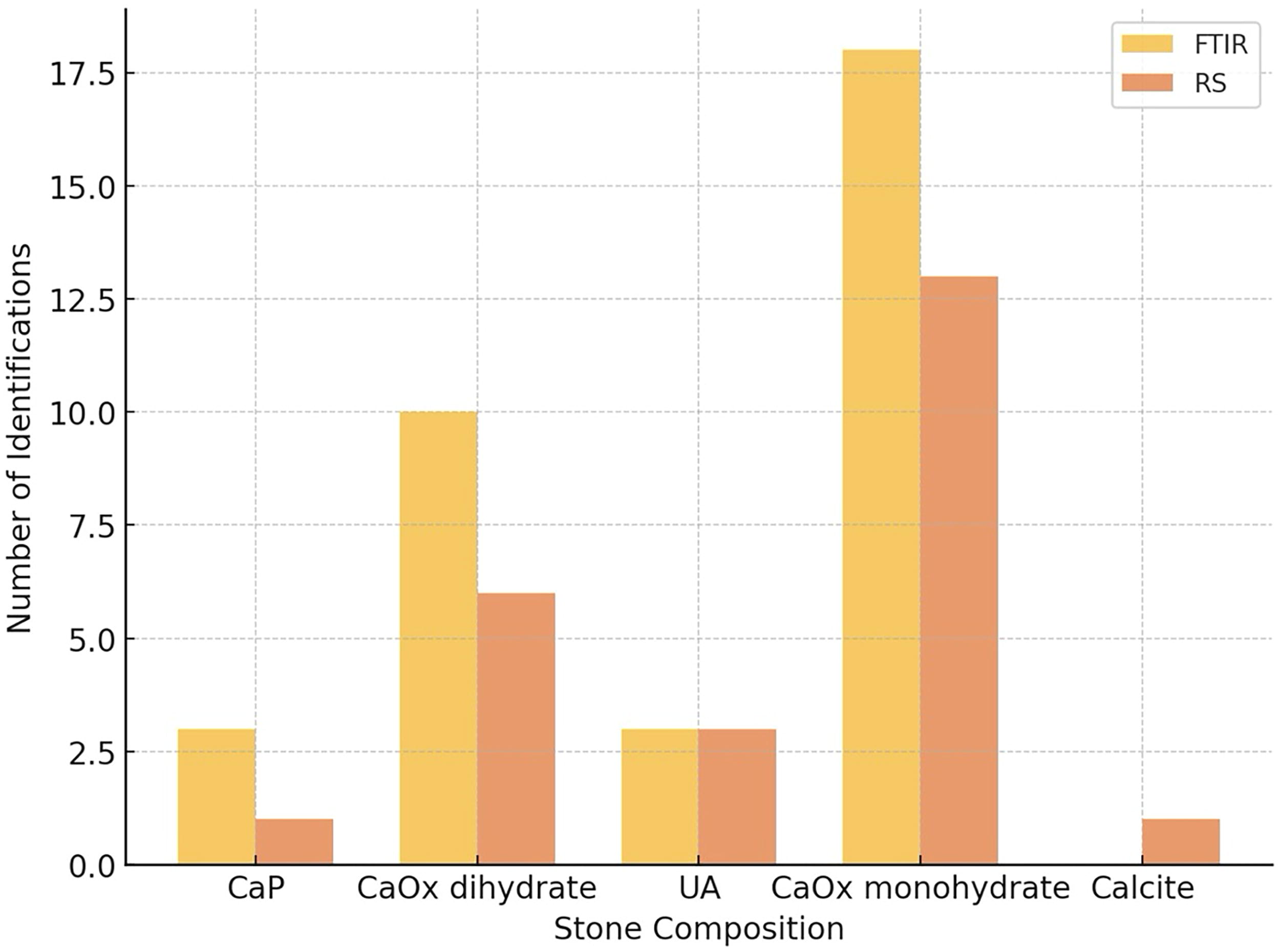
Comparison of FTIR and Raman results from the kidney stones. CaP = calcium phosphate; CaOx = calcium oxalate; FTIR = Fourier-transform infrared spectroscopy; RS = Raman spectroscopy; UA = uric acid.
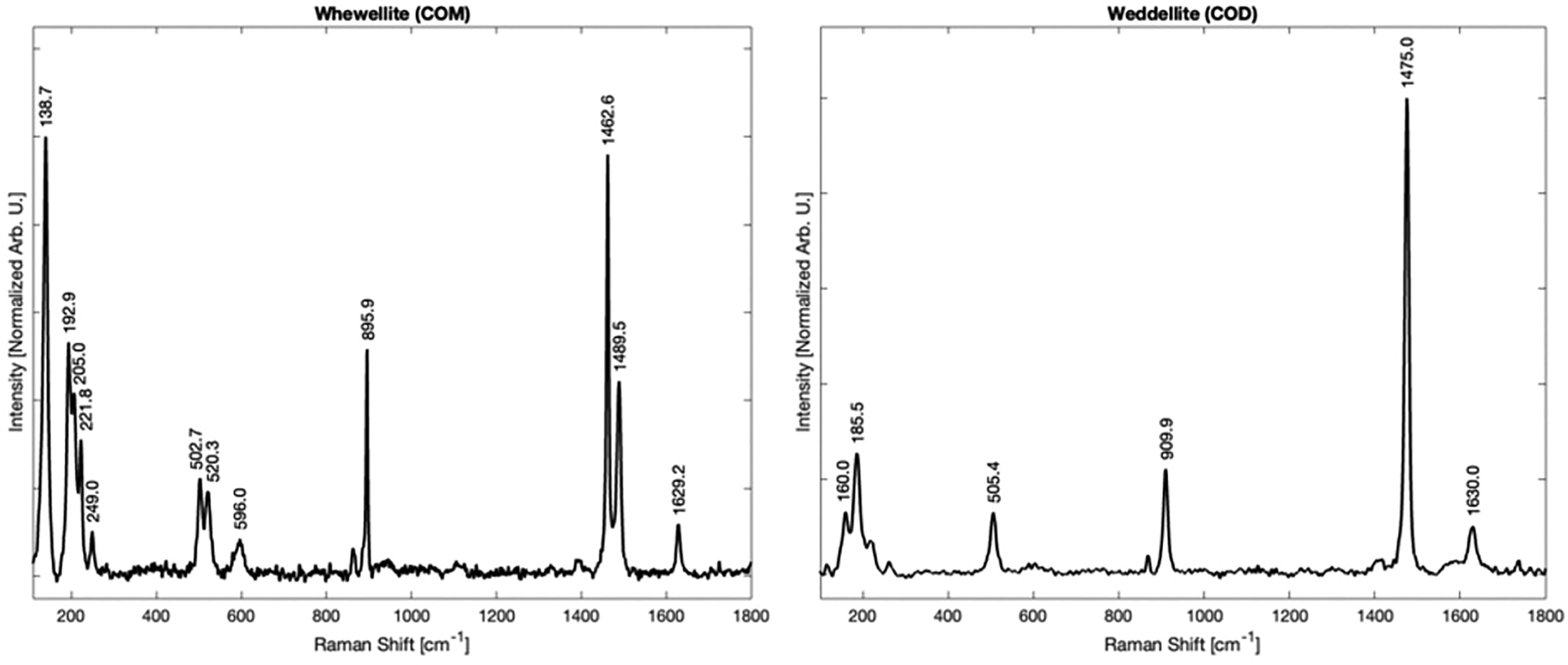
Raman spectra of calcium oxalate monohydrate and calcium oxalate dihydrate.
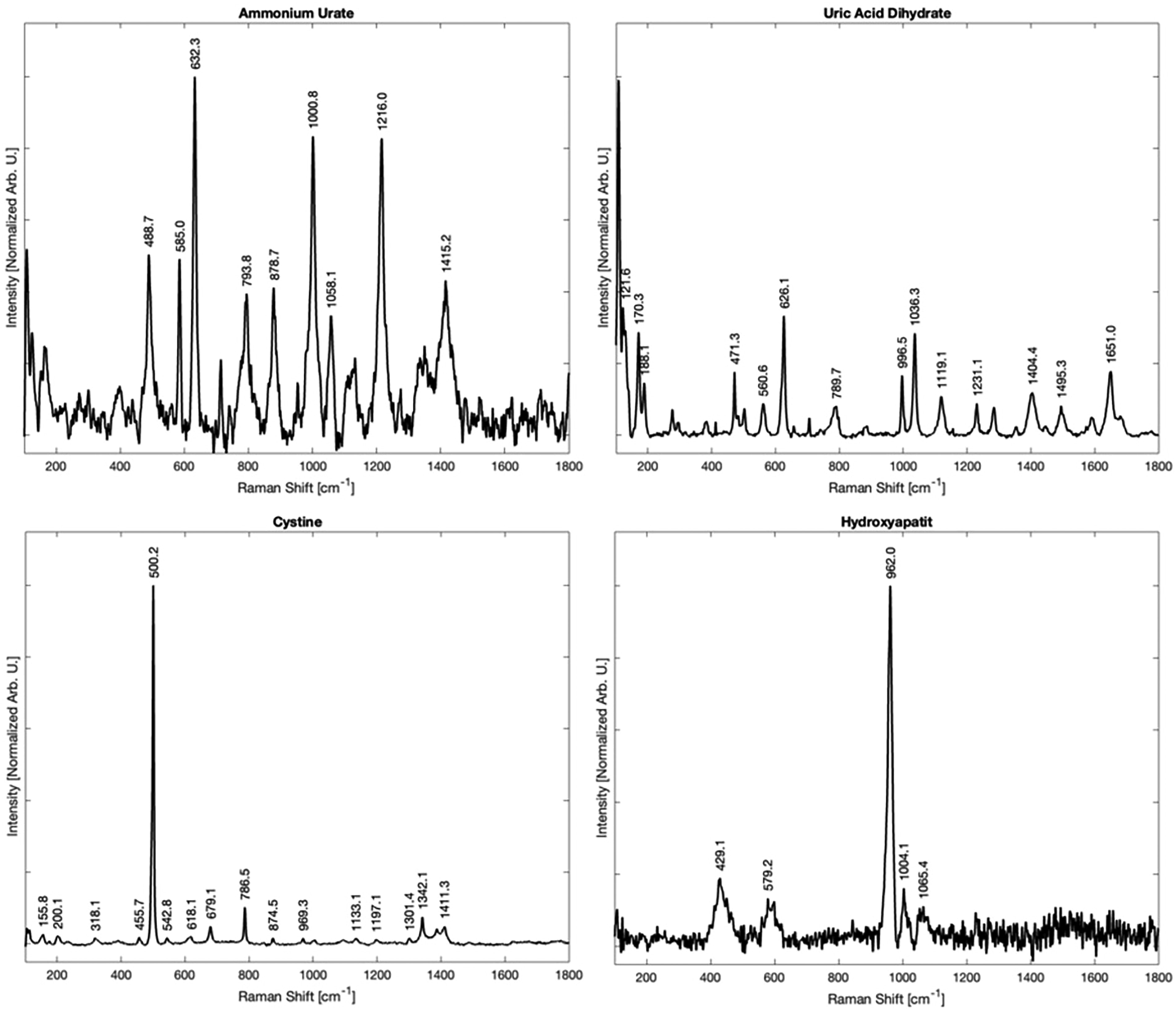
Raman spectra of stones formed of ammonium urate, uric acid, cystine, and hydroxyapatite.
Clinical Characteristics of the Patients
IQR = interquartile range; SD = standard deviation.
Comparing Stone Analysis Composition Utilizing FTIR Spectroscopy and RS
CaP = calcium phosphate; CaOx = calcium oxalate; FTIR = Fourier-transform infrared spectroscopy; RS = Raman spectroscopy; UA = uric acid.
Discussion
The prevalence of urolithiasis has increased substantially over the years. 8 Minimally invasive surgical techniques, such as URS, shock wave lithotripsy, and percutaneous nephrolithotripsy, have replaced the necessity for open surgery in the treatment of urolithiasis. 9 Advances in digital and fiberoptic technologies have led to the miniaturization of endoscopes used in these procedures. URS with laser lithotripsy has gained popularity because of these technological refinements. The widespread use of high-powered lasers has enabled stone dusting without removal, presenting an alternative to fragmentation and removal.
However, with the increasing preference for dusting, fewer stones are being sent for analysis, potentially leading to incomplete metabolic evaluations and suboptimal management strategies for stone formers. Currently, the primary modalities for stone composition analysis include XRD and FTIR, both of which require physical stone fragments for analysis. Understanding the composition of stones is instrumental in tailoring precise and targeted treatment strategies for patients. Different types of stones respond differently to various therapeutic interventions, such as medications or specific procedures such as lithotripsy. This information not only aids in devising effective preventive measures to reduce recurrence but also facilitates personalized patient care. Although metabolic investigations, including blood and urine biochemistry, help identify potential disorders, stone composition analysis provides essential insights into the specific lithogenic processes involved.
Chemical analysis (CA) has largely been supplanted by FTIR and XRD in the assessment of stone composition. Gilad et al. highlighted the limited information provided by CA, reporting that in 56% of cases, CA results did not correspond with those obtained through FTIR. Moreover, CA failed to identify the primary stone component in 16% of cases and overlooked the secondary stone component in 40%. 10 XRD is effective in identifying the crystalline components of stone material, but it has limitations in detecting nonrefractive amorphous materials when mixed with crystalline components, potentially causing issues when amorphous CaP and nonmineralogical components such as protein and matrix are present in the stone composition. 11 FTIR, in contrast, is a rapid and specific method based on the interaction of infrared light with molecules in stone components. 12 Unlike XRD, FTIR can identify noncrystalline materials, making it useful for identifying organic stone components, especially purines and drug metabolites, as demonstrated by Kasidas et al. 11
Stone composition has also been assessed utilizing RS. In a study by Kontoyannis et al., 13 the stone composition obtained through RS was compared with those from FTIR and XRD. The study reported that RS effectively identified the stone compounds present in renal calculi, with results consistent with those obtained through both XRD and FTIR. Similarly, Castiglione et al. 14 reported comparable findings, highlighting the good accuracy of RS in identifying the various components of ureteral calculi when compared with FTIR. These studies were conducted differently, analyzing stone fragments, whereas our study focused on microscopic stone fragments from the irrigation fluid.
In this study, we explored the use of irrigation fluid employed during URS with laser lithotripsy. Microscopic stone fragments within the fluid were subjected to analysis using RS. Our findings reveal that RS successfully identified stone composition in 82% of cases. Some samples of the irrigation fluid were devoid of stone fragments either because of their obstruction by organic matter or by the dilution of the fluid leaving nothing to measure. The comparison between FTIR and RS reveals that although both techniques are effective for identifying major components of kidney stones, FTIR provides a more detailed quantification of stone composition. This is because of the fact that in this study, the samples were measured using a submicron spot size, which is a very local measurement. Had the samples been separated from the organic matter it would have been easier to spot the microscopic stone fragments that were covered in mucus even after centrifugation with distilled water and the removal of the natent leaving the denser material for study. RS often confirms the presence of the primary stone components identified by FTIR but may not detect all components, especially in complex or mixed stones.
A key consideration in this study is the dual comparison being made: not only between RS and FTIR but also between different sample types—stone fragments vs microscopic particles obtained from irrigation fluid. Although RS has been validated for stone analysis in previous studies, a direct comparison between the RS method used in our laboratory and the FTIR method used by the reference laboratory remains an important factor in assessing the reproducibility and reliability of our findings. Variability in stone composition results across different laboratories, even when using the same analytical technique, has been well documented in the literature. Krambeck et al. reported significant discrepancies in stone composition identification across different institutions, highlighting that both sample preparation and spectral interpretation may contribute to inconsistencies. 15 These findings emphasize the importance of standardizing analysis protocols to minimize interlaboratory variation. Although our study demonstrated a high concordance rate between RS and FTIR for major stone components, further research should include direct intralaboratory comparisons of both techniques using identical specimens to validate the consistency and accuracy of RS in clinical practice.
Being able to have only stone microfragments in the sample would have allowed to use automated measurements and obtain statistical analysis of the samples. These methods allow using the optical microscope to scan the slide onto which the sample has been deposited, then identify the fragments, and direct the Raman excitation beam onto each microfragment. To the best of our knowledge, this study marks the first endeavor to assess stone composition by employing microfragments derived from the irrigation fluid collected during URS. Our findings reveal a close resemblance in stone composition results compared with those obtained through the conventional FTIR technique. When this technique is applied, there is no need for the active removal of stone fragments, while preserving the essential diagnostic information of stone composition. We believe the primary cause of the mismatch between FTIR and RS was suboptimal sample preparation before spectroscopy. This made it difficult to locate the stone fragments, particularly when blood residues in the fluid interfered with the accuracy of stone identification. Our findings align with prior research exploring the feasibility of stone analysis using microscopic fragments obtained during URS. Snicorius et al. conducted a study comparing the compositional analysis of dust specimens with retrieved stone fragments, both analyzed using FTIR. 16 Their results demonstrated a high degree of concordance between dust samples and retrieved fragments, supporting the idea that small stone particles can provide reliable compositional information. However, they also noted discrepancies in cases of mixed stones, where relative proportions of different components could not be fully assessed from dust samples alone. This is consistent with our findings, where RS accurately identified the major stone component in most cases but did not quantify relative component proportions. The study by Snicorius et al. underscores the potential utility of analyzing dust or microscopic stone fragments for stone composition assessment while highlighting the need for further refinement to ensure accuracy in mixed stone cases. Our study builds upon this concept by demonstrating the feasibility of RS as an alternative to FTIR for such analysis, providing a rapid, nondestructive method that does not require fragment retrieval.
Although this study demonstrates the potential of RS for real-time stone composition analysis, its practical implementation in clinical settings remains a challenge. Currently, RS requires meticulous sample preparation and manual identification of stone fragments, making it a labor-intensive process. In a health care environment where URS and stone analysis procedures are reimbursed at relatively low levels, the feasibility of integrating RS into routine practice depends on streamlining the workflow and automating key steps. Future developments, such as improved filtration methods, automated fragment detection, and standardized spectral analysis, could enhance efficiency and reduce the time and expertise required for RS analysis. Additionally, the inability to obtain RS results in three cases raises concerns about potential limitations in sample quality and fragment identification. In these cases, organic debris or excessive dilution of irrigation fluid likely hindered successful stone detection. This suggests that optimizing fluid collection and processing techniques will be crucial for maximizing the reliability of RS in a clinical setting. Although RS shows promise as a rapid, nondestructive method for stone analysis, further research is needed to refine its application before it can transition from a research tool to a widely adopted clinical practice.
The limitations of this study include the relatively small sample size, which may impact the generalizability of the findings to a broader population. Additionally, the study focused on a specific patient group undergoing URS with laser lithotripsy, and the results may not be directly applicable to other clinical scenarios. The use of microfragments from irrigation fluid for stone composition analysis is a novel approach and, although promising, further validation and replication studies are needed to confirm the reliability and reproducibility of this method. Additionally, the study did not explore potential variations in stone composition because of factors such as patient demographics or different laser settings during the lithotripsy procedure. Moreover, the comparison with FTIR, while showing almost similar results, may warrant a more comprehensive investigation to establish the equivalency of the two methods across a broader spectrum of stone types and compositions. A major limitation is the exclusive inclusion of calcium-based stones (CaOx and CaP) in the analysis. The lack of evaluation of UA, struvite, and drug-related stones restricts the generalizability of our findings to a broader population of stone formers. These stone types have distinct physicochemical properties that may influence their detection using RS, and further studies are required to determine the accuracy of RS in identifying such compositions. Additionally, our method does not quantify the relative proportions of each stone component. Although RS successfully identified the primary stone composition in most cases, it does not provide a comprehensive compositional breakdown as achieved with FTIR. This may limit its utility in cases of mixed stones, where precise compositional analysis is crucial for guiding metabolic evaluation and preventive strategies. Future research should aim to optimize sample preparation and improve detection techniques to address these limitations, ensuring broader applicability of RS in clinical practice.
Conclusions
This study represents an innovative attempt to evaluate stone composition using a novel approach that involves analyzing microfragments obtained from irrigation fluid during URS. The results showed significant concordance with those obtained using the standard FTIR method, indicating the potential effectiveness of this innovative technique for stone analysis. These encouraging findings suggest that further research is warranted to refine and enhance the method, taking into account variations in stone types, patient demographics, and procedural conditions.
Footnotes
Acknowledgment
I.P. is the incumbent of the Sharon Zuckerman research fellow chair.
Authors’ Contributions
The authors declare the following contributions to the study: O.R.: Conceptualization: designed the study methodology; data curation: collected patient samples and prepared them for analysis; and writing—original draft preparation: contributed to drafting the article. I.P.: Methodology: oversaw Raman spectroscopy (RS) procedures; formal analysis: conducted and interpreted RS data; and writing—review and editing: provided critical revisions to the article. A.C.: Investigation: performed ureteroscopy procedures and facilitated sample collection; resources: provided clinical resources for the study; and supervision: guided the study’s clinical implementation. D.G.: Project administration: coordinated the study at Assuta Ashdod University Hospital; methodology: oversaw RS procedures; supervision: supervised the clinical and research aspects; investigation: performed ureteroscopy procedures and facilitated sample collection; data curation: collected patient samples and prepared them for analysis; correspondence: handled article correspondence; and writing—review and editing: contributed to drafting the article and revised the article for intellectual content.
Author Disclosure Statement
No competing financial interests exist.
Funding Information
No funding was needed for this study.