
Abstract
Paratuberculosis, or Johne's disease, is caused by Mycobacterium avium subsp. paratuberculosis (Map), and it generates great economic losses for the dairy industry worldwide. In humans, Map has been associated with Crohn's disease. Mexico has unknown paratuberculosis prevalence, and yet, control programs have not been applied. This study aimed to determine the presence of Map in milk samples from seropositive goats and cows and bulk tank milk samples from herds previously designated Map-infected using indirect enzyme-linked immunosorbent assay. Map DNA was detected in 100% of the bulk tank milk samples of 14 bovine herds and 3 caprine flocks using a modified insertion sequence 900 polymerase chain reaction (PCR). Additionally, Map DNA was detected in 100% of the individual milk samples from 10 cows and 8 goats. Further, based on the findings of the experimental insertion sequence 900 PCR assessment, evaluation of bulk tank and individual milk samples through a type-specific PCR was performed, which confirmed our previous findings and revealed that 56.25% cow and 63.63% goat milk had concurrent infections of the C, I, and S types. Out of 14 bulk tank milk samples, 10 had viable mycobacteria. Paratuberculosis was detected at a high frequency in cow and goat milk, which suggests that raw milk ingestion represents a potential risk of Map infection.
Introduction
P
Materials and Methods
Samples
Animals from central Mexico were sampled from 14 dairy herds and 3 caprine flocks that were identified by reviewing formerly positive serology results in which 759 out of 3123 cows were randomly sampled. The sample size from each herd was determined taking into account the herd size and the estimated prevalence according to the U.S. Department of Agriculture National Animal Health Monitoring System. Regarding the goat flocks, 371 adult animals were sampled. Animals with positive serology were followed up until culling, to test for pathology and bacterial isolation to confirm paratuberculosis infection.
Ten seropositive cows were selected from herds with paratuberculosis seroprevalence higher than 21.82% determined by enzyme-linked immunosorbent assay (ELISA), from which 50 mL milk samples (composite of all four quarters) were collected by the operator during milking following the farm's usual disinfection and cleaning protocols. Eight seropositive goats were also selected from a flock with 9.67% ELISA-determined seroprevalence. At the same time, bulk tank milk samples from the 17 herds (14 bovine and 3 caprine) included in the former serological survey were aseptically collected.
Enzyme-linked immunosorbent assay
The serological statuses of the 18 animals studied were confirmed by the same indirect ELISA method using the PPA3 antigen (Allied Monitor, Fayette, MO), the ELISA cutoff value was 80%, which has 65.5% sensitivity and 84.0% specificity, as previously described (García-Marín et al., 1991).
Bacterial isolation
Culture for bacterial isolation was performed as follows: a 10 mL milk sample was centrifuged at 3100 g for 30 min. The resulting pellet and cream, within which the Map cells segregate, were resuspended in 10 mL of 0.75% hexadecylpyridinium chloride (JT Baker, Philipsburg, NJ), left at room temperature overnight, then centrifuged at 3100 g for 30 min, and the resulting pellet was resuspended in sterile phosphate-buffered saline. Each sample was spread onto three Herrold's egg yolk medium and/or Lowenstein–Jensen slants with or without mycobactin J (2 mg/L; Allied Mo). Culture media were supplemented with penicillin, chloramphenicol, and amphotericin B. Cultures were read after 8 weeks of incubation at 37°C and maintained for at least 5 months to be confirmed as negative (Ellingson et al., 2005; Gao et al., 2005). The identities of the microorganisms in the samples were verified as Map by growth rate, colony morphology, and Ziehl–Neelsen staining.
DNA extraction and polymerase chain reaction
For DNA extraction, a 10 mL milk sample was centrifuged at 3100 g for 30 min, and the resulting pellet and cream were resuspended in phosphate-buffered saline–Tween 20 and inactivated at 94°C for 10 min. The QIAmp DNA Mini Kit (Qiagen, Ventura, CA) was used, following the manufacturer's instructions. DNA was stored at 4°C until use. The IS900 polymerase chain reaction (PCR) primers used were P3N (5′-GGGTGTGGCGTTTTCCTTCG-3′) and P5N (5′-ATTTCGCCGCCACCGCCACG-3′) according to Ayele et al. (2005), which amplified a 314-bp fragment from the IS900 sequence. The PCR conditions were 94°C for 5 min, followed by 35 cycles at 94°C for 40 sec, 67°C for 40 sec, and 72°C for 40 sec, and a final extension of 72°C for 5 min. Type-specific PCR using DMC529, DMC531, and DMC533 primers was also performed (Collins et al., 2002) with the following modifications: 1 cycle at 95°C for 5 min, 35 cycles of 94°C for 30 sec, 60°C for 30 sec, and 72°C for 30 sec, and a final extension cycle at 72°C for 5 min. Expected products of 310 bp, from the C type, and 162 bp, from the S or I type, were identified using electrophoresis on a 2% agarose gel stained with ethidium bromide.
Results
Serological status and bacteriological isolation
The paratuberculosis seroprevalence of the 1130 sampled animals was determined using indirect ELISA, with values ranging between 8.0% and 24.07% in the dairy herds and 8.29% and 9.67% in caprine flocks. Map isolation from individual milk samples was not possible. During the 5-month incubation period, 10 out of 14 bovine bulk tank milk samples developed Map-compatible colonies, which were definitively identified using IS900 PCR and type-specific PCR. In caprine bulk tank milk samples, no colonies were observed during the 5-month incubation period.
PCR identification
Map DNA detection was performed using primers P3N and P5N based on the IS900 sequence in all individual cow and goat milk samples. Additionally, Map DNA presence was detected in the 14 bulk tank milk samples from the same number of infected bovine herds and from the 3 bulk tank milk samples from the caprine flocks (Fig. 1). Type-specific PCR confirmed these findings and revealed that the C type was present in all milk samples (cows and goats), regardless if it was an individual or a bulk tank sample (Fig. 2). Additionally, 30% (3/10) bovine and 50% (4/8) goat samples amplified the S or I type. Moreover, 50% bovine (7/14) and 100% (3/3) of the caprine bulk tank milk samples included either the S or I type, and 10 bovine bulk tank milk samples were positive for bacterial isolation, in which type-specific PCR demonstrated that six isolates were C type, and four tanks had the S or I type.
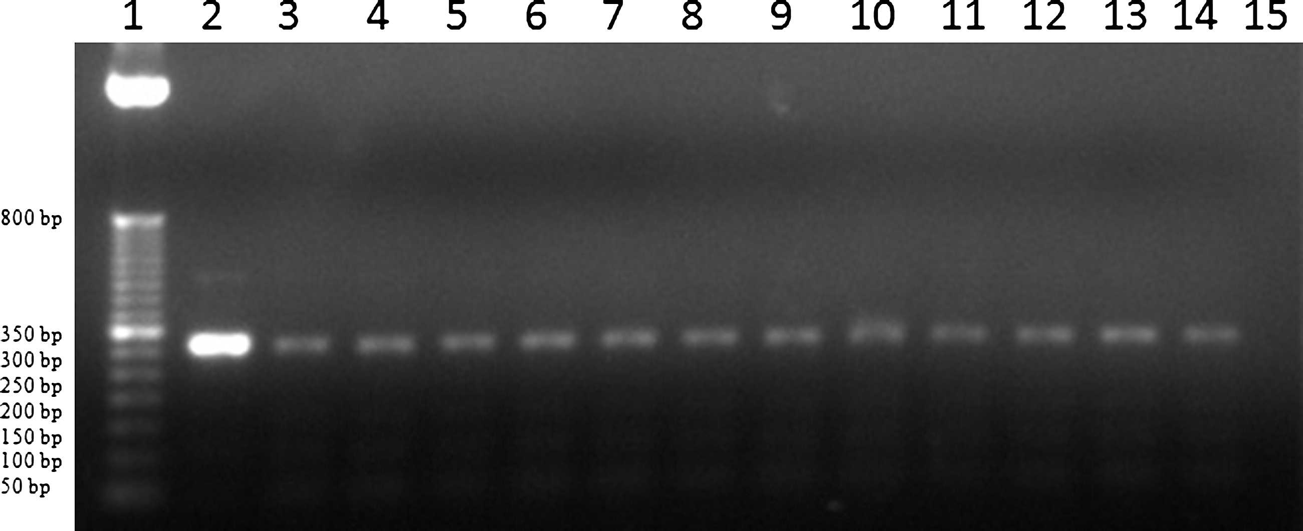
Polymerase chain reaction detection of Mycobacterium avium subsp. paratuberculosis from bulk tank milk samples of infected bovine herds and caprine flocks. Lane 1: 50-bp DNA ladder (Invitrogen, Carlsbad, CA); lane 2: positive control; lanes 3–11: bulk tank milk samples from infected bovine herds; lanes 12–14: bulk tank milk samples from caprine flocks; lane 15: nontemplate control.
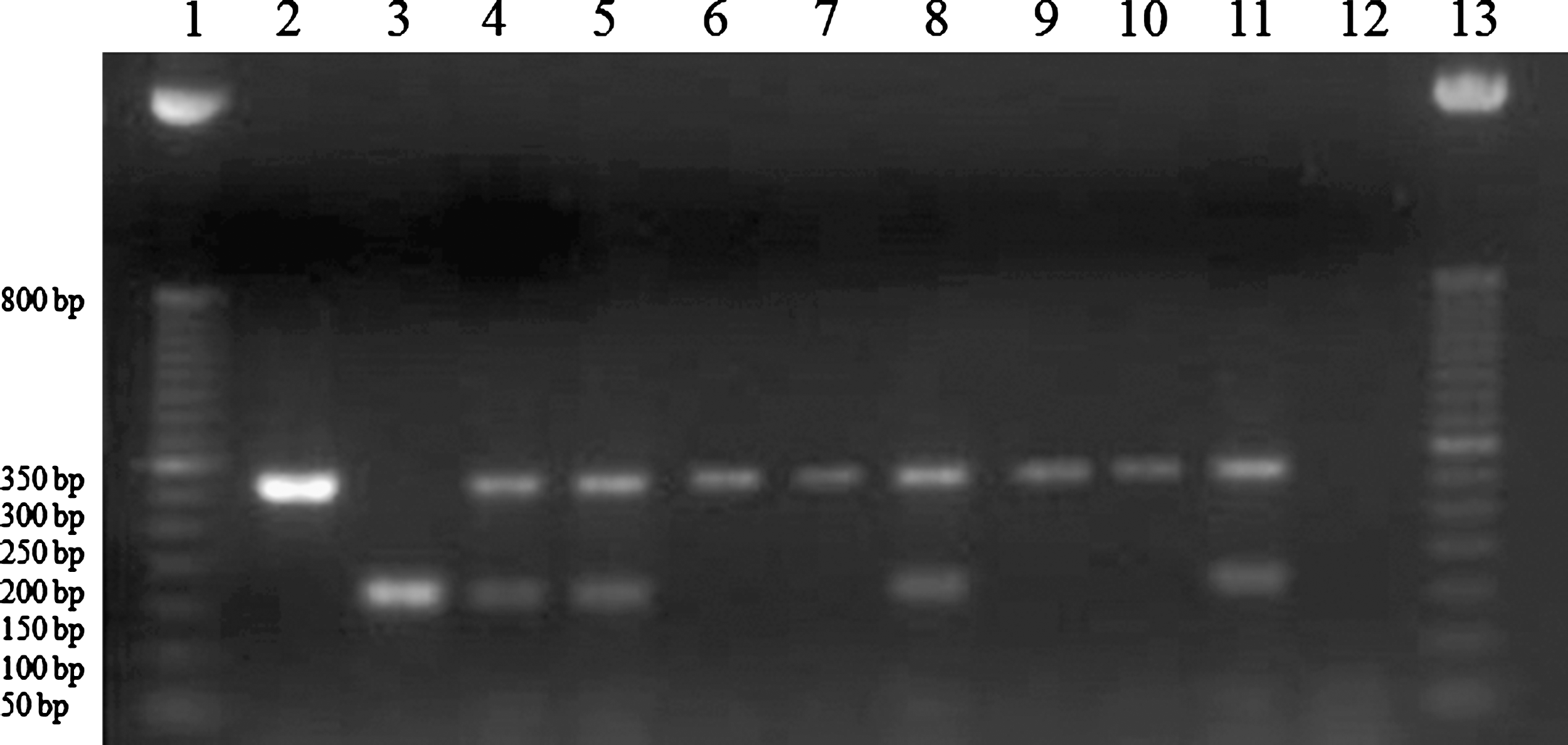
Type-specific polymerase chain reaction of Mycobacterium avium subsp. paratuberculosis from bulk tank milk samples of infected bovine herds. Lanes 1 and 13: 50-bp DNA ladder (Invitrogen); lane 2: C-type control; lane 3: S-type control; lanes 4, 5, 8, and 11: samples with C and S types; lanes 6, 7, 9, and 10: samples with C type; lane 12: nontemplate control.
Discussion
The economic impact of paratuberculosis has encouraged the creation of voluntary disease control programs in many countries (Ayele et al., 2005; McKenna et al., 2006). In the United States alone, it is estimated that nearly 68.1% of the U.S. dairy herds are Map-infected (NAHMS, 2008), and the resultant dairy losses may reach US$200 to US$250 million per year (Ott, 1999). Therefore, control programs tend to be mandatory and governmentally implemented in other countries, directing animal mobilization restriction policies and culling of the infected animals. Consequently, an accurate paratuberculosis diagnosis is currently demanded.
PCR analysis enables the identification of those animals that have active Map milk excretion. This is a useful tool in control programs and can be used to reduce the disease prevalence in dairy herds and caprine flocks. Additionally, PCR is a valuable technique for Map detection in tank milk samples and represents an easy, economic, and rapid identification of Map-positive herd and flock (Grant et al., 2002a; Pillai and Jayarao, 2002; Stabel et al., 2002; Muehlherr et al., 2003; Donaghy et al., 2008). However, it is worth mentioning the controversial Map detection limits of the IS900 PCR, which have been reported to be variable and questionable, because of the equipment used or the detection methods employed (Giese and Ahrens, 2000; Pillai and Jayarao, 2002; Grant, 2006; Stratmann et al., 2006; Donaghy et al., 2008).
Map recovery difficulties have been widely noted, and only a small proportion of viable Map present in a sample is recovered if chemical decontamination due to use of hexadecylpyridinium chloride occurs (Gao et al., 2005).
The fact that viable Map was detected in the bulk tank milk samples from infected herds and flocks, and not from individual samples, was not as striking as other results that have been reported (Stabel et al., 2002; Djønne et al., 2003). PCR is unable to determine if the microorganism was viable or not. Further, this technique is more sensitive than Map isolation, which explains the discrepancy between PCR and culture results (Pinedo, 2008). On the other hand, during individual milk sampling the udder cleaning procedures were supervised to minimize fecal contamination of the samples, which is known to be the major source of Map (Nauta and van der Giessen, 1998). Regarding bulk tank milk samples, the tank and milking machines and lines cannot be discarded as contamination sources. Hence, Map shedding in the milk of asymptomatic infected ruminants (Sweeney et al., 1992; Streeter et al., 1995) uncovers the pertinence of the disease control and surveillance programs as well as good milking practices.
Some authors suggested that a positive IS900 PCR result should be confirmed by DNA sequencing of the amplified products or by a PCR assay targeting genes other than IS900 (Englund et al., 2002; Stabel and Bannantine, 2005; Tasara and Stephan, 2005; Taddei et al., 2008). PCR primers developed by Collins et al. (2002) are considered species- and type-specific, enabling Map identification and confirmation. Moreover, type-specific PCR can be performed with DNA extracted directly from clinical samples to distinguish the cattle, sheep, or intermediate types. On the other hand, the restriction fragment length polymorphism (RFLP) method cannot be carried out with milk samples, barring bacterial isolation, which makes it a slower and more expensive method. Nevertheless, this technique is ideal for traceability purposes. In this study, the simultaneous presence of the C and S/I types was observed in several herds and also in individual milk samples. Mixed patterns have been previously described in sheep and bovine bulk tank milk samples from herds in which bovine and caprine populations coexist (Pavlík et al., 1995; Estévez-Denaives et al., 2007). Very few Map polymorphism studies have been published worldwide. Types C1 and C33 have already been identified in goats from Mexico (Chávez-Gris et al., 2004; Estévez-Denaives et al., 2007), although, to our knowledge, there have been no polymorphism studies of Mexican bovine strains published. Further molecular epidemiology studies are required to identify the polymorphisms in different regions of the country to understand their distribution.
So far, the association between Map and Crohn's disease in humans remains unclear and not enough epidemiological support is available. The detection of Map genetic elements in raw milk samples investigated in this study supports that the consumption of raw milk should continue to be regarded as a Map infection vector.
Footnotes
Acknowledgments
L.C. Favila-Humara received a fellowship from CONACYT. This work was partially supported by Universidad Nacional Autónoma de México PAPIIT IN208203-1 and IN208203-2 Grants. The authors acknowledge Unidad de Síntesis del Instituto de Biotecnología de la Universidad Nacional Autónoma de México for help with oligonucleotide synthesis.
Disclosure Statement
No competing financial interests exist.