
Abstract
We report here the results of the survey of antimicrobial resistance in 148 serotype Typhimurium strains isolated from cattle in France from 2002 to 2007 and displaying more than two antimicrobial resistances. Salmonella enterica serotype Typhimurium of definitive phage type 104 strains that are commonly resistant to ampicillin–amoxicillin, chloramphenicol–florfenicol, streptomycin–spectinomycin, sulfonamides, and tetracycline (ACSSuT phenotype) harbored resistance genes clustered on a complex class 1 integron In104 of the Salmonella genomic island 1 (SGI1). In our isolates, the most common antimicrobial resistance pattern was ACSSuT (77.7%) or ACSSuT combined to additional resistances. SGI1 was detected in 143 strains and constituted thus the main structure involved in resistance to antimicrobials in these strains. In spite of the high recombination potential of In104, SGI1 variability was quite limited among these strains since only two SGI1 variants, SGI1-B and SGI1-C, were identified. One hundred and thirty-eight out of the 143 SGI1-positive isolates belonged to the DT104 complex. Pulsed-field gel electrophoresis profile A was the most prevalent in 135 SGI1-positive isolates, confirming the diffusion of the DT104 clone. However, changes in phages susceptibility have occurred in three serotype Typhimurium strains of phage type DT12, as they displayed the same pulsed-field gel electrophoresis profile as the SGI1-positive serotype Typhimurium DT104. No variant harboring an additional resistance gene was identified, but the risk of recombination between SGI1 and any other mobile structure carrying other antimicrobial resistance genes is still an issue in serotype Typhimurium.
Introduction
M
SGI1 and variants classified in SGI1-A to SGI1-R have been described in a wide variety of S. enterica serotypes, including Agona, Albany, Cerro, Derby, Dusseldorf, Emek, Haifa, Infantis, Kentucky, Kiambu, Meleagridis, Newport, Paratyphi B, Tallahassee, Typhimurium, and Virchow, as well as in Proteus mirabilis (Cloeckaert et al., 2000; Boyd et al., 2002, 2008; Meunier et al., 2002; Doublet et al., 2003, 2004a, 2004b, 2007, 2008, 2009a, 2009c; Ebner et al., 2004; Levings et al., 2005, 2007; Ahmed et al., 2007; Chiu et al., 2007; Djordjevic et al., 2009). SGI1 variants were generated after chromosomal recombination events, by antibiotic resistance gene cassette replacement or by transposition of In4-type integrons and large IS26-composite transposons (Doublet et al., 2008, 2009c), but in all cases were found integrated within the last 18 bp of the thdF gene (Boyd et al., 2001, 2008). All these genetics events contribute to the diversification of the resistance phenotypes in S. enterica.
In this study, we characterized resistance genes implicated in MDR profiles of serotype Typhimurium isolates recovered from diseased cattle in France from 2002 to 2007. The detection and the characterization of SGI1 were performed to estimate the proportion and the variability of SGI1 in these isolates and to raise the question of SGI1 being still the major determinant of multidrug resistance in serotype Typhimurium.
Materials and Methods
Bacterial isolates
From 2002 to 2007, 255 serotype Typhimurium isolates recovered from diseased cattle were collected through the antimicrobial resistance monitoring scheme set up for diseased animals in France, Resapath network. Resapath network monitors resistance to antimicrobial agents in the main pathogenic bacteria isolated from diseased animals in France. The aims of the Resapath network are the detection of antimicrobial resistance emergence and the monitoring of its evolution over time and space. The network collects data and strains from voluntary veterinary diagnostic laboratories distributed in France and where identification of bacteria and antimicrobial susceptibility testing are performed upon veterinarians requests. During this period, serotype Typhimurium was the most prevalent serotype with 255 isolates recovered followed by serotypes Dublin (59 isolates), Mbandaka (32 isolates), Montevideo (19 isolates), and Anatum (15 isolates). The present study was conducted on the subset of 148 out of the 255 serotype Typhimurium isolates displaying resistance to at least two antimicrobial classes.
Control strains
Escherichia coli strain ATCC 25922 was used as a control in antimicrobial susceptibility testing by the disc diffusion method. S. enterica serotype Braenderup strain H9812 was used as a molecular size marker in the pulsed-field gel electrophoresis (PFGE) experiments. S. enterica serotype Typhimurium strain BN9181 carrying SGI1 was used as a control for polymerase chain reaction (PCR) and Southern blotting (Arcangioli et al., 1999).
Serotyping and phage typing
Isolates were serotyped on the basis of somatic O and phase 1 and phase 2 flagellar antigens by agglutination tests with antisera (Bio-Rad, Marnes-la-Coquette, France) according to the White–Kauffmann–Le Minor scheme (Popoff, 2001). Phage typing of serotype Typhimurium isolates was performed at the Pasteur Institute, Brussels, Belgium, according to (Threlfall and Frost, 1990).
Antimicrobial susceptibility testing
Antimicrobial susceptibility testing was determined by the disc diffusion method on Mueller–Hinton agar according to the guidelines of the Antibiogram Committee of the French Society for Microbiology (
Minimum inhibitory concentrations for nalidixic acid were performed using E-Test® strips (AES Chemunex, Bruz, France).
Detection of SGI1 by PCR mapping and Southern blot hybridization
The presence of SGI1 and its location in the chromosome of serotype Typhimurium isolates were detected by PCR with primers corresponding to the left and right junctions of SGI1 in the S. enterica chromosome (Table 1) (Boyd et al., 2001). The presence of entire SGI1 was also confirmed by Southern blotting of XbaI-digested genomic DNA with the p1–9 probe. This probe corresponds to a 2 kb EcoRI central region of SGI1, comprising parts of the S023 and S024 open reading frames (Boyd et al., 2001).
PCR, polymerase chain reaction; SGI1, Salmonella genomic island 1; CS, conserved sequence; MDR, multidrug resistant.
Detection of the complex class 1 integron In104 by PCR mapping and Southern blot hybridization
PCR mapping of the typical In104 integron associated with SGI1 was performed by using conditions and primers described previously (Table 1) (Cloeckaert et al., 2000). The antibiotic resistance genes organization was also assessed by Southern blotting of genomic DNA cut by HindIII by using as a probe the XbaI fragment of recombinant plasmid pSTF3, comprising nearly the entire In104 complex class 1 integron (Cloeckaert et al., 2000).
Pulsed-field gel electrophoresis
PFGE of genomic DNA digested with XbaI (Amersham Biosciences, Little Chalfont, UK) was carried out with a CHEF-DRIII system (Bio-Rad, Richmond, CA). The running conditions were 6 V/cm at 14°C for 24 h with pulse times ramped from 10 to 60 s. XbaI-digested DNA of S. enterica serotype Braenderup H9812 was used as molecular size markers. BioNumerics 4.1 (Applied Maths, Sint-Martens-Latern, Belgium) was used for image normalization and the construction of similarity matrices. Clustering was carried out by the unweighted pair group method with arithmetic averages based on the Dice similarity index.
Results
Antimicrobial susceptibility testing
Antimicrobial susceptibility testing showed that the most common MDR type was resistance to amoxicillin, chloramphenicol–florfenicol, streptomycin–spectinomycin, sulfonamides, and tetracycline (ACSSuT phenotype) found in 115 isolates (77.7%). R type ACSSuT associated with resistance to nalidixic acid (ACSSuTNal; minimum inhibitory concentration for nalidixic acid >256 mg/L) or trimethoprim (ACSSuTTmp) was found in 14 (9.4%) and 8 (5.4%) serotype Typhimurium isolates, respectively. Other R types found in these serotype Typhimurium isolates are described in Table 2.
ACSSuT, resistant to ampicillin–amoxicillin, chloramphenicol–florfenicol, streptomycin–spectinomycin, sulfonamides, and tetracycline; ND, not determined; NT, nontypeable; RDNC, reacts but not conform to the scheme.
Detection of SGI1
SGI1 was detected by PCR in 143 serotype Typhimurium isolates and was shown, as previously described, to be located between the thdF gene and a retron element (Table 2) (Boyd et al., 2000, 2001). The presence and location of SGI1 in these 143 isolates was also confirmed by Southern blot hybridization of XbaI-digested genomic DNA with the p1–9 probe corresponding to a 2-kb EcoRI fragment comprising parts of the S023 and S024 open reading frames. Two XbaI fragments of 4- and 9-kb sizes hybridized with the probe in all SGI1-positive isolates (data not shown). It was concordant to the SGI1 nucleotide sequence (GenBank accession no. AF261825) and identical to what is observed in other SGI1-carrying S. enterica serovars (Boyd et al., 2001; Mulvey et al., 2006).
Characterization of resistance genes and class 1 integrons
Characterization of the typical antimicrobial resistance genes associated with the integron In104 was performed as described previously by PCR mapping (Table 1). One hundred and forty-one serotype Typhimurium isolates displaying the R type ACSSuT yielded fragments A to E and floR of sizes expected from the serotype Typhimurium DT104 control strain BN9181 harboring SGI1. Classical gene cassettes of 1.0 and 1.2 kb were found at the two attachment sites of In104 in these isolates. Among these 141 serotype Typhimurium isolates, 26 displayed the R type ACSSuT combined with one or more additional resistances to nalidixic acid, gentamicin, or trimethoprim. Southern blotting hybridization of HindIII-digested genomic DNA with the pSTF3 probe showed that the organization of the integron In104 in these 26 isolates was the same as in serotype Typhimurium DT104 control strain BN9181. Thus, no insertion of genes conferring resistance to gentamicin or trimethoprim within the integron In104 was demonstrated, which suggests that these resistance genes were located elsewhere on the chromosome or on a plasmid.
In one of the serotype Typhimurium isolates displaying the R type ACSSuTGm, PCR amplification of classical gene cassettes using 5′-CS and 3′-CS primers yielded one fragment of 1.2 kb, whereas PCR A and PCR E, specific of the genes contained at the two attachment sites of In104, failed. Moreover, sequencing of the entire integron revealed the presence of the gene cassette bla PSE-1. To assess the gene cassette array at the second attI1 site, a PCR with the forward primer of PCR A, targeting the intI1 gene, and the reverse primer of PCR E targeting the bla PSE-1 gene was performed. The positive result for this PCR indicated the presence in this isolate of a variant of SGI1, called SGI1-B, which has been shown to harbor a single complete integron with the bla PSE-1 gene and occurred after recombination between 5′-CS of SGI1. Southern blotting of HindIII-digested genomic DNA with the pSTF3 probe revealed two fragments of 2.0 and 4.0 kb, indicating that the entire antibiotic resistance gene cluster was not present in this isolate (data not shown).
As tet(G) seemed not to be located within the In104, PCRs were performed with primers specific for tet(A), tet(B), tet(C), tet(D), and tet(E) corresponding to different tetracycline resistance genes mostly encountered in Salmonella. A positive result was obtained when primers specific for the tet(C) gene was used.
Florfenicol and chloramphenicol resistance was conferred by the floR gene in this isolate. Interestingly, no link between floR, tet(C), and the antibiotic resistance genes cluster was demonstrated by PCR mapping. Thus, to assess the presence of plasmid-borne floR and tet(C), mating experiments were carried out and antimicrobial susceptibility testing was performed on transconjugants, which all showed resistance to florfenicol and tetracycline associated with floR and tet(C) located on the same plasmid (data not shown).
One serotype Typhimurium isolate was resistant to amoxicillin, streptomycin–spectinomycin, and sulfonamides (R type ASSu). The PCR mapping results were only positive for fragment A specific for the aadA2 gene cassette. This SGI1 variant containing a single aadA2 gene cassette was previously characterized as SGI1-C (Boyd et al., 2002). Resistance to β-lactams in this isolate was shown to be conferred by bla TEM-1.
Resistance to β-lactams was mainly conferred by bla PSE-1 in serotype Typhimurium isolates harboring SGI1 or SGI1-B. In isolates in which SGI1 was absent or which harbored SGI1 without bla PSE-1, resistance to β-lactams was conferred by bla TEM-1 (Table 2). The plasmid-mediated quinolone resistance qnrA, B, and S genes were absent from all the serotype Typhimurium isolates resistant to quinolones.
Relation between phage typing, PFGE, and SGI1
Phage type 104 or related phage types (DT104L and DT104L/ad) were found in 140 MDR serotype Typhimurium strains. The eight other serotype Typhimurium isolates belonged to phage types DT12/ad (3), DT29/ad (1), DT208 (1), and RNDC133 (1), or were not typeable (Fig. 1) (Table 2).
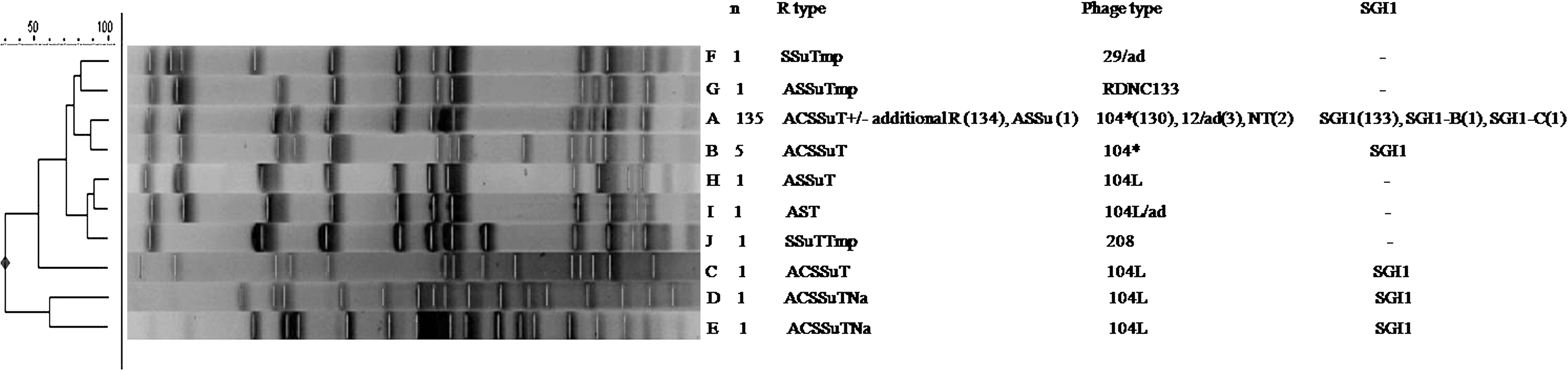
Cluster analysis generated by BioNumerics of the XbaI pulsed-field gel electrophoresis of the 148 serotype Typhimurium isolates. Comparison was performed using the Dice coefficient and clustering was by the unweighted pair group method with arithmetic averages. The different pulsed-field gel electrophoresis profiles, number of isolates, R and phage types, and presence or absence of SGI1 are indicated. *DT104 and related phage types DT104L and DT104L/ad were regrouped as DT104 when the number of isolates is greater than 1. SGI1, Salmonella genomic island 1. A–J correspond to the different pulsed-field electrophoresis profiles.
PFGE of XbaI-digested genomic DNA from the 148 serotype Typhimurium isolates generated 10 PFGE profiles (Fig. 1). PFGE profile A was the most prevalent and found in 135 SGI1 (or variants SGI1-B and -C)-positive isolates (91.2%). Among the 135 isolates displaying the profile A, 130 were of the phage type DT104 complex. Eight other SGI1-positive isolates of the DT104 complex displayed four different PFGE profiles (B, C, D, and E). These results confirmed that SGI1 is mainly encountered in the DT104 clone. Interestingly, in profile A, three serotype Typhimurium isolates harboring SGI1 belonged to phage type DT12/ad. Among the remaining isolates negative for SGI1, two were of the DT104 complex with specific PFGE profiles (H and I) and the other were of phage types DT29/ad, DT208, and RDNC133 and displayed also specific PFGE profiles (F, J, and G, respectively) (Fig. 1).
Discussion
MDR serotype Typhimurium DT104 was described for the first time in the United Kingdom in cattle in the mid-1980s, and has increased dramatically in the United Kingdom as in other countries in the 1990s (Ridley and Threlfall, 1998). Multidrug resistance in serotype Typhimurium DT104 has been shown to be associated with the presence of SGI1 (Boyd et al., 2001; Mulvey et al., 2006). SGI1 is the first genomic island containing an MDR gene cluster identified in S. enterica and may have been an important trait in the worldwide epidemic of the MDR serotype Typhimurium DT104 clone. On the other hand, in France, the annual incidence of salmonellosis in cattle has decreased from about 6 herds to 1 herd per 1000 between 1996 and 2000 (Chazel et al., 2005; Hendrikx et al., 2005). Although serotype Typhimurium is the most prevalent serotype recovered, it declined specifically in cattle since 1996. However, MDR serotype Typhimurium isolates still remain encountered in diseased animals. Thus, in the present study, we intended to investigate to what extent SGI1 may still account for this multidrug resistance in serotype Typhimurium isolated from cattle by studying 148 serotype Typhimurium isolates collected from 2002 to 2007 and that were resistant to ≥2 different classes of antimicrobials.
SGI1 was detected in 143 out of the 148 serotype Typhimurium isolates studied. These isolates displayed an ACSSuT type (115 isolates) or an ACSSuT type combined with one or more additional resistances (27 isolates), with the exception of one last isolate of the ASSu phenotype. PCR mapping of antimicrobial resistance genes of the In104 confirmed that the classical SGI1 element was present in all but two isolates and constituted thus the main structure involved in the resistance to antimicrobials in MDR serotype Typhimurium isolated from cattle. The only two SGI1 variants that were found out in this study were of the SGI1-B and SGI1-C type, with the notable and still undescribed finding of an ACSSuT phenotype conferred by SGI1-B and plasmid-borne phenicols and tetracycline floR and tet(C) genes. Thus, SGI1 variability was extremely limited among the serotype Typhimurium isolates studied, what stands in partial contradiction with the high potential of recombination of In104 (Mulvey et al., 2006). Also, as the remaining five isolates that did not harbor SGI1 were susceptible to florfenicol, this antimicrobial remains probably a good indicator for the presence of SGI1 in S. enterica isolates (Arcangioli et al., 1999; Briggs and Fratamico, 1999; Boyd et al., 2001, 2002).
Analysis of the phage typing results revealed that 138 (96.5%) out of the 143 isolates harboring SGI1 belonged to the DT104 complex (DT104 and closely related phage types, DT104L and DT104L/ad). Analysis of the distribution of PFGE profiles demonstrated that one profile was mostly prevalent and was encountered in 135 SGI1 positive out of the 148 MDR serotype Typhimurium isolates. Again, these results strongly demonstrate that the SGI1-carrying DT104 clone contributes still at first to the multidrug resistance of serotype Typhimurium strains isolated from cattle. Interestingly, 3 of the 135 SGI1 positive were of phage type DT12 and displayed a similar profile as the serotype Typhimurium DT104 strains positive for SGI1. SGI1 has previously been detected not only in MDR DT104 but also in related phage types DT120, U302, or DT12. In these phage types, the occurrence of R type ACSSuT may be explained by changes in phage susceptibility rather than by horizontal transfer of resistance genes (Lawson et al., 2002).
Resistance to third-generation cephalosporins and quinolones/fluoroquinolones was also monitored in this study, as extended spectrum cephalosporins and ciprofloxacin are the drugs of choice for the treatment of Salmonella infections in humans. Until now, resistance to third-generation cephalosporins has been only sporadically described in S. enterica isolated from animals in France, and only from poultry, poultry products, or cockles (Weill et al., 2004; Egorova et al., 2008; Doublet et al., 2009b). Also, high-level resistance to fluoroquinolones remains relatively uncommon in S. enterica (Baucheron et al., 2002). However, the global incidence of quinolones resistance in S. enterica isolates from humans and food animals seems to increase with recent isolates showing decreased susceptibility to fluoroquinolones (Carrique-Mas et al., 2008). Again in this study, resistance to third-generation cephalosporins or fluoroquinolones was not detected in the present panel of strains (data not shown).
In conclusion, almost two decades after the epidemic diffusion of the DT104 clone worldwide, multidrug resistance in serotype Typhimurium isolates from cattle was still mainly due to the resistance genes of SGI1 with the classical penta-resistance phenotype ACSSuT. In cattle and in contrast to what is observed in humans (Weill et al., 2006), the DT104 clone seems to have undergone little changes over the years. However, in animals, the growing spread of mobile genes conferring resistance to third-generation cephalosporins in other Enterobacteriaceae, and in particular in E. coli (Meunier et al., 2006; Girlich et al., 2007; Madec et al., 2008), raises the question of the ability of SGI1 to capture such genetic determinants. Should this occur, such a chromosomal support would probably confer additional selective advantages to their carriers and contribute to further enhance the animal reservoir of resistant foodborne pathogens such as S. enterica.
Footnotes
Acknowledgments
The authors would like to acknowledge the veterinary laboratories that participate in RESAPATH network. This work was supported by a grant from the French Ministry of Agriculture (Direction Générale de l'Alimentation) and by the EU-funded network of excellence MedVetNet (work package WP21).
Disclosure Statement
No competing financial interest exist.