
Abstract
Anisakids are nematodes whose larval stages are often present in fish, molluscs, and crustaceans. Members of the family Anisakidae belonging to the genera Anisakis and Pseudoterranova are implicated in human infections caused by the consumption of raw or undercooked fish. Adequate cooking will kill anisakid larvae, however, killed or inactivated larvae can still cause sensitization and immunoglobulin E-dependent hypersensitivity in human. This work describes the development of DNA-based tests to detect and quantify the presence of Anisakis spp. and Pseudoterranova spp. larvae in fish and fish-derived products, including fish fillets, surimi, fish sticks, canned fish, and baby food. Primers and TaqMan MGB probes recognizing only Anisakis spp. and Pseudoterranova spp. were designed on the first internal transcribed spacer 1 regions of rDNA for a real-time polymerase chain reaction assay. A commercial probe for 18S rDNA was used to detect and quantify the total eukaryotic DNA of the samples. The specificity and sensitivity of the assays were tested using reference samples prepared from mixtures made of Anisakis larvae in different quantity of codfish, and subsequent dilutions. Studies were performed to assess the ability of the test to detect and quantify anisakids in various products. Results showed that this test is able to detect anisakid DNA contained in a proportion of 1:105 in 1 ng of total DNA. The high prevalence of anisakids reported in main fishery species was confirmed by frequently detecting anisakids DNA in fish muscle and fish-derived products. A partial correlation was found between the number of larvae present in the viscera and the level of contamination of fish fillets. In conclusion, this molecular test is useful to detect the presence of Anisakis spp. and Pseudoterranova spp. in fish and fish-derived products and to quantify the level of contamination along the food chain, with potential applications for fish farms, fish markets, and food producers.
Introduction
A
Through the consumption of raw or undercooked fish food (e.g., sushi, sashimi, ceviche, and pickled herrings) humans can become incidental hosts for these parasites, and although larvae cannot progress their life cycles in humans, they pose a risk to human health either directly by causing debilitating infections (anisakidosis or anisakiasis) or by initiating immune hypersensitivity. These infections are most common in countries where raw fish is an important component of the traditional cuisine (among the 20,000 cases of anisakidosis reported in the literature, over 90% are from Japan); however, more and more cases are being reported in Western countries for several reasons, particularly the increase of fish consumption in the Mediterranean diet, known to prevent heart diseases, and the growing popularity of eating uncooked seafood (Audicana and Kennedy, 2008). The disease in humans may have increased when refrigeration was introduced on fishing boats in the mid-50s. Prior to this time, fishes were eviscerated immediately after capture, and the infective organisms were thus discarded (Jay et al., 2005). In Europe, approximately 2000 cases of anisakidosis have been documented, mostly in France, the Netherlands, and Spain, with cases also recorded in Belgium, Italy, and the United Kingdom (Pozio, 2008). In Western Europe, the majority of reported anisakidosis cases have been due to ingestion of herrings; in Spain, most cases have been related to the consumption of pickled anchovies; in the United States, Pacific salmon is the main species involved (Audicana and Kennedy, 2008).
Although adequate cooking and/or prolonged freezing kills anisakid larvae (Wharton and Aalders, 2002), dead or inactivated larvae can cause sensitization and immunoglobulin E (IgE)-dependent hypersensitivity in humans (Audicana and Kennedy, 2008). In addition, non-IgE–mediated mechanisms, such as the involvement of other immunoglobulin isotypes (IgG4) or nonimmunological events, have been described (Daschner and Pascual, 2005). Heat- and/or pepsin-resistant allergens from Anisakis simplex could explain reactions and symptoms after the ingestion of well-cooked or canned fish (Caballero and Moneo, 2004). A. simplex proteins have also been detected in sera from chickens fed with fishmeal and evidence has been provided, based on in vivo and in vitro tests, that highly sensitized allergic patients can respond to the presence of anisakid allergens in chicken meat (Armentia et al., 2006). Moreover, several case reports have shown allergy and anaphylactic reactions to Anisakis in domestic and occupational settings (Purello-D'Ambrosio et al., 2000; Scala et al., 2001; Nieuwenhuizen et al., 2006). To date, nine potential allergens of A. simplex have been identified (from Ani s 1 to Ani s 9) (Arrieta et al., 2000; Asturias et al., 2000; Pérez-Pérez et al., 2000; Moneo et al., 2005; Kobayashi et al., 2007a, 2007b, 2008; Rodriguez-Mahillo et al., 2007; Rodriguez-Perez et al., 2008).
Detection and identification of anisakid larvae from fish by visual inspection is often ambiguous for fresh fish and cannot be applied for the detection of these nematodes in frozen fish, let alone in processed food. This problem has triggered the development of molecular techniques for the better detection and taxonomic identification of anisakid species (reviewed by D'Amelio et al., 2009), most of which are DNA tests based on repetitive genomic regions. However, few methods are currently available to detect and quantify the presence of larvae residuals, still carrying allergen properties, in fish-derived products intended for human consumption. Indeed, although the use of real-time polymerase chain reaction (PCR) assays for the diagnosis of infections caused by protozoan parasites has been reported by several authors, its application with metazoan parasites of veterinary importance is limited (Gasser, 2006) or still at a preliminary stage in terms of sensitivity and reliability (A. simplex) (Lopez and Pardo, 2006).
The main aim of this work was to develop a sensitive real-time PCR test able to detect and quantify the presence of Anisakis spp. and Pseudoterranova spp. larvae in fish and fish-derived products, including fish fillets, surimi, fish sticks, canned fish (tuna), and baby food.
Materials and Methods
DNA extraction
Reference samples from various species of nematodes (A. pegreffii, hybrids A. pegreffii/A. simplex sensu stricto (s.s.), A. typica, A. brevispiculata, A. physeteris, Pseudoterranova complex, Contracaecum spp., and Ascaris spp.) were homogenized using a Braun 600-W device and DNA was extracted using the REDExtraction-N-Amp tissue PCR Kit (Sigma-Aldrich, St. Louis, MO) according to manufacturer's instructions. DNA quality was assessed by both PCR and gel electrophoresis using a universal eukaryotic primer set (EUKA; Table 1). The same method of extraction was used for fish, either fresh or frozen fillets. After counting of larvae by visual inspection, fish were carefully washed with water to remove any viscera contamination, then heads, fins, and tails were removed, and fillets finely homogenized. About 10–20 mg of tissue homogenate was used for DNA extraction. The DNA concentration of these samples was determined using a ND-1000 spectrophotometer (Nanodrop Technologies, Wilmington, DE).
Human 18S rRNA 20 × Predeveloped TaqMan Assay (Applied Biosystems).
ITS1, internal transcribed spacer 1.
DNA from baby food, surimi, fish sticks, and canned tuna products was extracted using the kit Wizard Magnetic DNA Purification System for Food (Promega, Madison, WI) according to manufacturer's instructions. For baby food the extraction was performed on large scale (from 1 g of starting material), whereas for other products it was performed from 200 mg of starting material. The DNA concentration of these samples was determined using a ND-1000 spectrophotometer (Nanodrop Technologies) to check that DNA was present in solution and in sufficient amount to enable the real-time assays.
Design of amplicons and real-time probes
Primers and probes used to detect and quantify the presence of anisakids were designed on the first internal transcribed spacer 1 regions of rDNA to allow the identification of all the species belonging to Anisakis and Pseudoterranova complexes (ANIKIT; Table 1); neither the host organisms (fish) nor other nematodes that possibly contaminate foodstuff from other sources than fish (e.g., Ascaris spp.) were amplified using these primers. The Human 18S rRNA 20 × Predeveloped TaqMan Assay from Applied Biosystems (Foster City, CA), including two unlabeled primers and a VIC dye-labeled TaqMan MGB probe, was used for amplification and relative quantification of the total eukaryotic DNA present in the sample (EUKA; Table 1). In both cases the amplicons were located on redundant genomic regions (ribosomal DNA), thus enhancing the detection the PCR products in degraded samples. Both ANIKIT and EUKA primer pairs were tested on reference samples from various species of nematodes.
Setup of real-time PCRs
A number of reference DNA samples were prepared and used in the real-time PCR assays. Positive and negative control DNA (20 ng/μL) were extracted from anisakid larvae and cod muscle. Three dilution series (from A to C) were obtained by extracting DNA from an homogenate containing 1 mg of anisakid larva and decreasing quantities of fresh cod (ref. A: 100 g, ref. B: 10 g, ref. C: 0.5 g).
Reactions were prepared and performed in 384 optical well plates using a TECAN FREEDOM EVO-150 liquid handling workstation (Tecan Trading AG, Männedorf, CH) and an ABI 7900HT real-time PCR instrument (Applied Biosystems) using the GeneAmp 7900HT sequence detection system software (PerkinElmer Corp., Foster City, CA). The real-time reaction mixture (total of 20 μL) contained 1 × final TaqMan Master Mix, 4 μL of EUKA assay mix for the EUKA amplicon, 300 nM of each ANIKIT primer and 250 nM of ANIKIT probe for the ANIKIT amplicon, and 5 μL of DNA template. The reference samples were diluted 1:10, 1:100, and 1:1000, whereas water was used as negative control. DNA extracted from fresh fish or frozen fillets was used at concentrations of about 20 ng/μL (total DNA amount in reaction: about 100 ng), whereas all the DNA extracted by the Wizard Magnetic DNA Purification System for Food (Promega) was tested without dilution. The PCR cycled as follows: 1 min at 50°C, then 10 min at 95°C followed by 40 cycles of 95°C for 15 s and 60°C for 1 min. Each sample (reference sample, control sample, and unknown sample) was tested in triplicates.
Relative quantification of anisakid DNA
Calculations were performed using a “standard curve method” for relative quantification previously described for gene expression (Livak and Schmittgen, 2001). Briefly, the amount of anisakid DNA in each sample was first normalized versus the amount of total DNA, and then the reference sample used as a calibrator. It should be stressed that when the relative amount of contamination was estimated by this method, the anisakid DNA cannot be discriminated from the total DNA of the sample. However, the method relies on the assumption that the expected concentration of anisakid DNA in fish muscles (and other samples) is irrelevant compared with the total DNA amount detected by the EUKA probe. Moreover, the final quantification of larval mass in the fish sample was calculated versus a known reference.
Detection and relative quantification of anisakids in fish samples of unknown contamination from the fish market
Four mackerels (origin: Atlantic ocean), 10 anchovies, and 10 herrings (origin: Mediterranean sea) were purchased at the fish market. After evisceration and counting of anisakid larvae, fishes were either processed as single fish or were pooled, according to their content of larvae. DNA samples obtained were subjected to real-time PCR in two different experiments (Exps. 1 and 2; Table 2). Each DNA sample was tested in triplicate and without dilution (5 μL per reaction); DNA from a noncontaminated fish (cod) was included as negative control; ref. B was included as positive control and used with serial dilutions (1:10, 1:100, and 1:1000).
The last column reports an estimation of the larvae concentration in fillets based on ref. B.
Detection and relative quantification of anisakid larvae residues in baby food and other fish-derived products
Fish-derived food products were purchased at various stores and chosen to represent the main commercial fish species (labels declare the fish species and their percentage in the product, typically 20%) and different commercial brands (listed from A to J in Table 3). Each DNA sample was tested without dilution and in triplicate (5 μL per reaction); DNA from cod was included as negative control.
Sample retested in a second experiment (see Table 4).
nd, no amplification or doubtful result; Ct, cycle threshold.
A subset of these samples was retested in a second experiment, together with two new additional baby food samples (2A_CA and 2B_CA; Table 4). New DNA extractions were carried out in a separate laboratory where no animal samples had been previously processed to circumvent potential problems of false positive due to DNA contamination; moreover, new reference DNA samples were obtained to facilitate relative DNA quantification in baby food and other products, that is, to refer each signal to the most appropriate standard. For ANIKIT, two positive control series of pooled DNAs were prepared for both baby food and surimi and fish sticks, in which very small amounts of anisakid DNA (from 0.1 ng/μL to 1 fg/μL) were mixed with cod DNA. For testing the EUKA probe, a preparation of either baby food or surimi and fish sticks pooled DNA was used both undiluted and diluted (1:10 and 1:100).
The last column reports the estimation of anisakid DNA concentration in the sample based on ref. BF.
BF, baby food.
DNA from cod was included as negative control. Each unknown DNA sample was tested without dilution in four triplicates for the ANIKIT probe and in three replicates for the EUKA probe (5 μL per reaction). All standard samples were amplified in triplicate.
Results
Setup of real-time PCRs
The ANIKIT primers and probe detected Anisakis spp. and Pseudoterranova spp. complex, as predicted from sequence alignment (data not shown), without cross-amplification of other nematodes species (e.g., Contracaecum spp. and Ascaris spp.). The EUKA primers and probe enabled the detection of components of eukaryotic origin (Fig. 1), which allowed the most objective relative quantification of anisakids.
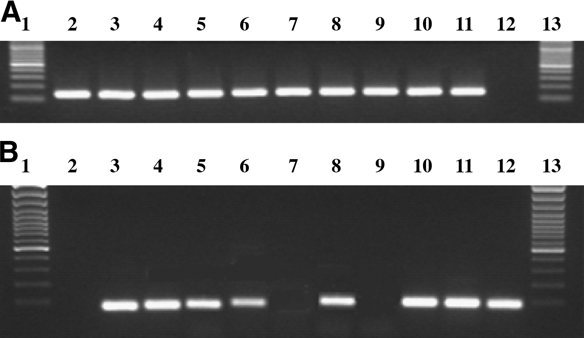
(
ANIKIT cycle threshold (Ct) mean values obtained by real-time PCR assays were proportional to the dilutions of each sample (2.0, 0.2, and 0.02 ng/μL). The first detectable amplification (Ct values of approximately 37) was obtained for ref. B (0.02 ng/μL) and ref. A (0.2 ng/μL), thus showing that the ANIKIT probe is sensitive enough to detect anisakid DNA diluted 1:10,000 in fish DNA (i.e., about 10 fg of larvae DNA in the PCR reaction) (Table 5).
The limit of sensitivity of ANIKIT probe corresponded to a Ct value of approximately 37, that is, for dilution 1:100 (log ng/μL = − 0.7) of series A or 1:1000 (log ng/μL = − 1.7) of series B (in bold).
Detection and relative quantification of anisakids in fish samples of unknown contamination from the fish market
The standard curves for both ANIKIT and EUKA probes were calculated for the specific reaction plate on serial dilutions of the positive control (ref. B). The presence of anisakid DNA was detected in all the four mackerel samples, in two out of four herring samples and in two out of three anchovy samples (Table 2). By reference to sample B, it is inferred that the contamination of mackerel samples varies between 10–260 mg of anisakid larvae per kg of fish. In the case of sample Mk 2, the large number of anisakid larvae counted in the viscera of a single fish (22) corresponded to the highest values of fillet contamination (0.26 g/kg), which is more than double that of ref. B. For both herrings and anchovies there was an approximate correspondence between the number of larvae and the Anisakis DNA test. In the case of herrings, the DNA test only gave a positive result for the samples with four or more larvae, either as a pool (Herr 1) or as an individual fish (Herr 4). For anchovies, only the most contaminated pooled sample (Anch 1) gave a positive DNA test result. In the case of Herr 1 and Herr 4 samples, the inferred values of fillet contamination were 10 and 15 mg, respectively, of anisakid larvae per kg of fish (about 1/10 compared with ref. B), while for Anch 1 the inferred value was much lower (2.4 mg of anisakid larvae per kg of fish).
Detection and relative quantification of anisakid larvae residues in baby food and other fish-derived products
The standard curves for both ANIKIT and EUKA probes were calculated for the specific reaction plate on serial dilutions of the positive control (ref. B).
The results of each sample and the mean Ct values (and standard deviations) of EUKA and ANIKIT probes are reported in Table 3. In some baby food samples the EUKA Ct values were above a threshold of about 31–32. This may be due to various causes: the product may have undergone extensive degradation, the percentage of fish DNA was lower than declared on label, and/or the specific DNA extraction had a lower than expected yield. In these cases, it was impossible to assess if larvae were absent from the product. Noncontaminated samples, however, showed no signal of anisakid DNA in all replicates, but also medium-high concentrations of total DNA. Four baby food samples (out of the 19 for which EUKA Ct was lower than 30) showed clear evidence of contamination. Four out of the seven minced fish products analyzed were found contaminated, with ANIKIT Ct values below 30. Only two of the contaminated products carried informative labels identifying the fish species they contained (i.e., cod).
The presence of anisakids in 7P and 8P (containing European hake) was also confirmed in the second experiment, albeit at a low relative larvae DNA concentration. One of the two new samples (2B_CA) containing the same fish species was found positive. After the new DNA extraction, the samples 5P (containing plaice) and 9P (containing European hake) gave a strongly positive result (about 100–150 fg of larvae DNA), whereas 3P and 4P (containing salmon) and 6P (containing sea bream) were confirmed as negative. Sample 2P (surimi) was confirmed positive, with extreme high values of anisakid DNA detected (about 1–2 pg) (Table 4).
Discussion
The infestation of fish by anisakid nematodes has increased considerably over the last 20 years, often involving fish species intended for human consumption. Indeed, a very large number of fish, cephalopod, and crustacean species worldwide act as hosts for anisakid nematodes belonging to the genera Anisakis (200 fish and 25 cephalopod species) and Pseudoterranova (75 fish species in the North Atlantic only) (Pozio, 2008). There is concern for public health which has brought about the introduction of specific laws to limit human infections, both in the United States and Europe. These include measures such as prolonged freezing at −20°C or below for at least 1 week, or blast freezing at −35°C or below for at least 15 h (Audicana and Kennedy, 2008).
This study describes a method able both to detect the presence of nematodes belonging to Anisakis and Pseudoterranova spp. in fish fillets and fish-derived food products and to perform a relative quantification of anisakid larvae content. The test relies on the design of PCR primers for a specific region of internal transcribed spacer 1 that amplify the DNA of both Pseudoterranova and Anisakis species, without any background, aspecific amplification of the related species of Ascaris and Contracaecum, which can contaminate food stuff through ingredients of various origin (e.g., vegetables in baby food). The selected region is small enough to allow PCR amplification and relative quantification in highly degraded samples (e.g., baby food). Tests of this assay showed that it had high sensitivity, being able to detect anisakid DNA contained in a proportion of 1:100,000 in 1 ng of total DNA.
Using the test to investigate the level of contamination in fish and fish products showed that anisakids were often present in fish muscle and fish-derived products, including fish baby food. The frequent presence of anisakids may reflect the low quality of fish stocks, and perhaps the use of fish that previously would have been discarded from the fresh and frozen fillets market, but which are regularly used for the preparation of these products. Remarkably, only the baby food products containing plaice and European hake, both species representing common fishery products and naturally prone to anisakid infection, gave a positive result to the test, whereas the baby food products containing sea bream and sea bass, which are typically produced by aquaculture (i.e., supposed not to be infected by Anisakis, unless caught in the wild), were not contaminated. This suggests that aquacultured could have advantages over wild fish as they may be guaranteed to be free from A. simplex and related parasites.
Values of larval concentration in fish fillets (reported in Table 2) showed a partial positive correlation with the number of larvae present in the viscera, suggesting that a migration of anisakid larvae from the viscera to muscles has occurred, which is consistently with the observations of other authors. The extent of postmortem migration of anisakid larvae from the mesenteries to muscles in their fish hosts remains controversial (Jay et al., 2005). Some studies have demonstrated postmortem migration of A. simplex larvae in herring, mackerel, and greater forkbeard, but not in blue whiting, whiting, walleye Pollock, or Chilean hake. This difference in the behavior of the larvae is thought to be related to the fat content of the fish, with more migration occurring in fatty fish (Farjallah et al., 2006). However, when such migration occurs, the immediate evisceration of captured fish could reduce the risk as could timely and prolonged freezing (Bouree et al., 1995).
Conclusions
The method presented here will enable the monitoring of the levels of anisakid contamination in fish fillets and foodstuff. This represents a very useful tool to perform periodic quality checks throughout the production chain and for food producers (e.g., baby food factories). Controls could be performed to define thresholds of maximum anisakid content (as maximum allergenic load) in final products. For extensive and semiextensive fish farms (potentially prone to anisakid contamination), timely testing would allow the quality of commercialized stocks to be assessed.
Footnotes
Acknowledgment
This work (project no. 6D42) was supported by the “Ministero delle Politiche Agricole e Forestali” (MIPAF, Rome, Italy).
Disclosure Statement
No competing financial interests exist.