
Abstract
Bacillus cereus can cause diarrheal and emetic types of food poisoning but little study has been done on emetic type of food poisoning in Korea. The objective of this study was to report on the emetic type of food poisoning associated with B. cereus in Korea. The toxin gene profile, toxin production, and antibiotic resistance of B. cereus isolates were investigated in this study. B. cereus was detected in three out of four samples, while the other food poisoning bacteria were not detected. All isolates (KUGH 10, 11, and 12) presented nhe A, B, and C diarrheal toxin genes (755, 743, and 683 bp), detected using NHA, NHB, and NHC primers, and ces emetic toxin gene (1271 bp), detected using CES primer, and produced nonhemolytic enterotoxin and emetic toxin (cereulide), detected using immunochemical assay and high performance liquid chromotography/mass spectrometry (HPLC/MS) analysis. All emetic-associated isolates were resistant to β-lactam antibiotics. Most important finding in this study was that the risk of emetic-type B. cereus food poisoning has existed in Korea. This suggested that the food poisoning caused by B. cereus producing emetic and diarrheal toxins should be constantly evaluated to prevent misdiagnosis between emetic and diarrheal types of food poisoning.
Introduction
B
In recent years, there has been increased awareness of the potential risk of emetic food poisoning caused by B. cereus in Korea. Seventy-three of 293 grains including rice, which is the staple food for Koreans, were contaminated with B. cereus (Park et al., 2009) and the dietary life of Koreans is very similar to Japanese. The potential risk of emetic type of food poisoning associated with B. cereus has existed in Korea. However, Korea Food and Drug Administration (
Materials and Methods
Outbreak investigation
Thirty-seven persons were infected after the consumption of cooked rice and fried rice on the 4th of October in 2005 at a small cafeteria in Paju, Korea. Twenty-four people who ate fried rice as a main dish presented acute symptoms (vomiting, headache, and abdominal pains) of food poisoning at 1–2 h after a lunch. The other people who ate cooked rice did not experience any symptoms of food poisoning. All patients recovered at 12–18 h after presenting acute symptoms. For detection of bacteria causing food poisoning, a microbiological investigation was performed with four vomit samples of patients treated at the Paju Public Health Center, the stool sample of the cook who did not present any symptoms of food poisoning, a kitchen towel, and kitchen utensils (a knife and a board; swabbed with sterilized cotton in the cafeteria). The other patients and fried rice were not investigated because vomiting ceased in 20 patients before visiting the health center, and the small cafeteria had been cleaned before inspection.
Isolation and identification of B. cereus and other food poisoning bacteria
To detect B. cereus in each sample, 25 g of vomit, 10 g of stool and towel, and 3 g of swabbed cottons were mixed with 10-fold volume of buffered peptone solution (Oxoid, Cambridge, England) and homogenized by a stomacher (Bagmixer 400; Interscience, Paris, France) for 5 min. The homogenized mixture was streaked onto Mannitol–Egg Yolk–Polymyxin (MYP; Difco, Detroit, MI) agar and incubated for 24 h at 37°C. During culture on MYP agar, the colonies with pink color were selected for culture on tryptone soya agar (Oxoid) for 24 h at 37°C. The biochemical identification of the isolates was determined using the API 50 CHB (bioMèrieix, Durham, NC) and the API 20E (bioMèrieix) according to the manufacturer's instructions and confirmed with the Apiweb® software (bioMèrieix). The pathogens such as Salmonella spp., S. aureus, Vibrio parahemolyticus, Clostridium perfringens, Listeria monocytogenes, Escherichia coli O157:H7, and Yersinia enterocolitica were tested for their presence according to the Korean Food Code (KFDA, 2005). B. cereus ATCC 12480, B. cereus ATCC 14579, and B. cereus F4810/72 were used as reference strains.
Assay for detection of B. cereus toxin genes
For detection of the toxin genes of B. cereus isolates, enterotoxin genes and emetic toxin gene were amplified by polymerase chain reaction (PCR). The primer pairs and PCR reaction conditions (Table 1) tested in this study were in accordance with previous reports (Guinebretiere et al., 2002; Stenfors et al., 2002; Ehling-Schulz et al., 2005b). The PCR reaction used for the thermal cycler (PTC-100; MJ Research, Watertown, MA) was performed in 20 μL reaction volumes following a protocol reported previously (Guinebretiere et al., 2002; Stenfors, et al., 2002; Ehling-Schulz et al., 2005b). The amplicons were separated by electrophoresis on 2% agarose gel in 0.5× Tris-borate EDTA (TBE) buffer. The gels were stained with ethidium bromide visualized using a UV transilluminator (Gel Doc 2000; Bio-Rad, Hercules, CA). The diarrheal and emetic-type reference strains used as positive controls were B. cereus ATCC 12480, ATCC 14579 and B. cereus F4810/72, respectively.
Assay for detection of B. cereus enterotoxin
The enterotoxin produced by B. cereus isolates was detected using two commercial immunoassay kits according to the manufacturer's instructions. The B. cereus Enterotoxin-Reversed Passive Latex Agglutination (BCET-RPLA) kit (Oxoid, Hampshire, UK) was used to detect the HBL enterotoxin. The Bacillus Diarrhoeal Enterotoxin Visual Immunoassay (BDE-VIA) kit (Tecra International, Frenchs Forest, NSW, Australia) was used to detect the NHE. B. cereus ATCC 12480, ATCC 14579, and B. cereus F4810/72 were used as reference strains.
Assay for detection of B. cereus emetic toxin (cereulide)
High performance liquid chromatography/mass spectrometry (HPLC/MS) analysis was performed to detect emetic toxin. Cereulide was extracted following a protocol reported previously (Häggblom et al., 2002; Jääskeläinen et al., 2003) with a minor modification. The biomass (two or three colonies) of B. cereus isolates was lysed by three repeated freeze–thaw cycles after growth for 4 days at 28°C on tryptone soya agar and extracted with 200 μL of methanol overnight. The filtered (0.45 μm) extract was used for HPLC/MS analysis of cereulide. HPLC/MS analysis was performed according to a protocol reported previously with a minor modification (Andersson et al., 1998). HPLC/MS analysis was performed by ULTRA 3000 HPLC (Dionex, Sunnyvale, CA) equipped with a XeHera C18 column (100 × 2.1 mm, 3-μm particle size) using an ESI electrospray ion trap mass analyzer (LCQ Advantage Max; Thermofinigan, Waltham, MA). The solvent was a mixture of 95% (vol/vol) acetonitrile, 4.9% (vol/vol) H2O, and 0.1% (vol/vol) trifluoro acetic acid at a flow rate of 0.25 mL/min. The sample injection volume was 5.0 μL and A 258 was monitored with a UV detector. The source parameters were as follows: capillary, −3700 V; end point offset, −500 V; nebulizer, 40.0 mL/min; dry gas, 7.0 L/min; and dry temperature, 300°C. The values of mass spectrum from 1100 to 1200 m/z were collected. For identification of molecular cereulide ions, the 1153 (M+H+ adduct) and 1170 (M+NH4 + adduct) m/z values were detected. B. cereus F4810/72, emetic-type reference strain, was used as positive control.
Antimicrobial susceptibility testing
Antimicrobial susceptibility of B. cereus isolates was tested with 15 antimicrobials using the Kirby–Bauer disk-diffusion method (Bauer et al., 1966; Drew et al., 1972). All isolates were grown in Muller–Hinton broth (Oxoid) for 18 h at 37°C. Antimicrobial disks were plated on Muller–Hinton agar (Oxoid) at 37°C for overnight after spreading of culture. B. cereus ATCC 12480, ATCC 14579 and B. cereus F4810/72 were used as reference strains.
Results and Discussion
Isolation and identification of B. cereus and other food poisoning bacteria
All samples were evaluated for the presence of B. cereus and other food poisoning pathogens according to Korean Food Code (KFDA, 2005). The biochemical identification of B. cereus with pink colony during culture on MYP agar was determined using API 50 CHB. B. cereus was detected in three out of four vomit samples. The other food pathogens such as Salmonella sp., S. aureus, V. parahemolyticus, C. perfringens, L. monocytogenes, E. coli O157:H7, and Y. enterocolitica were tested for their presence, but none were detected. B. cereus can cause diarrheal and emetic type of food poisoning (Schoeni and Wang, 2005). The symptoms of diarrheal type of food poisoning such as abdominal pain and diarrhea appear at 8–16 h after consumption of contaminated food including poultry, cooked meats, soups, and desserts (Kramer and Gilbert, 1992; Koneman et al., 1997). The symptoms of emetic type of food poisoning such as vomiting and nausea appear within 1–5 h after ingestion of contaminated food such as fried rice and cooked rice (Lee et al., 1995; McKillip, 2000). Our investigation revealed that this case of outbreak might be caused by emetic type of B. cereus based on the symptom (vomiting), ingested meal (fried rice), and bacterial finding (B. cereus).
Detection of enterotoxin and emetic toxin genes
B. cereus can cause diarrheal and emetic type of food poisoning and the major virulence factors of food poisoning were the HBL, NHE, and CytK enterotoxins encoded by hbl, nhe, and cytK genes (Granum et al., 1999; Ngamwongsatit et al., 2004). To detect the diarrheal toxin genes such as hbl, nhe, and cytK, three B. cereus isolates and reference strains (ATCC12480, ATCC 14579, and F4810/72) were analyzed in accordance with previous studies (Guinebretiere et al., 2002; Stenfors et al., 2002; Ehling-Schulz et al., 2005b). The hbl A (1154 bp), hbl C (740 bp), hbl D (829 bp), and cytK (505 bp) genes amplified by each of the primers showed diarrheal-type reference strains (B. cereus ATCC 12480 and ATCC 14579), but not the three isolates and emetic-type reference strain (B. cereus F4810/72) (Table 2). Three isolates (B. cereus KUGH 10, 11, 12) and all reference strains showed 755, 743, and 683 bp bands amplified from nhe A, nhe B, and nhe C genes using NHA, NHB, and NHC primers, respectively (Table 2). These results are in good agreement with previous reports in which most emetic type of B. cereus isolated from different sources possessed nhe gene (Anderson et al., 2001; Guinebretiere et al., 2002). B. cereus strains possessing hbl and cytK genes are frequently isolated from diarrheal type of food poisoning but rarely in emetic type of outbreaks (Guinebretiere et al., 2002).
B. cereus enterotoxin reversed passive latex agglutination was used to detect HBL enterotoxin of B. cereus.
Bacillus diarrhoeal enterotoxin visual immunoassay kit was used to detect NHE of B. cereus.
High performance liquid chromatography/mass spectrometry (HPLC/MS) with ion trap detector was performed to detect cereulide (emetic toxin).
Emetic-type B. cereus reference strains.
Diarrheal type B. cereus reference strains.
The ces gene target PCR was performed to detect cereulide (emetic toxin) synthetase gene in B. cereus. Three isolates (B. cereus KUGH 10, 11, 12) and emetic-type reference strain showed 1271 bp bands amplified from ces gene, whereas diarrheal-type reference strains did not (Fig. 1). Weber and Marahiel (2001) reported that the β-lactam antibiotics and cyclosporine, important bioactive peptides, are synthesized via nonribosomal mechanism employing nonribosomal peptide synthetase (NRPS). The heterocyclic structure of cereulide and the presence of
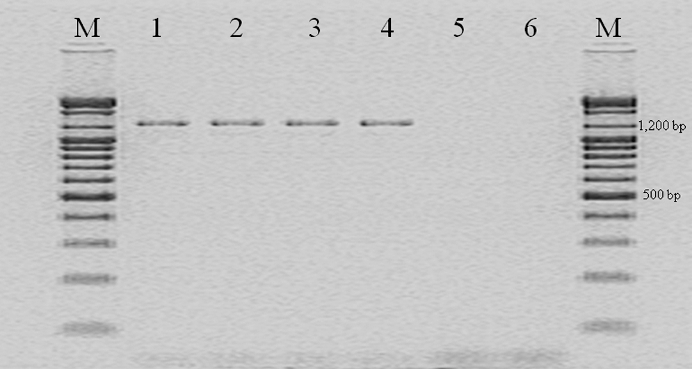
Polymerase chain reaction analysis for ces gene to detect cereulide (emetic toxin)-producing Bacillus cereus isolates and reference strains. Lane M: size marker; lane 1: B. cereus KUGH 10 isolated from vomit; lane 2: B. cereus KUGH 11 isolated from vomit; lane 3: B. cereus KUGH 12 isolated from vomit; lane 4: B. cereus F 4810/72 used as emetic-type B. cereus reference strain; lane 5: B. cereus ATCC 12480 used as diarrheal-type B. cereus reference strain; lane 6: B. cereus ATCC 14579 used as diarrheal-type B. cereus reference strain.
The presence of diarrheal (nhe) and emetic toxin (ces) genes was tested positive, and this is in good agreement with previous reports in which emetic type of B. cereus isolated from a wide range of food environment and clinical source included nhe gene and ces gene (Ehling-Schulz et al., 2005a). Three isolates were identified as emetic type of B. cereus and the results indicate that emetic type of B. cereus has a potential risk of diarrheal and emetic type of food poisoning simultaneously.
HPLC/MS analysis of enterotoxin and emetic toxin
BCET-RPLA and BDE-VIA commercial immunoassay kits were used to confirm the production of HBL and NHE, respectively. BCET-RPLA test, which detects HblC protein-encoded hblC gene, showed negative results. The HBL enterotoxin complex consist of B, L1, and L2 components and the enterotoxic activity is only expressed when all three components of the HBL complex are present together in B. cereus strains (Beecher et al., 1995; Schoeni and Wong, 1999). HBL enterotoxin, which causes a major diarrheal type of food poisoning, was not produced by the three isolates and emetic-type reference strain (B. cereus F4810/72) and also hblA, hblC, and hblD genes were not detected. NHE complex consists of NheA, NheB, and NheC proteins encoded by three genes nheA, nheB, and nheC, respectively (Granum et al., 1999). BDE-VIA test, which detects NheA protein, showed positive results in all isolates and reference strains. This result is in good agreement with previous reports in which the ability to produce NHE was evaluated in 92–100% of B. cereus isolates (Buchanan and Schultz, 1994; Day et al., 1994) and it was found that emetic type of B. cereus contained nhe gene complex and usually produced NHE (Ehling-Schulz et al., 2005a).
HPLC/MS analysis was performed with ion trap mass analyzer to confirm the cereulide (emetic toxin) produced by B. cereus isolates. HPLC/MS spectrum exhibited ion ranges (m/z) of 1153 (H+ adduct) and 1170 (NH4 + adduct), showing the same mass spectrum as that of reference strain (B. cereus F4810/72) (Fig. 2) and that previously reported (Andersson et al., 1998; Mikkola et al., 1999). Cereulide was detected in all isolates (B. cereus KUGH 10, 11, 12), which identified the isolates as emetic toxin-producing B. cereus.
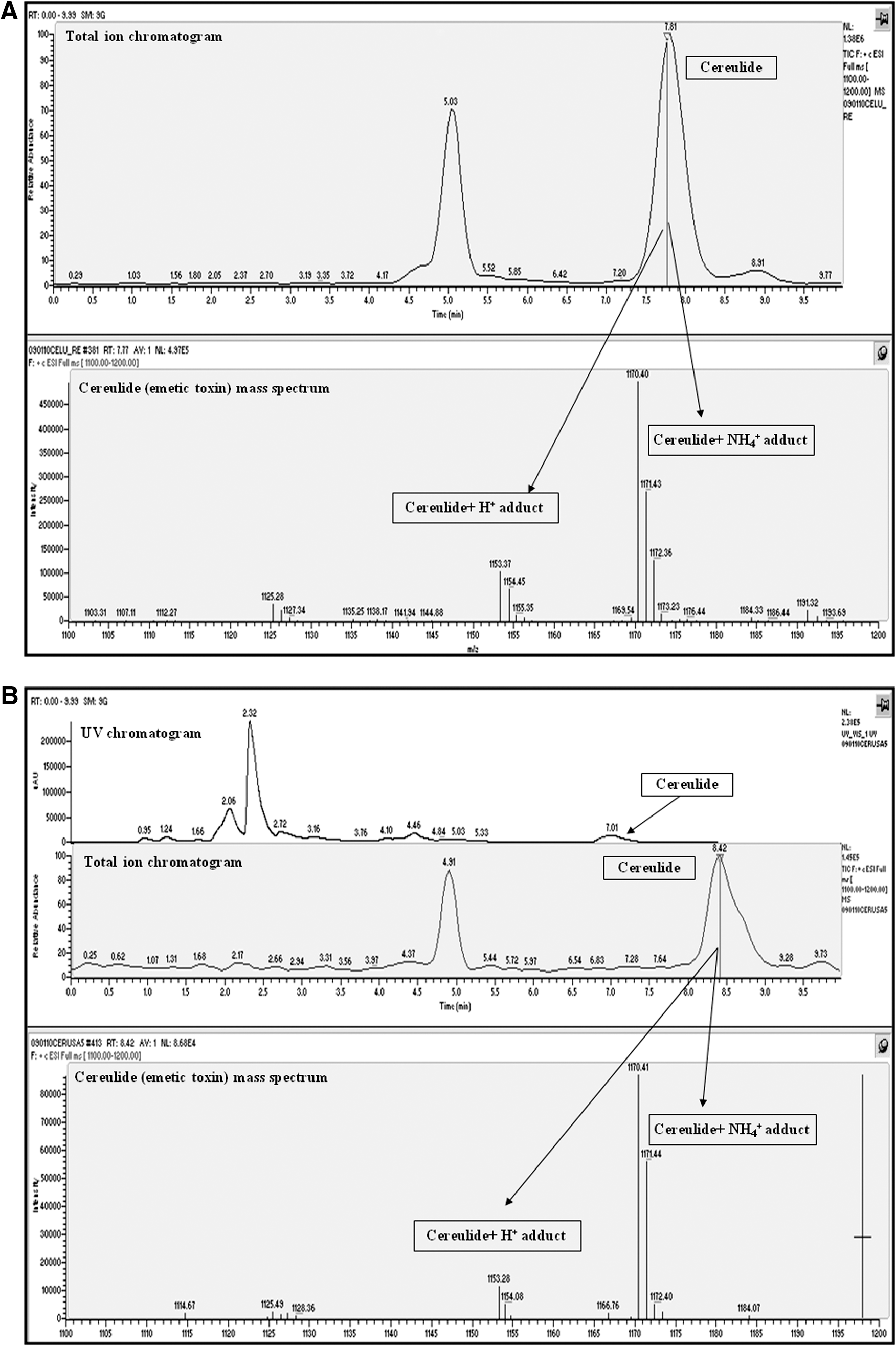
High performance liquid chromatography/mass spectrometry (HPLC/MS) analysis for cereulide (emetic toxin) in methanol extracts of tryptone soya agar cultures of B. cereus F4810/72 as reference strain (

Korean Food Drug Administration has reported that 13 outbreaks of B. cereus food poisoning (
Antimicrobial resistance
Three emetic type of isolates were tested for antibiotic resistance. All isolates were resistant to β-lactam antibiotics such as penicillin, cefepime, and ampicillin and susceptible to gentamicin, imipenem, chloramphenicol, and vancomycin. All isolates and diarrheal- and emetic-type reference strains showed similar antimicrobial patterns. Two of three isolates (KUGH 10, KUGH 11) were intermediately susceptible to rifampin and oxacillin (Table 3). The resistance to cefepime and penicillin in three isolates was due to the synthesis of β-lactamase (Chen et al., 2004), which is a clinically important cause of β-lactam antibiotic resistance (Kotiranta et al., 2000). Drobniewski (1993) and Jensen et al. (2001) reported that B. cereus strains were resistant to penicillin, cephalosporin, and ampicillin and susceptible to gentamicin, tetracycline, chloramphenicol, and vancomycin. B. cereus strains isolated from grains were susceptible to imipenem, vancomycin, and chloramphenicol and resistant to ampicillin and penicillin (Park et al., 2009). Roy et al. (2007) reported that B. cereus strains isolated from Indian fermented foods presented multiantibiotic resistance. Comparing the abovementioned reports, the results presented similar antimicrobial patterns.
Emetic-type B. cereus reference strains.
Diarrheal-type B. cereus reference strains.
R, resistance; S, susceptibility; I, intermediate.
In conclusion, this study revealed that the risk of food poisoning caused by emetic type of B. cereus has existed in Korea and all B. cereus isolates in this study produced emetic toxin (cereulide) and diarrheal toxin (NHE). Thus, analyzing the food poisoning caused by B. cereus, emetic and diarrheal toxin should be constantly evaluated to prevent misdiagnosis between emetic and diarrheal types of food poisoning.
Footnotes
Acknowledgment
This research was supported by a grant (09072KFDA030) from the Korea Food and Drug Administration in 2009.
Disclosure Statement
No competing financial interests exist.