
Abstract
In the present study, the efficiency of a broad-spectrum pulsed ultraviolet (UV)-light for the inactivation of Listeria monocytogenes Scott A, L. monocytogenes CNL 895807, and Pseudomonas fluorescens MF37 populations as agar seeded or suspended cells was investigated. The bacterial populations were treated by pulsed UV-light at different number of pulses (1 to 3), dose of energy (162, 243, or 324 J), and distance from the strobe (4, 9, or 12 cm). After pulsed UV-light treatment, the bacterial reduction was determined by standard plate count. The results showed that there was a significant reduction of population along with an increase of light energy and number of pulses. Decreasing the distance between the Petri dishes and the xenon lamp demonstrated an increase in bacterial reduction. Decontamination efficacy decreased significantly with the increase in level of contamination. This study demonstrates that pulsed UV-light can be used as an effective sterilizing method for the bacteria.
Introduction
F
This procedure is capable of reducing the microbial populations (bacteria, fungi, yeasts, and viruses), both vegetative cells and spores (Lagunar-Solar et al., 2006; Turtoi and Nicolau, 2007; Uesugi et al., 2007; Bialka et al., 2008). Today the literature on pulsed light is rapidly expanding, but a gap remains between basic and applied research with respect to food decontamination. Studies in laboratory conditions by MacGregor et al. (1998), Rowan et al. (1999), and Ghasemi et al. (2003) showed the capability of pulsed UV-light to achieve high lethality on bacteria between 6 and 9 log reductions. Gómez-López et al. (2005a) reported a 3.7 and >5.9 log reductions of Bacillus circulans and Bacillus cereus spores, respectively, when micro-organisms were treated on agar surface with 50 pulses with 7 J/pulse. After 5 pulses of pulsed light treatment at 0.7 J cm−2, Saccharomyces cerevisiae cells suspended in a potassium phosphate buffer were reduced by 6 log (Takeshita et al., 2003). The inactivation of viruses by pulsed UV-light treatment was investigated by Roberts and Hope (2003). In phosphate-buffered saline, a total dose of 1 J cm−2 was sufficient to effectively inactivate between 4.8 and 7.2 log of viruses.
However, microbial inactivation on foods has been focused on specific applications. Pulsed UV-light has been reported to be effective in reducing microbial contamination up to 8 log in Salmonella enteritidis from the surface of eggshells treated at 0.5 J cm−2 with 8 pulses (Dunn, 1996). The treatment of carrot slices with 2 pulses at 0.7 J cm−2 reduced yeast cells of Sac. cerevisiae with up to 6 log (Kaack and Lyager, 2007). After treating several vegetables (spinach, celeriac, radicchio, iceberg lettuce, white cabbage, carrots, green bell pepper, and soybean sprouts) at an intensity of 7 J, log reductions were between 0.56 and 2.04 at 180 sec/side (Gómez-López et al., 2005b).
More extensive basic research is needed to understand the microbial inactivation by this technique. Therefore, the aim of the present study was to investigate the effect of number of pulses, the pulse energy, the distance from the light source, and the inoculum size on the inactivation of Listeria monocytogenes and Pseudomonas fluorescens on solid (agar-seeded cells) and liquid (suspended cells) media, emphasizing the implications of the results for the food-processing industry.
Materials and Methods
Bacterial strains and culture conditions
L. monocytogenes Scott A and L. monocytogenes CNL 895807 kindly provided by Institut Pasteur (Paris, France) and Ps. fluorescens MF37 isolated from raw milk were used in this work. L. monocytogenes strains were grown in heart infusion broth (Merck, Darmstadt, Germany) at 37°C for 24 h. Ps. fluorescens MF37 was grown in nutritive broth (Merck) at 28°C with an agitation speed of 180 rpm for 24 h.
Pulsed UV-light equipment
The pulsed UV-light generator developed by the society Claranor (La colombière, France) was employed in the present study. In this apparatus, the treatment chamber was made in a rectangular parallelepiped unit (width: 60 cm, height: 45 cm, depth: 25 cm,) containing one cylindrical xenon lamp and air cooling system (Fig. 1). This system generated a broad spectrum of pulsed UV-light in the range of 180–1100 nm with pulse duration of 300 μsec. The electrical unit contains the capacitor that stores an adjustable pulse intensity of 162, 243, or 324 J, for an input voltage of 3000 V. A manual mechanical device allowed to change the distance between sample and the lamp.
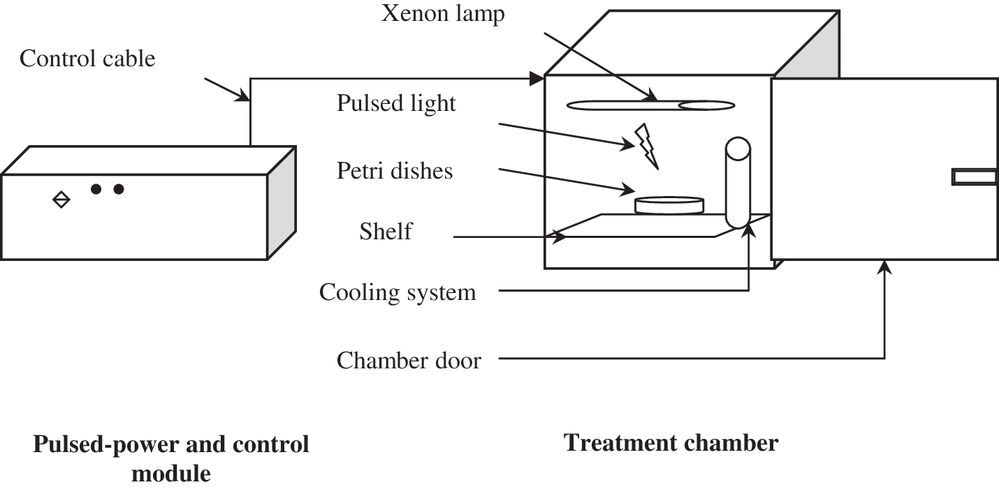
Schematic diagram of pulsed light treatment system.
To study the effect of distance from the lamp on inactivation of bacterial populations, the open Petri dishes were placed on the tray in the treatment chamber at three different distances from the xenon lamp: 4 (top), 9 (intermediate), and 12 cm (bottom). At each distance, the effect of number of pulses and energy intensities was investigated.
Sample preparation
The cells to be treated by pulsed light were prepared as agar-seeded cells and suspension cells, which represent solid and liquid food systems.
For agar-seeded cells, the 24 h bacterial culture in the broth medium was serially diluted. A 0.1 mL sample of the L. monocytogenes Scott A, L. monocytogenes CNL 895807, and Ps. fluorescens MF37 culture and from each dilution was uniformly surface-plated onto trypticase soy agar supplemented with 0.6% yeast extract (TSA-YE) (Merck).
For suspended cells, the 24 h bacterial cultures were centrifuged at 10,000 g for 10 min at 4°C. The supernatant was discarded, and the pellet was washed in phosphate buffer (pH 7) and resuspended in phosphate buffer. The resulting inoculum solution had ∼9 log colony forming units (CFU) mL−1, and the cell suspension was serially diluted up to 10−2 dilution. A 1 mL sample of cell suspension from each dilution and inoculum was transferred to a sterile three-well microplate (Nunc, Roskilde, Denmark). The thickness of the liquid layer in the glass slides was 6 mm.
Pulsed UV-light treatment and microbiological analysis
Agar-seeded plates were positioned in the treatment chamber on shelf at three different distances from the xenon lamp (4, 9, and 12 cm). At each distance, the pulsed UV-light treatment was performed at different light energetic densities per pulse (162, 243, and 324 J) with 1, 2, or 3 pulses. The energy of the pulse also expressed in J cm−2 according to the distances, as shown in Table 1. After which, the control and treated plates were incubated for 24–72 h at 37°C and 28°C for L. monocytogenes and Ps. fluorescens, respectively, and the colonies enumerated.
Distance.
Similarly, the suspended cell samples transferred to a sterile three-well microplate were treated under pulsed UV-light at a distance of 9 cm from the xenon lamp, at an energy of 324 J (0.95 J cm−2) with 1, 3, or 5 pulses. We have treated bacterial suspensions of 7, 8, and 9 log CFU mL−1 to determine the influence of the bacterial population size on the decontamination efficacy of pulsed UV-light. Untreated samples and samples immediately after pulsed UV-light treatment were analyzed for surviving populations by serial dilutions in phosphate buffer, and subsequently by surface plating of 0.1 mL sample on TSA-YE. After incubation for 24 to 72 h at 37°C and 28°C for L. monocytogenes and Ps. fluorescens, respectively, the colonies were enumerated. The log reduction was calculated by subtracting the log value of control from that treated sample.
Statistical analysis
All experiments were performed in triplicate using the same culture for the same treatment at the same time for all three trials. Statistically significant differences between treated and untreated cells were tested by analysis of variance using the STAT-VIEW software system.
Results and Discussion
In the present study, the bacterial inactivation by pulsed UV-light treatment on solid surface and liquid media has been evaluated. Various factors influence the effectiveness of pulsed UV-light to inactivate micro-organisms.
Agar-seeded cell treatment
To demonstrate the effectiveness of pulsed UV-light on solid surfaces, agar-seeded bacterial cells were treated by pulsed UV-light. The energy of pulse, the distance from the xenon lamp, and the number of pulses were investigated. For each set of parameters, the log reduction was calculated. The results from each experiment are presented in Figure 2A–C.

Log reduction in populations of () Listeria monocytogenes Scott A, ( ) L. monocytogenes CNL 895807, and (
 ) Pseudomonas fluorescens MF37 on agar-seeded plates with pulsed light (
Pulsed UV-light that generated at an input voltage of 3000 V was used to treat seeded surface agar at different energies corresponding to 162, 243, and 324 J. The tendency for increased reduction when the energy was increased can be seen by comparing the data at a constant shelf height of 12 cm with 1 pulse. At 162 J (0.45 J cm−2) of energy, 3.00 ± 0.11, 3.96 ± 0.46, and 2.83 ± 0.25 log reductions were shown for L. monocytogenes Scott A, L. monocytogenes CNL 895807, and Ps. fluorescens MF37, respectively (Fig. 2A). These values were significantly higher (p < 0.0001) with an increase of energy. In fact, at an energy of 324 J (0.95 J cm−2), the corresponding log reduction was >7 log for all species tested, which contributed to an effective inactivation. However, for the weak energy, the increase in number of pulses must be required to achieve complete elimination of the pathogen. An ∼7 log reduction was obtained at 162 J (0.45 J cm−2) within 2 or 3 pulses (Fig. 2C).
At the same number of pulses and at the same intensity of energy, the impact of the shelf height was examined. A single pulse with 162 J of energy achieved >7, 5–6, and ∼3 log reductions at shelf height of 4, 9, and 12 cm, respectively, for the three studied bacteria (Fig. 2B). The inactivation of bacterial populations on the agar surface reduced significantly (p < 0.0001) when the distance between the plate and the xenon lamp increased.
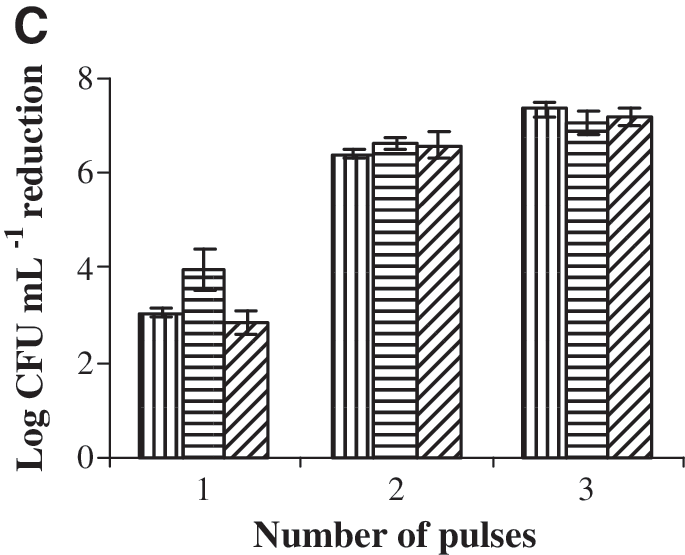
By examining the data at a shelf height of 12 cm and 162 J (0.45 J cm−2) of energy, it can be noted that increasing the number of pulses will increase significantly (p < 0.0001) the bacterial inactivation. The initial inoculum population of about 9 log CFU mL−1 was decreased to 5–6 and ∼2 log CFU mL−1 after 1 and 2 pulses, respectively, for the studied bacterial strains (Fig. 2C). At the highest number of pulse (3 pulses), the inactivation of the bacterial strains was >7 log CFU mL−1.
From Figure 2A, we observe that there was a significant increase in the reduction of population along with an increase of the energy intensity for the same number of pulse and the same distance from the xenon lamp. The level of inactivation achieved in the present work is similar to the observations reported by Fernández et al. (2009) in TSA-YE plates superficially inoculated with L. monocytogenes. They showed that the pulsed UV-light treatment at a higher energy provided the maximum bacterial inactivation. Roberts and Hope (2003) showed that the viral inactivation was 1.4–3.8 and 4.8–7.2 logs at 0.25 and 1 J cm−2, respectively.
According to our results, microbial reduction increased with the decrease of the distance from the xenon lamp, which contributed to an effective inactivation of the bacterial populations on the agar surface (Fig. 2B). As described by Ryer (1997), the intensity of pulsed UV-light reduced with the radial distance from the central axis of lamp. The lower bacterial reductions were observed at greater distances, and this might be due to the increase in energy dissipation as the light pulse travel from the xenon lamp to the Petri dishes. Since the light pulses need to travel a longer path, lesser energy is available for bacterial inactivation on the agar surface at 12 cm distances compared with those at 4 cm. Sharma and Demirci (2003) also observed that inactivation of Escherichia coli O157:H7 on alfalfa seeds reduced significantly as distance increased. In fact, when seeds were subjected to pulsed UV-light, the population reduction at 8 cm distance was 3.14 log CFU g−1, whilst at 13 cm distance, the reduction was 1.14 log CFU g−1.
At a specific distance and energy, the reduction in population increased significantly with increase in the number of pulses (Fig. 2C). Choi et al. (2010) showed also that the inactivation of L. monocytogenes at cultivated plates increased with increasing treatment time and electric power. Our results showed that pulsed UV-light treatment with 1 to 3 pulses were sufficient to effectively inactivate the bacterial populations (>7 log CFU mL−1) on the agar surface. These results are in agreement with the results obtained by Takeshita et al. (2003) and Feuilloley et al. (2006), whereas other studies showed that a great number of pulsed UV-light flashes (100 to 1000 pulses) used for microbial inactivation (MacGregor et al., 1998; Rowan et al. 1999; Ghasemi et al., 2003; Krishnamurthy et al., 2004). Various mechanisms have been proposed to explain the lethal effect of pulsed UV-light, all of them related to the UV part of the spectrum and its photochemical effect (Wang et al., 2005; Elmnasser et al., 2007). Likewise, the study of Woodling and Moraru (2007) demonstrated that the total elimination of UV light portion resulted in no lethal effects on Listeria innocua. In fact, McDonald et al. (2000) treated Bacillus subtilis spores in aqueous solutions and surfaces with pulsed UV-light and concluded that the inactivation was significantly higher than continuous wave UV light.
In this work, the experiment on the effect of these parameters (energy, distance from the xenon lamp, and the number of pulses) had on the inactivation efficiency of pulsed UV-light treatment was performed to evaluate the contribution and optimization of conditions for food product decontamination. Therefore, to use it for food application, the choice of treatment conditions is necessary to achieve a complete decontamination.
Suspended cell treatment
To demonstrate the effectiveness of pulsed UV-light on a liquid medium, the bacterial cells were suspended in phosphate buffer and treated by pulsed light with 1, 3, and 5 pulses at a distance of 9 cm from the xenon lamp and at energy of 324 J (0.95 J cm−2). Figure 3A–C shows the changes in the level of inactivation as a function of inoculum size in the buffer.
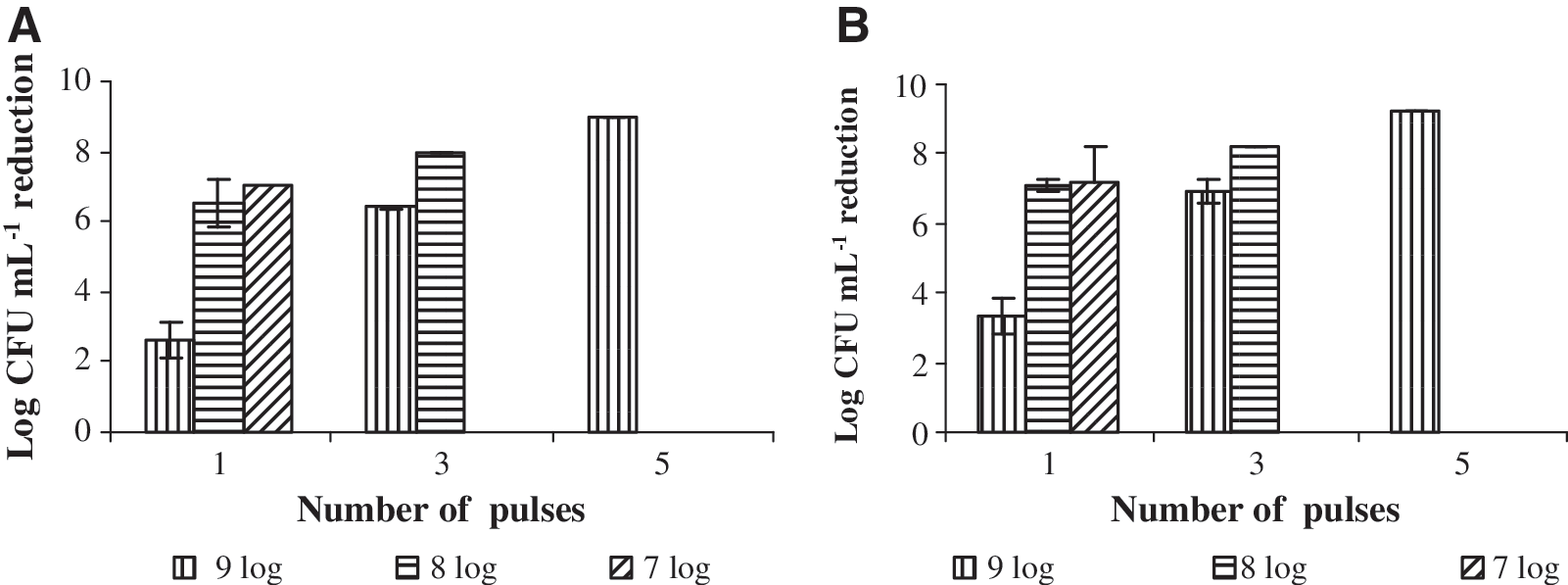
Log reduction in populations of (
For L. monocytogenes Scott A and L. monocytogenes CNL 895807, 2.64 ± 0.53 and 3.30 ± 0.07 log reductions were obtained after the treatment of an inoculum of 9 log CFU mL−1 with 1 pulse. More than 6 log reduction was obtained for the two strains of L. monocytogenes after a 3-pulse treatment. However, after the treatment of an inoculum of 8 and 7 log CFU mL−1 with 1 pulse, we showed that log reductions were between 6 and 7 for L. monocytogenes Scott A, and >7 for L. monocytogenes CNL 895807 (Fig. 3A, B).
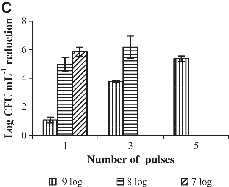
For Ps. fluorescens MF37, 1.09 ± 0.21, 3.77 ± 0.07, and 5.37 ± 0.19 log reductions were obtained after the treatment of an inoculum of 9 log CFU mL−1 with 1, 3, and 5 pulses, respectively. However, a single pulse demonstrated ∼5 and 6 log reductions for an inoculum of 8 and 7 log CFU mL−1, respectively (Fig. 3C).
In our experiment, the inactivation efficiency strongly decreased when the bacterial concentration had increased from 7 to 9 log CFU mL−1 because of the poor penetration capacity of pulsed UV-light. Therefore, the relatively low rate of cell reduction for the 9 log CFU mL−1 inoculum is most likely due to the initial very high bacterial number, which induces a fall-off in pulsed UV-light. In fact, the direct target of pulsed light is the upper layers of bacteria, which are easily inactivated. However, bacteria on the bottom layers are protected by those on the upper layers; therefore, the bacterial inactivation is lower. Because of the shadowing effect (cell-to-cell protection), the high bacterial number requires treatment at higher number of pulses (5 pulses) than low bacterial number (1 pulse) (Fig. 3A–C). We showed that food with low inoculum is efficiently decontaminated. The pulsed UV-light process is efficient when the food product treatment occurs immediately after the transformation steps. Similarly, Ghasemi et al. (2003) observed the same shadowing effect in high inoculum when they studied the pulsed UV-light treatment of Esherichia coli and Sal. enteritidis in a liquid medium. In the case of solid products, Gómez-López et al. (2005a) showed that the inactivation efficiency was strongly decreased with the increase in counts of L. monocytogenes.
Conclusion
The present study clearly shows the potential of pulsed UV-light for inactivating bacterial strains in solid and liquid systems. The fact that a complete microbial inactivation can be achieved after a very short treatment time (1 to 3 pulses) indicates much promise for the use of pulsed UV-light as an alternative to thermal decontamination processes. For an industrial implementation, the dose of energy per pulse and the position of xenon lamp will determine the lethality, and products should be flashed as soon as possible after contamination occurs. Moreover, optimization of these factors could increase the inactivation and thus obtain degrees of inactivation close to what is recommended by the authorities concerned with food safety. Nevertheless, further research on the influence of pulses on foods with a more complex composition is needed to compare their effectiveness and understand the mechanisms by which they act.
Footnotes
Acknowledgments
This work was financially supported by the “Agence Universitaire de Francophonie.”
Disclosure Statement
No competing financial interests exist.