
Abstract
Q fever is a widespread zoonosis caused by the obligate intracellular micro-organism Coxiella burnetii. The objective of this study was to determine the prevalence rate of C. burnetii in bulk milk samples from dairy bovine, ovine, caprine, and camel herds in Isfahan province, Iran. In the present study, 567 bulk milk samples from 186 dairy bovine, ovine, caprine, and camel herds were tested for C. burnetii using a nested polymerase chain reaction assay. The animals whose milk samples collected for this study were clinically healthy. In total, 8 of 247 (3.2%) bovine milk samples were positive; the positive samples originated from 6 of 90 (6.7%) dairy herds. Eight of 140 (5.7%) ovine bulk milk samples from 42 sheep breeding farms and 5 of 110 (4.5%) caprine bulk milk samples from 32 goat breeding farms were positive for C. burnetii. One of 70 (1.4%) camel bulk milk samples from 22 camel breeding farms was also positive for C. burnetii. Although no extensive prevalence study was undertaken, the results of this study indicate that clinically healthy dairy animals are important sources of C. burnetii infection in Iran. To the authors' knowledge, this study is the first report of direct identification of C. burnetii using polymerase chain reaction in bulk milk samples from dairy ovine herds in Iran and the first report of direct identification of C. burnetii in bulk milk samples from dairy camel herds. Further intensive prevalence studies on Coxiella infection and on possible risks of dairy products will be needed to elucidate the epidemiology of Q fever in Iran.
Introduction
Reproductive disorders such as abortion, stillbirth, and delivery of weak and unviable newborns have been reported in livestock infected with C. burnetii (Bildfell et al., 2000). In people, infection is often asymptomatic, and in its mild form can be mistaken for other flu-like illnesses. Chronic infection, however, is a severe disease that requires prolonged antibiotic therapy, because the infection can result in endocarditis (Zhang et al., 1998).
Serological methods have been used to detect antibodies to C. burnetii (Addo, 1980; Soliman et al., 1992; Thomas et al., 1995; Rodolakis et al., 2007; Khalili and Sakhaee, 2009). These assays may not be useful for the diagnosis of acute infection due to the delay in antibody development. Further, it is difficult to discriminate between current and past infection because antibodies often persist after the organisms disappear from the blood (Zhang et al., 1998). Polymerase chain reaction (PCR) assay has become a useful tool for the detection of C. burnetii in clinical samples because of the low detection limit and high diagnostic sensitivity and specificity (Zhang et al., 1998; Berri et al., 2003; Öngör et al., 2004; Fretz et al., 2007; Guatteo et al., 2007). This assay has been described as the most sensitive and rapid means to identify shedder animals (Arricau-Bouvery and Rodolakis, 2005).
Currently, there is limited information on the prevalence of Q fever in human in Iran. A high seroprevalence of C. burnetii (24%–36%) has been recently reported in febrile patients in southeast Iran (Khalili et al., 2010). The objective of the present study was to determine the prevalence rate of C. burnetii in bulk milk samples from dairy bovine, ovine, caprine, and camel herds in Isfahan province, Iran, using a nested PCR assay.
Materials and Methods
Collection of samples
Overall, 186 bovine, ovine, caprine, and camel herds were randomly selected in Isfahan province, Iran. Isfahan is a major industrial center and the second largest city in Iran with about 4.5 million inhabitants. From January to December 2009, a total of 247 bovine bulk milk samples from 90 commercial dairy herds (3–8-year-old Holstein cows), 140 and 110 ovine and caprine bulk milk samples from 42 and 32 sheep and goat breeding farms (2–10 year-old Lori-Bakhtiari breed), and 70 camel bulk milk samples from 22 dairy camel herds (Camelus dromedarius, 3–11 years old) were collected (Table 1). Bovine, ovine, caprine, and camel herds were strictly separated from each other, and the animals were kept in stall-free housing. C. burnetii infection status of the herds was unknown before this study, and there was no established surveillance or management targeting C. burnetii control. The animals whose milk samples collected for this study were clinically healthy, and the milk samples showed normal physical (color, pH, and density) characteristics. The samples were immediately transported to the laboratory in a cooler with ice packs and were processed within an hour of collection.
PCR detection of C. burnetii
To detect C. burnetii in milk samples, the samples were first centrifuged and the cream layers were removed using the method described previously (Berri et al., 2003). DNA was purified using a Genomic DNA purification kit (Fermentas, GmbH) according to the manufacturer's instruction, and total DNA was measured at 260 nm optical density according to the method described by Sambrook and Russell (2001).
All oligonucleotide primers were obtained from a commercial source (Cinna Gen, Iran). The nested PCR assay used to screen for C. burnetii was designed from the nucleotide sequence of the com1 gene encoding a 27-kDa outer membrane protein (OMP) as previously described (Zhang et al., 1998), and the amplification was carried out according to the method described elsewhere (Fretz et al., 2007). For the nested PCR assay with primers OMP1-OMP2 and OMP3-OMP4, the first amplification was performed in a total volume of 25 μL containing 5 μL of DNA sample, 0.5 mM MgCl2, 0.2 mM (each) dNTPs, 1 μM primer OMP1, 1 μM primer OMP2, and 0.5 U/reaction of Taq DNA polymerase (Roche Applied Science).
The PCR assay was performed at 94°C for 4 min and then for 30 cycles of 94°C for 1 min, 56°C for 1 min, and 72°C for 1 min in a DNA thermal cycler (Master Cycler Gradiant). In the second amplification, the reaction was performed in a total volume of 25 μL containing 2 μL of DNA sample, 0.5 mM MgCl2, 0.2 mM (each) dNTPs, 0.8 μM primer OMP3, 0.8 μM primer OMP4, and 0.5 U/reaction of Taq DNA polymerase. The PCR assay was performed at 95°C for 4 min and then for 30 cycles of 94°C for 1 min, 57°C for 1 min, and 72°C for 1 min. The PCR-amplified products (OMP1-OMP2, 501 bp; OMP3-OMP4, 438 bp) were examined by electrophoresis in a 1.5% agarose gel, stained with a 1% solution of ethidium bromide, and examined under UV illumination.
To avoid the risk of false-positive results, a sterilization protocol to prevent amplification of contaminants was followed. Further, PCRs were performed in duplicates along with positive and negative controls. In the present study, C. burnetii DNA (Serial Number: 3154; Genekam Biotechnology AG) and DNase-free water were used as the positive and negative controls, respectively.
Statistical analysis
Data were transferred to Microsoft Excel spreadsheet (Microsoft Corporation) for analysis. Using SPSS 16.0 statistical software (SPSS, Inc.), chi-square test analysis was performed and differences were considered significant at values of p < 0.05.
Results
In the present study, a total of 567 bulk milk samples from 186 dairy bovine, ovine, caprine, and camel herds in Isfahan province of Iran were tested for C. burnetii using a nested PCR assay (Fig. 1). In total, 8 of 247 (3.2%) bovine milk samples were positive (Table 1). The positive samples were from 6 of 90 (6.7%) dairy herds. Three of 8 positive bulk milk samples were from 1 dairy herd (herd number 29) (Table 2). Eight of 140 (5.7%) ovine and 5 of 110 (4.5%) caprine bulk milk samples were positive for C. burnetii. Five of 8 positive ovine bulk milk samples were from 2 dairy herds (herd number 10 and 19), and 2 of 5 goat bulk milk samples were from 1 dairy herd (herd number 4) (Table 2). Only 1 of 70 (1.4%) camel bulk milk samples collected from 22 camel breeding farms was positive for C. burnetii. There were not significant differences in the level of contamination with C. burnetii between raw milk samples from different species.
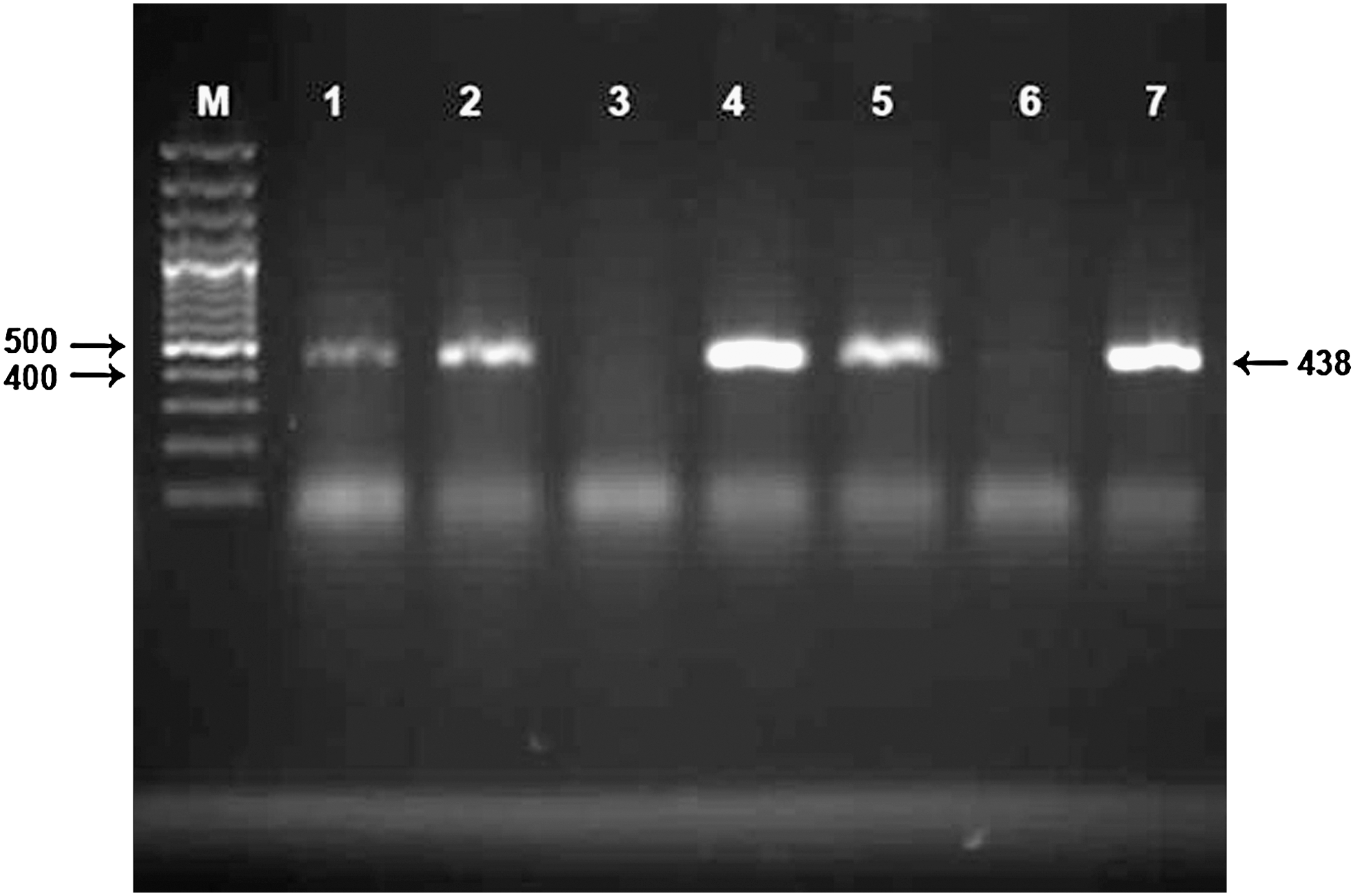
Electropherogram of the amplification products of the nested polymerase chain reaction assay. M, 100 bp DNA ladder; lanes 1, 2, 4, and 5, Coxiella burnetii–positive milk samples from dairy bovine herds; lane 3, negative dairy bovine milk sample; lane 6, negative control; lane 7, positive control.
Overall, the highest prevalence of C. burnetii occurred in the milk samples taken between July and September (1.8%) followed by in the milk samples taken between April to June (1.2%) and January to March (0.9%). No significant differences in the prevalence rates of C. burnetii were observed for bovine, caprine, and camel milk samples taken in different periods of the colleting samples; however, significantly higher prevalence rates of C. burnetii (p < 0.05) were found in ovine milk samples taken between July and September (5%).
Discussion
The prevalence of C. burnetii in bovine milk samples observed in this study (3.2%) is similar to a recent report in Switzerland that showed a prevalence of C. burnetii of 4.7% in bovine bulk tank milk samples using a nested PCR assay (Fretz et al., 2007). In a study conducted in the United States a very high prevalence of C. burnetii (>90%) in bulk tank milk samples from dairy herds has been reported (Kim et al., 2005). In another study conducted in Chaharmahal va Bakhtiari province of Iran, C. burnetii was identified in 6.2% of 210 bovine bulk milk samples (Rahimi et al., 2009).
In this study, eight and five ovine and caprine bulk milk samples from sheep and goat herds were positive for C. burnetii, respectively. In a recent study in Switzerland, all 81 ovine and 39 caprine bulk milk samples were negative for C. burnetii using a nested PCR assay (Fretz et al., 2007). In another study conducted in Turkey, 3.5% of single milk samples from 400 sheep of 22 flocks were positive for C. burnetii by a PCR assay. In that study, all positive results were obtained from flocks with a history of abortion (Öngör et al., 2004). In a study conducted in Chaharmahal va Bakhtiari province of Iran, 1.8% of caprine bulk milk samples were positive for C. burnetii; however, no C. burnetii was isolated from 110 ovine bulk milk samples (Rahimi et al., 2009).
In this study C. burnetii was identified in 1 of 70 (1.4%) camel bulk milk samples taken from 22 camel breeding farms. Several investigators have reported antibodies against C. burnetii in camel sera (Addo, 1980; Schelling et al., 2003; Mazyad and Hafez, 2007), and a high seroprevalence of C. burnetii of 71% has been reported in camels (Soliman et al., 1992); however, no previous report could be found on the direct isolation of C. burnetii from camel milk. Although the results of this study show that milk of camels can be a source for C. burnetii infection, further studies are required to establish the prevalence of C. burnetii infection in camel milk.
Testing animal based only on bulk milk sample can lead to a misclassification of the status of the animal because C. burnetii may be shed by other routes such as vaginal mucus, feces, urine, placenta, or birth fluids, (Guatteo et al., 2006). The differences between the prevalence of C. burnetii in bovine, ovine, caprine, and camel milk samples found in this study may be due to the different routes of shedding C. burnetii present in these animals. For example, while ovine's shed C. burnetii mainly in feces and vaginal mucus, milk is the main shedding route in bovines. It seems that caprines excrete C. burnetii in their vaginal discharges, feces, and milk (Rodolakis et al., 2007). Further, the infected animals may not persistently shed C. burnetii. Infected animals shed C. burnetii mainly during parturition and lactation. Therefore, detection of C. burnetii in bulk tank milk greatly depends on the sampling time. The use of repeated sampling can reduce the likelihood of falsely classifying a herd as C. burnetii negative (Guatteo et al., 2007). The risk of transmission of C. burnetii within a herd is highly dependent on the prevalence of shedder animals (Guatteo et al., 2007). It has been suggested that in herds with PCR-positive bulk tank milk, pools of 10 milk samples can be tested by a PCR assay to identify the shedding animals (Rodolakis et al., 2007).
The findings of the present study are limited to PCR-based detection of C. burnetii DNA in bulk milk samples, so we are unable to speculate on the viability of organisms in milk samples, or on the sensitivity and specificity of the nested PCR assay compared to other diagnostic methods.
The results of this study indicate that clinically healthy dairy animals are important sources of C. burnetii infection in Iran. Therefore, to prevent the spread of infection in animal and human populations, control of bovine, ovine, caprine, and camel coxiellosis should be instituted. Although governmental regulations of milk pasteurization and sanitation in dairy processing plants have been established in Iran for many years, direct sale of unpasteurized milk and dairy products from producers to the consumer is not uncommon in many regions, including Isfahan province. In fact, the consumption of fresh, unpasteurized milk from camels is a traditional practice in some rural areas. The present results also suggest that testing bulk tank milk as an easy and inexpensive method could be used to assess the efficiency of control schemes aimed at controlling and/or preventing C. burnetii infection in dairy herds.
To the authors' knowledge, the present study is the first report of direct identification of C. burnetii by PCR in bulk milk samples from dairy ovine herds in Iran and the first report of direct identification of C. burnetii in bulk milk samples from dairy camel herds. Further intensive prevalence studies on Coxiella infection among farmers, milk-processing workers, veterinarians, and slaughterhouse workers and on possible risks of dairy products will be needed to elucidate the epidemiology of Q fever in Iran.
Footnotes
Acknowledgments
The authors would like to thank H. Momtaz and M. Momeni for the sincere help in performing technical parts of the project. The authors are also grateful to M. Moosavian, S. Kazemzadeh, I. Azadkhah, A. Rasooli, and A. Parvish for assistance with sampling.
Disclosure Statement
No competing financial interests exist.