
Abstract
A potential factor leading to the spread of antimicrobial resistance (AR) in bacteria is the horizontal transfer of resistance genes between bacteria in animals or their environment. To investigate this, swine fecal samples were collected on-farm and cultured for Escherichia coli, Salmonella enterica, Campylobacter spp., and Enterococcus spp. which are all commonly found in swine. Forty-nine of the samples from which all four bacteria were recovered were selected yielding a total of 196 isolates for analysis. Isolates were tested for antimicrobial susceptibility followed by hybridization to a DNA microarray designed to detect 775 AR-related genes. E. coli and Salmonella isolated from the same fecal sample had the most AR genes in common among the four bacteria. Genes detected encoded resistance to aminoglycosides (aac(3), aadA1, aadB, and strAB), β-lactams (ampC, ampR, and bla TEM), chloramphenicols (cat and floR), sulfanillic acid (sul1/sulI), tetracyclines (tet(A), tet(D), tet(C), tet(G), and tet(R)), and trimethoprim (dfrA1 and dfh). Campylobacter coli and Enterococcus isolated from the same sample frequently had tet(O) and aphA-3 genes detected in common. Almost half (47%) of E. coli and Salmonella isolated from the same fecal sample shared resistance genes at a significant level (χ 2, p < 0.0000001). These data suggest that there may have been horizontal exchange of AR genes between these bacteria or there may be a common source of AR genes in the swine environment for E. coli and Salmonella.
Introduction
One problem that needs to be addressed is the potential for the development and amplification of AR genes via horizontal transfer of genetic elements encoding AR (Michael et al., 2004; Boerlin and Reid-Smith, 2008). This type of transfer may occur from relatively innocuous commensal strains where AR develops, and can then be transferred to pathogens resulting in an MDR pathogen. This can take place in a variety of environments, including on-farm environments where animals are treated with antimicrobials, thus selecting for AR bacteria that could be transmitted to humans via contaminated food products. However, studies supporting these hypotheses have relied solely on observations of phenotypic resistance to imply the amplification and transfer of AR, whereas other studies have identified only a few genes potentially transferred between donor and recipient bacteria (Aarestrup, 1999; Akwar et al., 2007; Frye and Fedorka-Cray, 2007; Aarestrup et al., 2008b; Jakobsen et al., 2010). Recent advances in gene detection technology, such as DNA microarrays, enable the identification of large numbers of AR genes in a single assay and can contribute to a better understanding of gene transfer (Chen et al., 2005; Perreten et al., 2005; Bruant et al., 2006; Frye et al., 2006, 2009; Ma et al., 2007; Batchelor et al., 2008; Zou et al., 2009).
In this study, these new methods were used to investigate AR genes in isolates from the on-farm environment. A previously described DNA microarray designed to detect 775 AR-associated genes was used to identify AR genes and genetic elements in bacteria isolated from animal samples (Frye et al., 2006, 2009). This microarray method was applied to bacteria isolated from swine feces collected on-farm during the Collaboration in Animal Health Food Safety and Epidemiology (CAHFSE) pilot study in swine (
Materials and Methods
Swine farm sampling and feces collection
Isolates were obtained from the CAHFSE swine program conducted by USDA from 2003 to 2006, as described on the Web site
Bacterial isolation and identification
Generic Escherichia coli
One-hundred-microliter aliquots of fecal dilutions (1:9 wt/vol, in phosphate-buffered saline [PBS, 0.1 M, pH 7.2]) were streaked for isolation onto CHROMagar EEC™ (Hardy Diagnostics, Santa Maria, CA) plates. The plates were incubated for 18–24 h at 42°C, after which colonies indicative of E. coli were selected.
Salmonella
Feces (1 g) were incubated in 10 mL of GN Hajna (Difco Laboratories, Detroit, MI) for 18–24 h at 37°C, and Tetrathionate broth (Difco Laboratories) for 40–48 h at 37°C. After initial enrichments, aliquots (100 μL) were transferred to 10 mL of Rappaport-Vassiliadis R10 broth (Difco Laboratories), which were incubated for 18–24 h at 37°C. Ten- microliter aliquots of Rappaport-Vassiliadis R10 broth were then streaked onto Xylose-Lysine-Tergitol-4 (Difco Laboratories) and Brilliant Green Sulfa (Difco Laboratories) agar. Plates were incubated for 18–24 h at 37°C. Isolated colonies characteristic of Salmonella were inoculated into triple sugar iron and lysine iron agar slants for biochemical confirmation. Presumptive positive isolates were serogrouped using serogroup-specific antisera (Difco Laboratories) and sent to National Veterinary Services Laboratories, APHIS, USDA (Ames, IA), for serovar determination.
Campylobacter spp
Fecal samples were diluted (1:9 wt/vol, in PBS) and 100 μL aliquots were inoculated onto Campy-Cefex agar plates (Stern et al., 1992) and into 1 mL Bolton Broth enrichment media in 24-well tissue culture plates (Falcon 353047; Becton Dickinson Labware, Franklin Lakes, NJ). Agar plates and enrichment broth were incubated for 36–48 h at 42°C under microaerobic conditions (5% O2, 10% CO2, and 85% N2). Presumptive Campylobacter colonies were selected by observation of cellular morphology and motility using a wet mount under phase-contrast microscopy. Isolates were identified by species using a commercial multiplex PCR (BAX® PCR; DuPont Qualicon, Wilmington, DE).
Enterococcus spp
One-hundred-microliter aliquots of fecal dilutions (1:9 wt/vol, in PBS) were inoculated into 24-well tissue culture plates (Becton Dickinson Labware) containing 1 mL of Enterococcosel broth (Becton Dickinson, Sparks, MD) per well. The plates were incubated for 18–24 h at 37°C, followed by streaking for isolation onto Enterococcosel agar (Becton Dickinson). Isolates were identified by species using a multiplex PCR (Jackson et al., 2004a).
Bacterial growth conditions and antimicrobial susceptibility testing
E. coli and Salmonella were grown in Luria Bertani (LB) media, on LB agar or blood agar plates at 37°C as indicated. Campylobacter coli were grown on Campy-cefex plates or in Bolton Broth and incubated at 42°C for 48 h under microaerobic conditions (5% O2, 10% CO2, and 85% N2) in zip-top storage bags. Enterococcus spp. were grown in LB, brain heart infusion, or on blood agar plates at 37°C with standard methods. All bacteria were stored by addition of glycerol (10% final volume) to cells grown in liquid media and freezing at −80°C.
Susceptibility testing for E. coli, Salmonella, and Enterococcus spp. was performed using custom-made broth microdilution plates for the Sensititer™ system (TREK Diagnostic Systems, Inc., Westlake, OH) as described previously (
DNA extraction and labeling
Genomic DNA from E. coli, Salmonella, and Enterococcus spp. isolates was extracted from 5 mL of overnight cultures grown in LB (Gram negative bacteria) or brain heart infusion (Gram positive bacteria) broth using the GenElute Bacterial Genomic DNA kit (Sigma, St. Louis, MO) as described previously (Frye et al., 2006). Camplyobacter genomic DNA was isolated from colonies collected from Campy-cefex plates using the Puregene DNA isolation kit (Gentra Systems, Minneapolis, MN) according to manufacturer's directions. DNA was labeled with Cye dye-labeled dCTP (Amersham, Piscataway, NJ) via random priming and extension with Klenow fragment (New England Biolabs, Beverly, MA), followed by purification with a Qiagen PCR clean up kit (Qiagen, Valencia, CA) as previously described (Porwollik et al., 2001).
DNA microarray design and construction
The array consists of probes designed to detect 775 AR-associated genes and was constructed as described previously (Frye et al., 2006, 2009; Zou et al., 2009). Controls included three positive control probes designed to detect bacterial 16S rDNA sequences (Liu et al., 2005), two Cy3- and Cy5-labeled lambda DNA controls, 12 buffer (50% dimethyl sulfoxide) only spots, and 130 empty (background) spots. Twelve probes were also synthesized in duplicate or triplicate as controls. All probes were synthesized (Operon Inc., Huntsville, AL), diluted in printing buffer (35 μM in 50% dimethyl sulfoxide), and arrayed in triplicate onto UltraGaps slides (Corning Inc., Life Sciences, Acton, MA) with an Omnigrid robot (Genemachines, San Carlos, CA) and post-processed as described previously (Porwollik et al., 2003; Frye et al., 2006).
Hybridization and scanning
Dye-labeled DNA was dried and resuspended in 80 μL of hybridization buffer (25% Formamide, 5 × SSC, 0.1% SDS, and 1% BSA), boiled for 5 min, and applied to the microarray under a LifterSlip (Erie Scientific, Portsmouth, NH). Hybridization was performed overnight in a hybridization chamber (Corning Inc., Life Sciences) submerged in a 42°C water bath in the dark. Protocols suggested by the manufacturer (Corning, Inc., Life Sciences) for hybridizations in formamide buffer were used for prehybridization, hybridization, and posthybridization wash processing (step 1: 2 × SSC, 0.1% SDS, 5 min at 42°C; step 2: 0.1× SSC, 0.1% SDS, 10 min at room temperature; step 3: 4× 0.1× SSC, 1 min at room temperature). Microarrays were scanned with a ScanArray Lite Laser scanner (PerkinElmer Life and Analytical Sciences, Waltham, MA) using ScanArray Express 3.0 software or with a GenePix 4100A (Molecular Devices, Sunnyvale, CA) and GenePix Pro software.
Hybridization analysis and scoring
Hybridizations were analyzed as previously described (Frye et al., 2006, 2009; Zou et al., 2009). Briefly, images were analyzed and quantified using ScanArray Express 1.1 software (PerkinElmer Life and Analytical Sciences). Hybridization signal intensities were measured in arbitrary intensity units by adaptive quantification mode followed by local background subtraction; the median values of the triplicate gene probes were recorded. Gene presence or absence was determined by setting a cutoff at two times the median intensity for all probes on the array. Genes with hybridization intensities greater than this cutoff were scored as detected (Frye et al., 2009; Zou et al., 2009). Hybridizations without detection of at least one 16S rDNA or with obvious hybridization anomalies (high background, streaks, spots, blotches, etc.) were considered failed and were repeated.
Data and statistical analysis
Genes detected in different bacteria species and samples were tabulated in Excel® spreadsheets and totaled, and genes shared by different bacteria isolated from the same sample were determined. The χ 2-test was used to compare the predicted frequency of resistance genes detected in different bacteria from the same sample to the actual frequency observed. Pairs of isolates where <10 genes were detected in one isolate were excluded from analysis. The χ 2-test was considered significant at p < 0.0000001.
Cluster analysis of isolates with resistance genes detected
Relationships between isolates were determined by hierarchical cluster analysis using open source CLUSTER 3.0 with Euclidean distance for genes detected (Eisen et al., 1998; de Hoon et al., 2004). The dendrogram was viewed using Java TreeView version 1.1.4r3 (
Plasmid replicon typing
Three panels of multiplex PCR were used to determine the presence of 18 plasmids commonly found in E. coli and Salmonella and to identify them based on incompatibility (Inc) groups. PCR primers and conditions were previously described (Carattoli et al., 2005; Lindsey et al., 2009).
Results
Bacteria recovered from swine fecal samples selected for analysis
A total of 7960 swine fecal samples were collected from 2003 to 2006 by the CAHFSE program. All of these samples were cultured for Salmonella and ∼40% (n = 3198) of the samples were also cultured for the other three bacteria. E. coli and Enterococcus spp. were isolated most often at 88.7% and 63.9% of samples, respectively. Campylobacter and particularly Salmonella were less frequently isolated at 61.0% and 9.1%, respectively. Phenotypic testing, including species determination, serovar determination, and antimicrobial susceptibility testing, was completed on all isolates (data available at
Clinical and Laboratory Standards Institute breakpoints were used to determine resistance phenotype when available.
Gen, gentamicin; Kan, kanamycin; Str, streptomycin; Bac, bacitracin; Amc, amoxicillin-clavulanic acid; Amp, ampicillin; Cef, cephalothin; Cro, ceftriaxone; Cep, cefepime; Fox, cefoxitin; Pen, penicillin; Tio, ceftiofur; Chl, chloramphenicol; Fla, flavomycin; Van, vancomycin; Cli, clindamycin; Lin, lincomycin; Ery, erythromycin; Tyl, tylosin; Met, metronidazole; Nit, nitrofurantoin; Linc, lincomycin; Ci, ciprofloxacin; Nal, nalidixic acid; Syn, Synercid; Sul, sulfisoxazole; Tet, tetracycline; Tri, trimethoprim-sulfamethoxazole.
Phenotypic data for isolates of each bacterial species from the 49 samples were collected. None of the E. coli isolates were O157:H7 based upon colony morphology observed on CHROMagar. All Campylobacter isolates were assayed by Bax PCR and found to be C. coli (Table 1). The S. enterica isolate serovars were Derby (n = 19), Typhimurium (6), Typhimurium var. 5- (8), Heidelberg (5), Bovismorbificans (1), Newport (1), Infantis (1), Worthington (1), Mbandaka (6), and Untypable (1). Enterococcus species isolated included E. faecalis (28), E. durans (2), E. hirae (10), E. solitarius (2), E. casseliflavus (2), E. faecium (3), E. mundtii (1), and E. gallinarum (1) (Table 1).
Antimicrobial susceptibility of isolates
AR phenotypes for each isolate are shown in Table 1. Antimicrobials tested and total resistance found for each genus or species of bacteria are summarized in Table 2. Most of the isolates were resistant to one or more of the antimicrobials tested. Many of the E. coli and Salmonella isolates were resistant to aminoglycosides, β-lactams, folic acid pathway inhibitors, chloramphenicol, and tetracycline. None were resistant to members of the quinolone class of antimicrobials. For this study, resistance to more than one class of antimicrobial was considered MDR. The most common MDR E. coli detected were resistant to tetracycline and streptomycin (n = 6); ampicillin and tetracycline (n = 5); and ampicllin, streptomycin, and tetracycline (n = 5). Only two Salmonella isolates were susceptible to all antimicrobials tested (pan-susceptible); however, resistance was detected to all antimicrobials tested except the quinolones. Most of the Salmonella isolates (n = 41) were MDR. Salmonella Typhimurium was resistant to five or more antimicrobials more often than any other serovar, and resistance to eight or more antimicrobials occurred among serovars Newport, Derby, Heidelberg, and one untypable (Table 1). The C. coli isolates had the least resistance detected, and four isolates were susceptible to all antimicrobials tested (Table 1). The most common MDR phenotype for C. coli was detected in 15 isolates and included resistance to azithromycin, erythromycin, clindamycin, and tetracycline (Table 1). All Enterococcus spp. isolates were resistant to at least two antimicrobials tested, and one was resistant to 10 drugs. The most prevalent MDR phenotype detected was resistance to bacitracin, erythromycin, lincomycin, tetracycline, and tylosin, all of which were E. faecalis. The most extensively resistant isolate was resistant to bacitracin, erythromycin, flavomycin, kanamycin, lincomycin, penicillin, streptomycin, synercid, tetracycline, and tylosin (Table 1).
Percent based only on bacteria tested with this antimicrobial.
NT, not tested for this bacteria.
AR genes detected in isolates
Hybridization data for all isolates are summarized in Table 3. A completed dataset is available in Supplementary Table S1 (Supplementary Data are available online at
Genes and gene families with multiple probes are summarized and presented only once; ND indicates that none of the gene probes in this table were detected in that isolate (full hybridization data are available in Supplementary Table S1).
Probes with positive hybridizations to more than one isolate from the sample are indicated in bold.
Shaded samples hybridized to the same gene probes at a significant level as determined by the χ 2-test (p < 0.0000001).
Salmonella isolates also had a large number of positive probe hybridizations with an average of 56 genes detected per isolate. The most numerous aminoglycoside resistance genes detected in the Salmonella isolates were from the family of aadA genes followed by the str family. Genes detected and number of isolates in which they had been found were aadA1 (n = 30), aadA1b (n = 20), aadA2 (n = 20), aadB (n = 17), and strAB (n = 22). β-lactam resistance genes were detected and included blaL2 (n = 18), ampR (n = 15), ampC (n = 11), per-2 (n = 12), and tem (n = 10). Sulfamethoxazole and sulfisoxazole resistance genes detected most often in the Salmonella isolates were the highly homologous sul1 (n = 16) and sulI (n = 20) genes, whereas trimethoprim resistance genes detected were dfrA1 in 18 isolates and dfh in 13 isolates. Chloramphenicol resistance genes floR (n = 10) and cat4 (n = 9) were also detected. Tetracycline resistance genes found in most isolates included tet(A) (n = 22), tet(R) (n = 17), tet(C) (n = 13), and tet(D) (n = 21). Heavy metal resistance genes were also detected in several of the isolates with at least one mer gene detected in 21 of the isolates. Genes encoding resistance to sanitizers such as qacE were detected in 23 isolates. Many genes often mobilized with AR were also detected the most numerous were the transposase gene tnpAIS26 (n = 30) and the class I integrase gene intI1 (n = 16).
C. coli isolates had the lowest number of hybridizations to AR gene probes with an average of 17 genes detected per isolate. Over half of the C. coli isolates had aminoglycoside resistance genes detected and included aph-A3 (n = 19) and several close homologs to this gene (n = 24). The endogenous cam-1 β-lactamase gene was found in 30 of the isolates. Only three isolates had chloramphenicol resistance genes detected including floR, cat, and cmlA5. The cmeB and cmeC efflux pump genes were detected in 25 and 30 isolates, respectively. The tet(O) tetracycline resistance gene was found in a large number of the C. coli isolates (39), with just a few hybridizations to tet(A), tet(M), tet(R), and tet(S). Very few mobilization associated genes were found in the C. coli isolates.
The Enterococcus genera isolates had a moderate number of hybridizations to the gene probes with an average of 23 genes detected per isolate. The most prominent aminoglycoside resistance genes detected were acc(3) (n = 10) and aphA-3 (n = 7). Several β-lactam resistance genes were also detected including the blaL2 gene (n = 11) and the type-II SCCmec (methicillin resistance) cassette-associated gene pre (n = 22). The linB, lincomycin resistance gene, was detected in six isolates. The macrolide resistance gene most often found was the ermB gene, which was detected in 26 enterococcus isolates. Chloramphenicol resistance genes identified included the cat gene (n = 5). Several tetracycline resistance genes were detected, including tet(O) (n = 27), tet(M) (n = 23), tet(W) (n = 10), and tet(A) (n = 6). Some vancomycin resistance genes detected, including vanC (n = 1) and vanH (n = 4); additionally, a gene encoding resistance to the glycopeptide, bleomycin, was detected in eight isolates. Efflux pump genes were detected, including one associated with the type-II SCCmec cassette that was found in 22 isolates. Sanitizer resistance genes were detected, including the qacE gene, in eight isolates. Finally, multiple transposon genes often mobilized with resistance genes were detected, including tpnA (n = 28), a Tn1546 homolog, and trans (n = 35), another transposon related gene.
Genes detected in different bacterial species isolated from the same sample
To understand the distribution of resistance genes detected in the isolates, genes detected in the bacteria were compared to determine which genes were shared by the different bacteria isolated from the same fecal sample. Predominant genes detected are summarized in Table 3, with shared genes indicated by bold font and samples that share genes at a significant level by the χ 2-test indicated by shading.
E. coli and Salmonella had the most genes detected in common from the same sample at 1048, with 224 genes detected at least once in both Salmonella and E. coli isolated from the same sample. Aminoglycoside resistance genes were some of these, including aac(3) (shared in five samples), the family of aadA1 homologs (12 samples), aadB (8 samples), and strAB genes (10 samples). β-lactam resistance genes were found in common, the most numerous of which were ampC (8 samples) and its regulator, ampR (11 samples), blaL2 (10 samples), and others included class A β-lactamases, bla SME-1 (five samples), bla PER2 (five samples), and bla TEM (two samples). Sulfamethoxazole and sulfisoxazole resistance genes were also shared, including the highly similar sul1 (one sample) and sulI (two samples) genes, as well as sulII (one sample). The most often shared trimethoprim resistance genes were dfrA1 (six samples) and dfh (eight samples). Chloramphenicol resistance genes were also detected in pairs of E. coli and Salmonella, including the cat gene in six samples and the floR gene in four samples. Tetracycline resistance genes were shared quite often and included tet(A) (17 samples), tet(D) (17 samples), tet(C) (six samples), and tet(R) (eight samples). Several members of heavy metal transport systems were also detected, including members of the mer family, the most common being merT, where it was detected in 13 pairs of isolates from the same sample. Quaternary ammonium resistance genes, qacE, were also detected in both E. coli and Salmonella from 8 samples. Genes associated with the transfer of resistance were detected in common, including insA (15 samples), intI1 (five samples), and tnpAIS26 (14 samples). For almost half of the samples (23), the correlation between genes detected in E. coli and Salmonella was determined to be significant (Tables 3 and 4, shaded samples). The most prevalent gene probes shared by pairs of E. coli and Salmonella isolated from each sample are summarized in Table 4 (complete data available in Supplementary Table S1).
Genes and gene families with multiple probes are summarized and presented only once; ND indicates that none of the gene probes in this table were shared in isolates from this sample (full hybridization data are available in Supplementary Table S1).
Shaded samples hybridized to the same gene probes at a significant level as determined by the χ 2-test (p < 0.0000001).
The next most frequent bacterial species isolated from the same sample to share genes was Salmonella and Enterococcus at n = 125, with 64 probes shared by at least one pair of bacteria from the same sample. Several aminoglycoside resistance genes were detected in both of these bacteria from the same sample, including aadA1 (two samples), aadA2 (three samples), aadB (one sample), and strB (one sample). The blaL2 gene was detected in four Salmonella and Enterococcus sample pairs and was the only β-lactamase gene detected in common. The tetracycline resistance gene probes tet(A) (two samples) and tet(R) (one sample) were also detected. The quaternary ammonium resistance gene probe qacE was also detected in both isolates from five samples. The transposase gene probe insA also had positive hybridizations with seven pairs of Salmonella and Enterococcus. One sample (ID number 15) had genes shared at a significant level between Enterococcus and Salmonella, each of which also shared genes at a significant level with the E. coli isolated from that sample (Table 3).
A similar number of E. coli and Enterococcus pairs from the same sample had shared genes detected at n = 117, with 71 shared by at least one pair. Some of these included aminoglycoside resistance gene probes aadA1 (one sample), aadA2 (two samples), aphA3 (one sample), and strB (two samples); β-lactamase resistance gene probes ampC (two samples), blaL2 (two samples), bla SME-1 (one sample); quaternary ammonium resistance gene probes qacE (two samples), qacF (one sample), and qacH (two samples); tetracycline resistance gene probes included tet(A) (two samples), tet(M) (one sample), tet(O) (three samples), tet(R) (one sample), and tet(W) (one sample); transfer-related gene probes included insA (five samples), intI1 (two samples), and transB (one sample). In addition, Salmonella, E. coli, and Enterococcus also had a few samples where all three bacteria shared a positive hybridization to the same probe (n = 62 for 33 probes). These included aadA1 (one sample), aadA2 (two samples), aadB (one sample), strAB (one sample), blaL2 (two samples), qacE (two samples), qacF (one sample), qacH (two samples), tet(A) (two samples), and insA (four samples).
The remaining pairs of bacteria had only a few genes found to be common, and all of these were pairs with Campylobacter, which had the lowest number of genes detected in the study. The total for Campylobacter and Enterococcus was 52, Campylobacter and Salmonella were 31, and Campylobacter and E. coli were 28. The tet(O) gene probe alone stood out as being shared by the Campylobacter with other bacteria isolated from the same sample in this study. The tet(O) gene was detected in 21 pairs of Campylobacter and Enterococcus and three pairs of Campylobacter and E. coli. Genes found in common with three or more bacteria isolated from the same sample were rare at 41 occurrences where three species shared the same hybridization. Some genes encoding resistance that fell into these categories were aadA1, aadA2, strB, blaL2, merR, qacE, tet(A), and tet(O). None of the probes hybridized to all four species from the same sample. None of the other pairs of isolates had genes in common from the same sample detected at a significant level.
Cluster analysis of isolates determined by resistance genes detected
The isolates in the study were grouped using cluster analysis based on presence or absence of resistance genes in each isolate. No isolates from the same sample grouped together, and almost all of the isolates grouped more closely to members of the same genus or species of bacteria than they did to different bacteria regardless of sample source (data presented in Supplementary Fig. S1). This was observed even when clustering analysis was done only on E. coli and Salmonella data or only on data from these bacteria with a significant number of genes shared (Supplementary Figs. S2 and S3). An exception to this was the E. coli isolate from sample 18 and the Salmonella isolate from sample 13 that grouped together and shared 94 out of 107 (87.9%) resistance genes detected (Table 3 and complete dataset in Supplementary Table S1). The remaining isolates of different bacteria that grouped loosely together by cluster analysis did so due to very few genes (<5) being detected in all of these isolates (Supplementary Figs. S1–S3, Table 3, and Supplementary Table S1). The genes detected in the isolates were also clustered. Probes for genes that shared sequence homology grouped together such as those for closely related alleles (e.g.bla CMY-2 and bla CMY-13). Genes associated with similar functions or pathways (e.g., mercury transport and conjugation) and genes often found in the same genetic element (e.g., integron and cassettes genes) also grouped loosely together (Supplementary Fig. S3).
Plasmid replicons detected
Plasmid replicon typing of E. coli and Salmonella isolates detected several replicons often associated with these species. Salmonella isolates had replicons detected that included IncA/C, B/0, FIA, FIB, FIIA, F, HI1, and P. E. coli isolates had IncB/O, FIC, FIIA, F, HI1, and Y plasmid replicons detected (Table 5). Isolates of E. coli and Salmonella from five samples had plasmid replicons detected in common. Sample 18 had the FIB replicon detected in both the E. coli and Salmonella isolates, and samples 36, 37, 44, and 45 had the IncF replicon detected in isolates of both species.
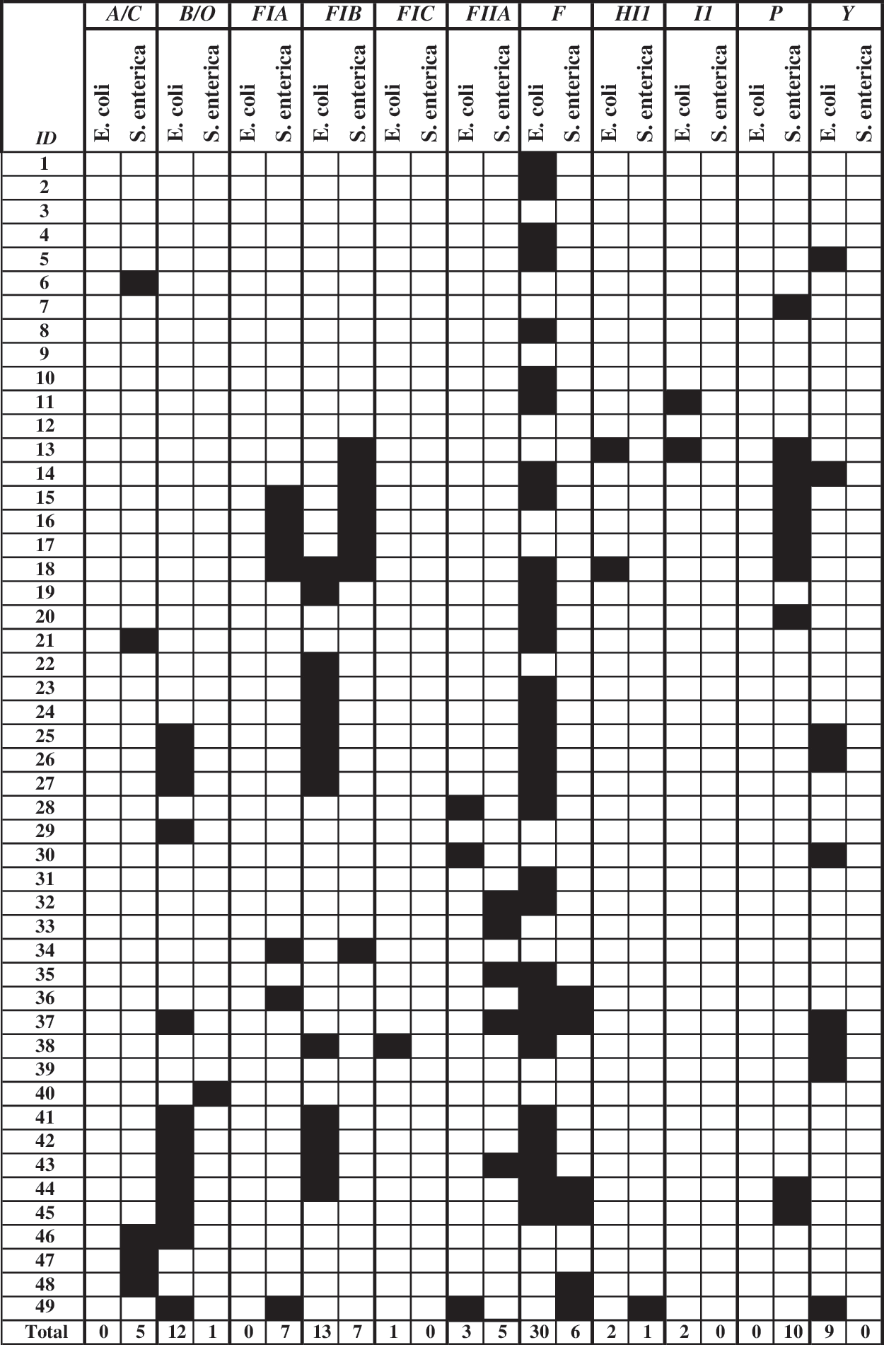
Black block indicates replicon detected; IncX, N, W, HI2, K, L/M, and T were not detected in any isolates.
Discussion
This study investigated genetic elements associated with AR shared by bacteria isolated from the same swine fecal sample. Samples were not randomly selected for this study and were chosen when E. coli, S. enterica, C. coli, and Enterococcus spp. were all successfully isolated, and were then selected to obtain the maximum number of AR phenotypes possible. Despite the selection criteria for the most resistances, overall the other phenotypic data are very similar to that found by the entire CAHFSE study (data available at
AR genes in the isolates were detected using a microarray allowing high-density analysis of each isolate with hundreds of AR gene probes (Frye et al., 2006, 2009; Zou et al., 2009). The genes detected were similar to those previously identified in swine isolates by other groups using PCR and sequencing methods as well as a few microarray studies (Gebreyes and Altier, 2002; Jackson et al., 2004b; Boerlin et al., 2005; Ma et al., 2007; Kozak et al., 2009). For example, E. coli isolates in the current study had positive hybridizations to probes for members of the aac, aad, aph, and str families of aminoglycoside resistance genes, including aac(3), aac(6), and aph3, which encode kanamycin resistance, and aadA1 and strA-strB, which encode streptomycin resistance. Many studies have established that these genes are often found in aminoglycoside-resistant E. coli isolated from pork, swine, or the swine environment (Sandvang and Aarestrup, 2000; Rosengren et al., 2009). β-Lactam resistance gene probes with positive hybridizations to the E. coli isolates included bla TEM, ampC, and bla CMY homologs, which is also similar to previous reports (Chen et al., 2005; Heider et al., 2009). One of the most often detected groups of genes encoded tetracycline resistance and included homologs of tet(A), tet(B), tet(C), and tet(R) resistance and regulatory genes (Boerlin et al., 2005). Genes for sulfamethoxazole and trimethoprim resistance were also detected in E. coli and included members of sulI/sul1, and sulII and dfrA1, and dhf families, respectively (Singh et al., 2005; Solberg et al., 2006; White et al., 2002). Also identified were integron, transposon, plasmid genes (e.g., intI1, tnpA, and repA), efflux genes, and sanitizer and metal resistance genes predominantly members of the qac, and mer families of efflux systems. Other studies of E. coli isolated from pork, swine, or swine production facilities found similar results (Bass et al., 1999; Walsh and Fanning, 2008). In cases where these studies investigated the genetic structure and specific mechanisms of resistance most were found to be associated with MDR plasmids, class I integrons, or a class I integron located on a plasmid (Bass et al., 1999; Sandvang and Aarestrup, 2000; White et al., 2002; Ajiboye et al., 2009; Fricke et al., 2009b). At least one plasmid replicon was also detected in 41 of the E. coli isolates. These were most often IncF plasmids, including FIB and F, and IncB/O was also detected in many isolates.
The Salmonella isolates in this study had resistance- and mobilization-associated genes detected similar to those found in E. coli. Some specific differences between Salmonella and E. coli were hybridizations of Salmonella DNA to a larger number and wider variety of β-lactam resistance genes. These included probes for bla CMY, bla SME, bla PSE, and bla SHV homologs. This is consistent with the greater number of β-lactam-resistant Salmonella isolates in this study as well as resistance to a wider variety of β-lactam antimicrobials in the isolates. These results are also comparable to other studies (Villa et al., 2000; Gebreyes and Altier, 2002; Gebreyes and Thakur, 2005; Frye and Fedorka-Cray, 2007; Heider et al., 2009). In previous studies when large numbers of resistances and resistance to extended-spectrum cephalosporins such as ceftiofur were detected, the resistant elements were often found to be encoded on IncA/C plasmids containing the bla CMY-2 gene, resulting in what has been termed the MDR-AmpC phenotype (Arlet et al., 2006; Frye and Fedorka-Cray, 2007; Zaidi et al., 2007; Zhao et al., 2007). The IncA/C replicon was detected in five Salmonella isolates, two of which also had the bla CMY gene detected. However, bla CMY was also detected in three other Salmonella that were not IncA/C replicon positive. These results and comparisons to our previous work suggest that the two bla CMY gene and IncA/C replicon-positive, ceftiofur-resistant isolates may harbor an MDR-AmpC IncA/C or related plasmid (Frye et al., 2006, 2009; Lindsey et al., 2009, 2010). The other three bla CMY-positive Salmonella had no plasmid replicons detected, and the gene may be located on an undetected plasmid or elsewhere in the genome. Some Salmonella isolates had other plasmid replicons detected, including FIA, FIB, FIIA, F, HI1, and P, several of which have been reported to carry AR (Carattoli et al., 2001, 2005, 2006; Debroy et al., 2010). In other swine studies, resistances were found to be located in Salmonella Genomic Island 1 (SGI1), which includes a class I integron encoding penta-resistance (Boyd et al., 2001; Doublet et al., 2003, 2004; Ebner et al., 2004; Fluit, 2005). Some Salmonella in the current study hybridized to gene probes found in many SGI1 variants, including aadA2, qacE, floR, tet(A) (which will also hybridize with the tet(G) gene of SGI1), bla PSE-1, sul1, intI1, and dfrA1 (Boyd et al., 2001; Doublet et al., 2003, 2004; Ebner et al., 2004; Fluit, 2005). Based on these results, many isolates in this study may harbor SGI1.
Many of the resistance genes were found to be shared among isolates from the same sample, but mostly among the closely related E. coli and Salmonella. Not only did these bacteria share many of the same resistance genes, but in almost half of the samples the genes were also shared at a significant level. These data confirm many other studies that have seen similar correlations; however, the large amount of information yielded by the current study is intriguing and may be evidence for a common source of resistances in E. coli and Salmonella, or for transfer of these MDR elements between these species. Genetic elements such as plasmids that could facilitate transferring AR between these bacteria were assayed for, but only detected in a few isolates. Common plasmids detected in both bacteria were F in four samples and FIB in one sample. Although these plasmids have been found to carry resistances, they are not as prevalent as others like IncA/C (Carattoli et al., 2001, 2005, 2006; Debroy et al., 2010). Many other plasmids were detected in both E. coli and Salmonella isolates, but not from the same sample. These data suggest that if exchange of the these genes occurred, in most cases the elements responsible for the transfer were not detected or were already lost in either the donor, the recipient, or both.
A recent study using similar methods to investigate dairy cattle isolates concluded that while common AR genes were detected in E. coli and Salmonella isolates, it was unlikely that multiple large groups of genes were exchanged between these bacteria. This was based upon hierarchical clustering of their AR microarray data that resulted in E. coli and Salmonella isolates forming separate clusters (Scaria et al., 2010). This is similar to the cluster analysis of our data where we also found that isolates grouped into clusters based upon species rather than sample. However, some probes on our array are relatively species specific; thus, even when many common resistance genes are detected in different species, the isolates will still be clustered by the species-specific genes detected because these will be the discriminating factors used by the cluster analysis to group the isolates. Additionally, in our study the microarray also detected common AR mobilization-associated elements such as class I integrons, transposons, and IS elements, which were not assayed by the microarray used in the dairy cattle study (Scaria et al., 2010). Although these elements cannot transfer between bacteria, they are often transferred by conjugal elements to other bacteria where they can integrate into their new host's DNA (Nemergut et al., 2008). Detection of these genes in conjunction with AR genes suggests that integrons in our isolates could have been mobilized by a transfer element between E. coli and Salmonella, followed by the loss of the transfer element. However, it is necessary to avoid over interpretation of the significance at which common genes were detected in E. coli and Salmonella. For example, many of the isolates from different samples also share genes that implicate a common source for resistance genes in the swine environment, or historical exchange of MDR elements rather than recent events or exchange within the animals from which the fecal droppings were collected.
Resistance genes detected in Campylobacter and Enterococcus were also similar to what was found in previous swine studies. The most prevalent resistance genes found in C. coli isolates were tet(O), aphA-3, and cam-1 encoding tetracycline resistance, aminoglycoside resistance, and an endogenous Camplyobacter β-lactamase, respectively (Aarestrup et al., 2000; Jackson et al., 2004b; Thakur and Gebreyes, 2005). The Enterococcus spp. isolates had a far greater number of resistance genes detected, including aad, aac, aphA-3, tet(M) and tet(O) homologs, ermB, vanC, and transposon-related mobilization elements, tnpA, insA, and trans-1, many of which have been previously described (Jackson et al., 2004b). Enterococcus and Campylobacter isolates both hybridized to probes for tet(O). However, in most cases Enterococcus isolates hybridized to both tet(O) and tet(M) probes, whereas C. coli isolates hybridized almost exclusively to the tet(O) probe (Aarestrup et al., 2000; Gibreel et al., 2004; Thakur and Gebreyes, 2005). This is evidence for sharing homologous but not identical tetracycline resistance genes in these bacteria. Moreover, comparison of the C. coli tet(O) sequence in GenBank to the Enterococcus spp. tet(M) sequence found 79% homology by BLAST analysis.
The effects of selective pressure on development, maintenance, and transfer of resistance and MDR elements are well documented in the literature. One of the observations in this study was that sanitizing agent and metal resistance genes were detected in E. coli and Salmonella isolates. These resistance genes are often found to be genetically linked to AR genes by residing on the same genetic element. It is possible that the utilization of sanitizers and metal containing compounds as well as other practices or natural events in the animal environment could be selecting for AR phenotypes in the absence of selection by antibiotics (Fricke et al., 2009a, 2009b; Lindsey et al., 2009; Welch et al., 2009). This could have profound effects on AR bacteria where production practices have begun to rely on metal compounds in feed as an alternative to antimicrobials or in swine facilities and processing plants where sanitizers like quaternary ammonium compounds are used to reduce bacterial contamination in pens, on processing surfaces, or on meat products (Hasman and Aarestrup, 2002; Aarestrup and Hasman, 2004; Pettigrew, 2006). If genetic resistance elements to these compounds are linked to AR genes, withdrawal of antimicrobials may not reduce AR in the bacteria of concern.
This study found that some common resistance genes were shared by isolates from the same swine fecal sample. This was especially true for E. coli and Salmonella, and more rarely for C. coli and Enterococcus spp. possibly indicating that exchange occurs more often between closely related bacteria. A few potential vehicles for resistance gene transfer in E. coli and Salmonella were detected as well as genetic elements associated with mobilization of AR, such as integrons and transposons. Further studies will be required to confirm this exchange, identify the transfer elements, and determine whether AR gene transfer took place recently in the swine environment. Additional analysis of MDR in Salmonella and E. coli animal isolates will allow us to investigate the development, evolution, and spread of MDR genetic elements in animal environments, processing facilities, and food products. This information will improve our understanding of the mechanisms driving AR and enable the development of strategies to reduce AR and promote human and animal health.
Footnotes
Acknowledgments
The authors would like to thank all CAHFSE partners, especially David A. Dargatz, Charles A. Haley, and Christine A. Kopral with the APHIS, USDA, without whom samples could not have been collected. The authors thank Georgina Hidalgo, Alice Wilcher, Mike Asher, Russ Turpin, Jonathan Cudnik, Jovita Haro, Jodie Plumblee, Sandra House, Lari Hiott, and Takiyah Ball for technical assistance.
Disclosure Statement
No competing financial interests exist.
Disclaimer
Mention of trade names or commercial products in this article is solely for the purpose of providing specific information and does not imply recommendation or endorsement by the U.S. Department of Agriculture.
References
Supplementary Material
Please find the following supplemental material available below.
For Open Access articles published under a Creative Commons License, all supplemental material carries the same license as the article it is associated with.
For non-Open Access articles published, all supplemental material carries a non-exclusive license, and permission requests for re-use of supplemental material or any part of supplemental material shall be sent directly to the copyright owner as specified in the copyright notice associated with the article.